2. species. As a conclusion, we propose a revised systematic classifi-
cation of the Cronobacter serotypes. We believe that our results
also unify current Cronobacter lipopolysaccharide and O-PS
chemical structure research.
MATERIALS AND METHODS
Bacterial strains. The bacterial cultures used in this study were provided by
theAmericanTypeCultureCollection(ATCC;Manassas,VA,USA)andThe
Czech Collection of Microorganisms (CCM; Brno, Czech Republic). Some
isolates ofCronobacter spp. were kindly donated by Carol Iversen (Univer-
sity of Dundee), Hana Drahovská (Comenius University, Bratislava, Slo-
vakia), and Igor Hochel (University of Chemistry and Technology,
Prague, Czech Republic). Details of the strains are summarized in Table 1,
and the strains are characterized in Table S1 in the supplemental material.
The provisional Cronobacter species identity was based on the species-
specific PCR system targeted to the rpoB gene (16) and confirmed using
partial sequence analysis of the elongation factor G gene (fusA) (17, 18).
DNA was extracted using a GenElute bacterial genomic DNA kit (Sigma-
Aldrich),viatheGram-negativebacterialprotocol.Inaddition,thebiochem-
ical characterization (data not shown) was performed by the use of an ID 32E
test system (bioMérieux), and more-precise biochemical testing was accom-
plished using conventional tube methods as described previously (1, 19).
Genomes. In silico analyses were carried out using (n ϭ 30) Cronobacter
genomes accessible at GenBank (http://www.ncbi.nlm.nih.gov/genome
/?termϭcronobacter) (20) and the PubMLST Cronobacter database (www
.pubmlst.org/cronobacter/) (21). The latter hosts newer assemblies of the
GenBank entries. The Cronobacter genomes included in the study were C.
sakazakii 680, 696, 701, 2151, 8399, HPB 5174, NCIMB 8272, SP291,
NBRC 102416, ATCC BAA-894, ES15, ES713, ES35, E764, and E899; C.
malonaticus 507, 681, LMG 23826, and CMCC 45402; C. turicensis z3032,
564, and z610; C. condimenti 1330; C. dublinensis 582 and 1210; C. dubli-
nensis subsp. dublinensis LMG 23823; C. dublinensis subsp. lactaridi LMG
23825; C. dublinensis subsp. lausannensis LMG 23824; C. muytjensii ATCC
51329; and C. universalis NCTC 9529 (Table 2).
Restriction fragment length polymorphism analysis of O-antigen
gene cluster amplicons by use of MboII (OAg-RFLP). Three pairs of
primers (see Table S2 in the supplemental material) for amplification of
the O-antigen gene cluster (10, 13, 15) were evaluated in silico using Fast
PCR software (PrimerDigital Ltd.). Their complementarity was tested on
a set of available genomes (Table 2).
RFLP analysis of the O-antigen gene cluster was performed with all 82
strains (10). Primers were targeted to the JUMPStart site and gnd gene.
The PCR products were digested with MboII. The resulting mixtures were
separated using a 1.5% agarose gel and analyzed by BioNumerics 6.4 (Ap-
plied Maths) (Table 3).
Serotyping PCRs. Eleven PCR protocols for Cronobacter serotyping
were applied. These methods were targeted to different serotypes and
performed according to previously published protocols as follows: (i) C.
sakazakii serotypes O1 to O7 (heptaplex, SHO1 to SHO7) (12), (ii) C.
sakazakii O4 (SO4) (14), (iii) C. malonaticus O2 (MaO2) (13), (iv) C.
muytjensii O2 (MuO2) (14), (v) C. turicensis O2 (TO2) (11), (vi) C. turi-
censis O3 (TO3) (14), (vii) C. dublinensis O1 (DO1) (14), (viii) C. dubli-
nensis O2 (DO2) (14), (ix) C. universalis O1 (UO1) (14), (x) C. turicensis
O1 and C. malonaticus O1 combined (TO1/MaO1) (13), and (xi) C. saka-
zakii O3 and C. muytjensii O1 combined (SO3/MuO1) (13) (see Table S2
in the supplemental material).
Lipopolysaccharide structures. In this work, lipopolysaccharides are
compared with their assigned serogroups. Structures of lipopolysaccha-
rides described in the literature were discussed. The structures of the avail-
able serotypes were as follows: C. sakazakii O1 (22), O2 (23–25), O3 (26),
O4 (27, 28), O6 (28), and O7 (29); C. malonaticus O1 (27), O2 (30), and
O4 (11); C. turicensis O4 (31); C. dublinensis O1 (32) and O3 (33); C.
muytjensii O1 (34); C. universalis O1 (35); and E. asburiae (36) (see Table
S3 in the supplemental material).
Isolation of lipopolysaccharides. LPS was prepared following the
method described by Jaradat et al. (37) with minor modifications. Briefly,
cells (250 ml) were harvested in the exponential phase of growth by cen-
trifugation (5,000 ϫ g, 10 min), washed 3 times, and resuspended in 5 ml
of 50 mM sodium phosphate buffer (pH 7.0). The cells were sonicated for
30-s intervals for 5 min at 9 to 12 W. The suspension was incubated first
with 20 g/ml of proteinase K in 0.1 M Tris-HCl (pH 8.0) at 60°C for 1 h
and second with pancreatic RNase and DNase (0.1 g mlϪ1
) in 20 mM
MgCl2 at 37°C for 10 min. Inactivation of enzymes was performed at 60°C
for 10 min, and the obtained suspension was mixed with an equal volume
of preheated phenol. After incubation (70°C for 15 min) with occasional
mixing was performed, the mixture was centrifuged (18,000 ϫ g, 1 h) and
the resulting aqueous layer was collected and dialyzed using dialysis tub-
ing with molecular weight (MW) cutoff values of 6,000 to 8,000 at room
temperature against distilled water until no detectable phenol odor re-
mained. The samples were then lyophilized and stored at Ϫ20°C until
used. The extracted LPSs from Cronobacter were separated using both
sodium dodecyl sulfate-polyacrylamide gel electrophoresis and sodium
deoxycholate-polyacrylamide gel electrophoresis (SDS-PAGE and DOC-
PAGE, respectively).
Software. For the in silico analyses, FastPCR 6.1 (27) and Genetyx 5.2
(Genetyx Corporation) were used. RFLP profiles were analyzed using the
TotalLab TL100 software (Nonlinear USA Inc.).
RESULTS
Restriction fragment length polymorphism of O-antigen gene
cluster. The primers (see Table S2 in the supplemental material)
previously described for the amplification of complete O-antigen
clusters (sequencing primers) (10, 13, 15) of Cronobacter strains
were tested in silico. The emphasis was put on the sensitivity of
sequencing primers. The primers were compared to the full-ge-
nome sequences of 30 Cronobacter strains representing the whole
genus (Tables 2 and 4).
From three pairs of the sequencing primers (10, 13, 15) (see
Table S2 in the supplemental material), the primer pair described
by Sun et al. (10) was predicted to amplify the targeted gene cluster
from most genomes (73%) (Table 2). However, seven strains of C.
sakazakii and one of C. condimenti were predicted to produce no
amplicon with this primer pair.
Subsequently, this set of primers was chosen for the restriction
fragment length polymorphism analysis of 82 Cronobacter strains.
Amplified O-antigen gene products, with a size range of from 7 to
12.5 kbp, were detected in all strains (Table 3; see also Fig. S1 in the
supplemental material). Amplicons were digested with MboII and
separated by gel electrophoresis. The restriction digest profiles
produced from the tested strains were clear and reproducible, giv-
ing from 8 to 18 restriction DNA fragments with sizes ranging
from 150 to 2,800 bp (Table 3; see also Fig. S1). The obtained
profiles were grouped into 17 distinguishable genotypes desig-
TABLE 1 Summary of strains used in this study
Bacterial
species
No. of
strains fusA allele(s)
No. of clinical
isolates
C. condimenti 1 86 0
C. dublinensis 12 20, 21, 30, 46, 48 1
C. malonaticus 9 7, 13 4
C. muytjensii 7 24, 35, 64 1
C. sakazakii 42 1, 3, 8, 9, 15, 17, 18 15
C. turicensis 8 22, 28 2
C. universalis 3 19, 32, 34 1
E. cloacae 1
E. aerogenes 1
Diversity of O Antigen within Genus Cronobacter
August 2015 Volume 81 Number 16 aem.asm.org 5575Applied and Environmental Microbiology
onAugust4,2015byguesthttp://aem.asm.org/Downloadedfrom
3. nated A to Q and Y (Tables 3 and 4; see also Table S1). Some of the
characteristic profiles (A, B, C, D, E, G, H, I, J, K, and M) were
identical to those previously reported (10, 13–15) (Table 4). Four
previously described profiles (C. sakazakii O5 and O7, C. ma-
lonaticus O1, and C. muytjensii O1) were not detected within our
strain collection. In contrast, we detected six new profiles, five of
which were as follows: C. turicensis (F), C. dublinensis (L and Q),
C. universalis (P), and C. condimenti (N). The sixth profile, that of
strain Cb43 (CDC9369-75), is very similar to that of C. sakazakii
O2 (B), with few significant differences (lacking 1,665-bp and
975-bp bands and possessing 1,545-bp and 870-bp bands). This
was probably caused by a mutation within the O-antigen gene
cluster and was therefore given a different profile designation (Y).
Serotype-specific PCRs. The primers (see Table S2 in the sup-
plemental material) previously described for the amplification of
the genes from the O-antigen cluster (10–14) of Cronobacter were
TABLE 2 In silico analysis of sequencing and serotyping primers carried out using publically available Cronobacter genome sequences
Bacterial species Isolate GenBank accession no.
MLST
sequence
type (ST/CC)
Size (bp) of amplified O-
antigen cluster according to: Serotyping PCR result(s)
Mullane
et al.a
Sun
et al.b
Jarvis
et al.c
Heptaplexd
Duplexe
Individualf
C. sakazakii ES15 NC_017933 125 12,660 10,684 11,418 SHO1
680 NZ_CALG01000025 8 SHO1
NBRC 102416 NZ_BAWU00000000 8 12,370 10,395 11,418 SHO1
ATCC BAA-894 NC_009778 1 12,371 10,395 11,771 SHO1
ES35 NZ_AJLC00000000 8 SHO1
NCIMB 8272 NZ_AWFW00000000 4 12,660 10,684 11,708 SHO2
SP291 NC_020260 4 12,660 10,684 11,708 SHO2
701 NZ_CALE01000616 4 12,723 10,716 11,771 SHO2
8399 NZ_AWSP00000000 4 12,660 10,684 11,708 SHO2
2151 NZ_AJKT00000000 4 12,660 10,684 11,708 SHO2
ES713 AJLB00000000 4
E764 NZ_AJLA00000000 73 SHO4 SO4
696 NZ_CALF00000000 12 22,276 SHO4 SO4
HPB5174 NZ_JNBN00000000 45 SHO4 SO4
E899 NZ_AFMO00000000 4
C. malonaticus 507 NZ_CALD00000000 11 14,356 12,378 13,404 SHO5
681 NZ_CALC00000000 7 9,608 7,614 8,656 SHO6 MaO2
CMCC 45402g
NC_023032 7 9,396 7,386 8,444 SHO6 MaO2
LMG 23826 NZ_AJKV00000000 7 7,387 SHO6 MaO2
C. turicensis z3032 NC_013282 24 14,484 12,463 13,532 TO1/MaO1
564 NZ_CALB00000000 5 10,792 8,814 9,840
C. condimenti 1330 NZ_CAKW00000000 98
C. dublinensis 582 NZ_CALA00000000 80 7,641 DO2
1210 NZ_CAKZ00000000 106 13,357 11,380 DO1
C. dublinensis subsp.
dublinensis
LMG 23823 NZ_AJKZ00000000 106 13,362 11,383 12,410 DO1
C. dulinensis subsp.
lactaridi
LMG 23825 NZ_AJKX00000000 79 12,374 10,396 11,423 DO1
C. dublinensis subsp.
lausannensis
LMG 23824 NZ_AJKY00000000 80 7,423 11,423 DO2
C. muytjensii ATCC 51329 NZ_AJKU00000000 81 12,305 13,328 MuO2
C. universalis NCTC 9529 NZ_CAKX00000000 54 11,781 9,828 10,829 UO1
NCTC 9529/2 NZ_AJKW00000000 54 11,782 9,829 10,830 UO1
a
2008 (15).
b
2011 (10).
c
2011 (13).
d
Probe targeted to SHO1 to SHO7 (heptaplex), Sun et al., 2011 and 2012 (10, 12).
e
Probe targeted to C. turicensis and C. malonaticus (TO1/MaO1) (duplex) and C. sakazakii and C. muytjensii (SO3/MuO1) (duplex), Jarvis et al., 2011 (13).
f
Probe targeted to C. malonaticus (MaO2), Jarvis et al., 2011 (13), and to C. dublinensis (DO1 and DO2), C. sakazakii (SO4), C. turicensis (TO3), C. muytjensii (MuO2), and C.
universalis (UO1), Jarvis et al., 2013 (14), and Sun et al., 2012 (11).
g
Originally identified as C. sakazakii.
Blažková et al.
5576 aem.asm.org August 2015 Volume 81 Number 16Applied and Environmental Microbiology
onAugust4,2015byguesthttp://aem.asm.org/Downloadedfrom
4. tested in silico using the full sequences of 30 Cronobacter genomes
(Tables 2 and 4). Twenty-six genomes gave predicted positive re-
sults. However, primers specific for C. sakazakii O3 and O7, C.
turicensis O2, and C. sakazakii O3/C. muytjensii O1 were predicted
to give negative results for all tested genomes. The published se-
rogroup-specific assays (11–15) (see Table S2) were then applied
to 82 Cronobacter strains (see Table S1). The specificity of each of
the primer pairs was cross-tested with strains in the corresponding
serotype (Table 3 and 4). Briefly, the majority (78/82) of strains
gave positive results, with only primers specific for C. sakazakii O5
and O7 and C. turicensis O2 giving negative results. The data from
the in silico analysis and experimental results were collated and are
discussed below.
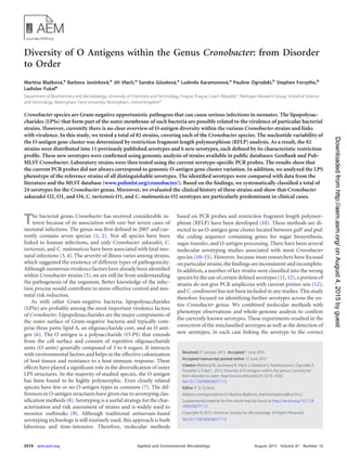
Analysis of LPS patterns. O-antigen variations are responsible
for the well-known ladder-like patterns of LPS molecules on poly-
acrylamide gels. The LPS patterns were determined for 37 selected
strains, including representatives of all serogroups that were dis-
tinguishable with respect to RFLP profiles (Fig. 1). Each group of
strains defined by RFLP profiles demonstrated one characteristic
LPS pattern. All strains showed typical smooth LPS patterns con-
taining lipid A and core (located toward the end of each gel) and
O-antigen sugar repeats, with the exception of one: strain Cb 43
produced two bands only in the lower part of the gel that were
characteristic of the lipid A-oligosaccharide core (lipid A-core)
and of the lipid A-core with only a short O-antigen polysaccharide
or single O-unit molecule (lipid A-core plus one O unit) without
any repetitive subunits in the upper part of the gel (Fig. 1).
For a polyphasic analysis of Cronobacter serotype groups, the
TABLE 3 Results of serotyping PCR and characteristic MboII restriction profiles of amplified O-antigen gene clusters
a
Serotypes in bold represent newly identified RFLP profiles.
b
PCR protocols were based on the literature as follows: C. sakazakii serotypes 1 to 7 (heptaplex, O1-SHO7) (12), C. sakazakii O4 (14), C. malonaticus O2 (13), C. muytjensii O2
(14), C. turicensis O2 (11), C. turicensis O3 (14), C. dublinensis O1 (14), C. dublinensis O2 (14), C. universalis O1 (UO1) (14), C. turicensis O1 and C. malonaticus O1 together (13),
and C. sakazakii O3 and C. muytjensii O1 together (13). S, sakazakii; Ma, malonaticus; Mu, muytjensii; D, dublinensis, T, turicensis, U, universalis.
c
Unexpected size of the PCR product (890 bp).
d
The restriction profile was created by Genetyx software using the sequence of C. malonaticus strain G2706.
FIG 1 Electrophoretic patterns of LPS from Cronobacter strains. (Left) Silver-stained DOC-PAGE of LPS patterns of the strains of genus Cronobacter. Lane 1,
marker (Page Ruler Plus prestained protein ladder); lane 2, Cb28 (C. sakazakii O1, D); lane 3, Cb24 (C. sakazakii O2, B); lane 4, Cb05 (C. sakazakii O2, B); lane
5, Cb43 (Y); lane 6, Cb08 (C. sakazakii O3, I); lane 7, Cb26 (C. sakazakii O4, C); lane 8, Cb01 (C. malonaticus O2, M); lane 9, Cb55 (C. muytjensii O2, K); lane 10,
Cb79 (C. muytjensii O2, K); lane 11, Cb78 (C. condimenti O1, N); lane 12, Cb53 (C. turicensis O1, G); lane 13, Cb73 (C. turicensis O5, F); lane 14, Cb75 (C.
turicensis O5, F); lane 15, Cb71 (C. turicensis O3, A); lane 16, Cb59 (C. universalis O1, H); lane 17, Cb70 (C. universalis O2, P); lane 18, Cb69 (C. dublinensis O2,
E); lane 19, Cb62 (C. dublinensis O1b, L); lane 20, Cb56 (C. dublinensis O1a, J); lane 21, Cb13 (C. dublinensis O4, Q). Corresponding serotypes and restriction
profiles are presented in the brackets. (Right) Silver-stained SDS-PAGE of LPS patterns of the strains of genus Cronobacter. Lane 1, marker (Page Ruler Plus
prestained protein ladder); lane 2, Cb43 (rough type of C. sakazakii O2); lane 3, empty; lane 4, standard of smooth type of LPS (E. coli E 0111); lane 5, standard
of rough type of LPS (E. coli EH 100).
Diversity of O Antigen within Genus Cronobacter
August 2015 Volume 81 Number 16 aem.asm.org 5577Applied and Environmental Microbiology
onAugust4,2015byguesthttp://aem.asm.org/Downloadedfrom
6. laboratory results and additional information from the literature
have been collated. The RFLP restriction digestion profiles were
compared with LPS patterns and PCR profiling results. These lab-
oratory results were then evaluated with existing metadata about
the strains: the incidence in the Cronobacter PubMLST database,
source, O-antigen chemical structures, genome sequences, and
O-antigen gene cluster description. On the basis of this polyphasic
investigation, we proposed a systematic classification of the cur-
rently known serotypes within the genus Cronobacter (Table 4).
DISCUSSION
Serotyping is a useful method for characterization of Gram-neg-
ative bacteria. Several studies focused on the O-antigen classifica-
tion within the Cronobacter genus have been reported, including
molecular methods (12–15) as well as structural studies (12, 22,
25, 34). Until now, there have been 17 recognized serotypes
among the Cronobacter species. However, some serotypes were
defined using culture collections with strains identified by 16S
rRNA and biochemical profiling, neither of which can accurate
identify Cronobacter species (10, 17). In contrast, the multilocus
sequence type (MLST) analysis of Cronobacter strains is supported
by whole-genome phylogeny (39–41).
In this study, different serotyping techniques were used to an-
alyze 82 different Cronobacter strains representing all currently
known species which have been identified based on DNA se-
quencing and reference to the open-access PubMLST Cronobacter
database (http://pubmlst.org/cronobacter/ [accessed 31 Decem-
ber 2014]) with over 1,000 described strains and metadata (39).
The genotypes of the O-antigen gene clusters of the strains were
evaluated by restriction analysis and PCRs using the O-antigen
processing genes, and LPS patterns were determined in chosen
strains (Fig. 1). The obtained results were compared with pub-
lished data, including the open-access Cronobacter PubMLST da-
tabase (Table 4).
Serotyping of C. sakazakii. Within the C. sakazakii species, C.
sakazakii O1 (SO1) and SO2 were the predominant serotypes;
SO3 and SO4 were seen less often. Furthermore, the percentage of
clinical isolates within particular serotypes was considered by ref-
erence to the Cronobacter PubMLST database. Almost 50% of C.
sakazakii O2 strains from the database originated in clinical sam-
ples, and 90% are sequence type (ST) 4. This particular sequence
type is of importance as it is the predominant C. sakazakii neonatal
meningitis pathovar (42). This implies the association of this se-
rotype with neonatal health risk. C. sakazakii O4 strains had a
higher incidence of clinical samples (35%) than C. sakazakii O1
strains (12%) and C. sakazakii O3 strains (0%).
SO5, SO6, and SO7 were not detected in the identified C. saka-
zakii strains in the DNA sequence base.
The structures of O antigen have been determined for C. saka-
zakii serotypes O1, O2, O3, O4, and O7 (Table 4). These contain
between five and seven saccharides in each O-antigen unit (12, 22,
24–26, 28, 29). They all produce a branched form of O antigen; the
most frequent serotypes, C. sakazakii O1 and O2, have two sac-
charides in the branching fragment of the O antigen, whereas the
rest have only one.
Serotyping of C. malonaticus. Within the C. malonaticus spe-
cies, two serotypes (MaO1 and MaO2) were previously described
and we propose two new ones: C. malonaticus O3 (MaO3) and O4
(MaO4). C. malonaticus O1 shares sequence similarities with C.
turicensis O1, Shigella dysenteriae D11, and Escherichia coli O29,
a
PCRprotocolswerebasedontheliteratureasfollows:C.sakazakiiserotypes1to7(heptaplex,SHO1toSHO7)(12),C.sakazakiiO4(SO4)(14),C.malonaticusO2(MaO2)(13),C.muytjensiiO2(MuO2)(14),C.turicensisO2(TO2)
(11),C.turicensisO3(TO3)(14),C.dublinensisO1(DO1)(14),C.dublinensisO2(DO2)(14),C.universalisO1(UO1)(14),C.turicensisO1andC.malonaticusO1together(TO1/MaO1)(13),andC.sakazakiiO3andC.muytjensiiO1
together(SO3/MuO1)(13).
b
Analysiswasperformedonthepublishedgenomes(38).
c
Analysiswasperformedonall82strainsofCronobacter.
d
Thenumbersofthestrainsofclinicaloriginareshowninparentheses.
e
Characteristicsfromtheliterature(10–15).
f
RFLPprofilesandserotypesinboldarenewlyidentified.
g
Datarepresentcharacteristicsreportedintheliterature(10–14,22–31,34–36).ThestructureshighlightedingraybelongtoC.dublinensisserotypeO1withoutrecognitionbetweenDO1aandDO1b.
h
qtyS,quantityofsugarsinO-antigenunit.
i
qtyB,quantityofsugarsinthebranchingpartoftheO-antigenunit.
j
On,previouslyunknownserotype.
k
O?,previouslyunknownserotype,proposedserotype.
l
RFLPprofileofthestrainwithroughtypeoflipopolysaccharides.
m
X,cancelledserotype.
n
Incorrectlyidentifiedstrain;theMLSTdatabaseidentifieditasC.malonaticus.
o
RFLPprofilecreatedbytheuseofGenetyxsoftware.
p
L,linear,B,branched.
Diversity of O Antigen within Genus Cronobacter
August 2015 Volume 81 Number 16 aem.asm.org 5579Applied and Environmental Microbiology
onAugust4,2015byguesthttp://aem.asm.org/Downloadedfrom
7. suggesting related ancestries (31). The branched type of O-antigen
structure has been previously reported for the C. malonaticus O2
serotype (27). Approximately half of the C. malonaticus O2 strains
are of clinical origin (n ϭ 58) (39).
Discrepant results were observed between the species desig-
nation and serotype C. sakazakii O6. Only three strains in the
PubMLST database are categorized as C. sakazakii O6. All three
are C. sakazakii isolates from deli meat in China. In silico analysis
showed positive results with two genomes from the species C.
malonaticus (LMG 23826 and 681), which was consistent with
obtained experimental data. All of our C. sakazakii strains gave
negative results with C. sakazakii O6-specific primers; moreover,
all of our C. malonaticus strains (identified reliably as C. malonati-
cus O2) showed the same O-antigen gene cluster profile (M) and
gave positive results with this serotyping PCR. Hence, C. sakazakii
serotype O6 should be regarded as identical to C. malonaticus O2.
The reassignment of C. sakazakii O5 and O6 to C. malonaticus
is due to the apparent initial incorrect species identification by
Sun et al. (10). The original G2706 and G2708 strains used to
define the C. sakazakii O5 serotype were originally identified to the
species level using 16S rRNA sequencing and biochemical profil-
ing; both procedures are now recognized as being prone to errors
(17). According to MLST analysis, both strains are C. malonaticus.
With respect to these findings, we propose to reclassify these C.
sakazakii O5 strains as C. malonaticus O3 based on the C. ma-
lonaticus 507 reference strain (restriction profile Z generated in
silico; Cronobacter PubMLST genome sequenced strain ID74).
Furthermore, we propose the new serotype C. malonaticus O4
(MaO4), as specified by the G3882 reference strain. This strain was
previously assigned as C. turicensis O2 (11); however, according to
the Cronobacter PubMLST database, strain G3882 is C. malonati-
cus (ID949, sequence type 371). The O-antigen gene sequence,
specific PCR results, and even the structure of the O-polysaccha-
ride for this strain have already been described (11) (http:
//pubmlst.org/cronobacter/). Both C. malonaticus O3 and O4
have the linear type of O antigen (11, 12).
Serotyping of C. turicensis. The species C. turicensis contains
at least three serotypes. It is noteworthy that the LMG 23827T
reference strain for C. turicensis O1 (TO1) was isolated from blood
from the fatal infant meningitis case and also that two of our three
C. turicensis O1 strains were clinical isolates. C. turicensis O3
(TO3) was detected in three strains, which were also positive with
C. sakazakii SHO5. This can be explained by the reclassification of
strain G4105 (identified by Sun et al. [10] as C. sakazakii O5) as a
C. turicensis strain as given on the MLST database (http://pubmlst
.org/cronobacter/). Though the described O-antigen gene clusters
of C. malonaticus O3 and C. turicensis O3 share high sequence
similarity (12, 14), they have different restriction profiles (Z and
A, respectively) (10). To make this clear, the chemical structures of
the corresponding O-antigens would be supportive, but unfortu-
nately they have not been reported as yet.
Recently, Czerwicka et al. (31) published the only O-antigen
structure of three strains of C. turicensis. This is a branched type of
O-antigen with only four saccharides. Based on their results, the
new serotype C. turicensis O4 (TO4) should be recognized. The
new serotype is specified by the reference strain C. turicensis 564
(genome sequenced; Cronobacter PubMLST strain ID82) and has
the structure published by Czerwicka et al. (31). A further C. tu-
ricensis serotype (O5) is proposed for reference strain E676, with a
defined RFLP profile (profile F) and LPS pattern (Table 3 and
Fig. 1).
In the literature, one more O-PS structure was reported for C.
turicensis HPB 3287 (36). It was, however, later identified as En-
terobacter asburiae, as given in the Cronobacter PubMLST database
(http://pubmlst.org/cronobacter/).
Serotyping of C. dublinensis. For C. dublinensis, two sero-
types, C. dublinensis O1 (DO1) and DO2, have already been re-
ported (14). Both serotypes were identified among our C. dublin-
ensis strains. The C. dublinensis O1 serotyping PCR recognized
strains with two types of O-antigen gene profiles (profiles J and L).
Since those strains produced different lipopolysaccharide profiles
(Fig. 1, lanes 20 and 19), this serotype should be subdivided as
DO1a and DO1b, respectively. Two similar O-PS structures have
been published for C. dublinensis O1 (32). Interestingly, the C.
dublinensis serotypes mentioned above corresponded to the pre-
vious distinction of C. dublinensis subspecies: C. dublinensis O1a
correlates with C. dublinensis subsp. lactaridi, C. dublinensis O1b
with C. dublinensis subsp. dublinensis, and C. dublinensis O2 with
C. dublinensis subsp. lausannensis (1).
MacLean et al. characterized the O-antigen structure of C. dub-
linensis strain HPB 3169 (33). This O antigen is structurally similar
to that recently reported for Cronobacter malonaticus O3 strain
G2706 (28). Due to the significant differences in the O-antigen
gene sequence, it should be a new serotype, C. dublinensis O3
(DO3), specified by its O-antigen structure (33).
One of our C. dublinensis strains (Cb13) did not give a positive
result in any serotyping PCR and had its own restriction profile
(Q) and LPS pattern. This suggests that it could be a member of
another C. dublinensis serotype, provisionally designated DO4
here; however, we do not have enough information to further
classify this serotype.
Serotyping of C. muytjensii. Two serotypes (13, 14) have been
described within the C. muytjensii species: O1 (MuO1) and O2
(MuO2). All C. muytjensii strains in this study were MuO2 and
were assigned RFLP profile K.
Serotyping of C. universalis and C. condimenti. There were
two RFLP restriction profiles (profiles H and P) for C. universalis.
The two strains had previously been reported as C. universalis O1
(UO1) (35). The new serotype was classified as C. universalis O2
(UO2). The only strain of C. condimenti has its specific O-antigen
gene profile (N) and LPS pattern, supporting assignment of its
own serotype, C. condimenti O1 (CO1).
Occurrence of complete and incomplete LPSs within Crono-
bacter strains. In the literature, there is limited information on
the occurrence of strain variants in the genus Cronobacter. Mul-
lane et al. (15) reported two rough-colony strains: strain CFS1001
of C. sakazakii O1 and strain E892 of C. sakazakii O2. These strains
produced restriction fragments of the O-antigen gene cluster
identical to those of other (smooth) strains, namely, C. sakazakii
O1 and O2, respectively. The mutations responsible for the rough
LPS phenotype are often localized outside the O-antigen gene
cluster (30, 38). Therefore, the RFLP of the O-antigen gene cluster
may not indicate whether the isolate exhibits smooth or rough
LPS, as was the case with strains CFS1001 and E892 (both exhib-
iting rough LPS) reported by Mullane et al. (15).
In our study, all strains except one (CDC9369-75) had full-
length LPSs (Fig. 1). The LPS pattern indicated a defect in the
O-antigen polymerization in this strain (43, 44). Correspond-
ingly, its O-antigen RFLP profile (Y) was very similar but not
Blažková et al.
5580 aem.asm.org August 2015 Volume 81 Number 16Applied and Environmental Microbiology
onAugust4,2015byguesthttp://aem.asm.org/Downloadedfrom
8. identical (see Results and Table 3) to the profile characteristic of C.
sakazakii O2 (B), suggesting the existence of a mutation in the
O-antigen cluster. This finding was supported also by the results
from serotyping heptaplex PCR. The Cb43 strain gave positive
results with the primer pair specific to C. sakazakii heptaplex O2
(SHO2); however, the size of the product was 900 bp instead of 152
bp (specific size for all other C. sakazakii O2 strains). Remarkably,
this primer pair is targeted to the wzy gene for the O-antigen
polymerase that mediates the transfer of nascent polymer to the
new O unit. Strains with mutations in this gene are observed to
produce semirough LPS (38). It is therefore probable that the
Cb43 strain shows a semirough LPS phenotype. To confirm our
observations, molecular studies of the genes involved in the LPS
biosynthesis, especially studies of the O antigen and the core,
would be appropriate.
Conclusion. To the best of our knowledge, this report provides
the first polyphasic description of serotype diversity across the
entire Cronobacter genus. Several genotyping and phenotyping
techniques were successfully combined to separate Cronobacter
strains into serotype clusters. We compared our results with those
reported in the literature and with the data from the curated open-
access Cronobacter PubMLST database. From the information ob-
tained, 24 Cronobacter serotypes are now described. This includes
7 new serotypes. These serotypes have been characterized by the
RFLP profiles of the O-antigen gene clusters and LPS patterns.
ACKNOWLEDGMENTS
This work was supported by the Czech Grant Agency (project no. P503/
12/P704 and project no. 13-23509S).
We thank C. Iversen from University College Dublin, H. Drahovská
from Comenius University in Bratislava, Slovakia, and I. Hochel from the
University of Chemistry and Technology in Prague, Czech Republic, for
providing some bacterial strains used in this study.
This publication made use of the Cronobacter MLST website (http:
//pubmlst.org/cronobacter/) developed by Keith Jolley and sited at the
University of Oxford (21). The MLST scheme was developed by Adam
Baldwin (Warwick University, United Kingdom) in close collaboration
with Stephen Forsythe (Nottingham Trent University, United Kingdom).
The development of this site has been funded by the Wellcome Trust.
REFERENCES
1. Iversen C, Mullane N, McCardell B, Tall BD, Lehner A, Fanning S,
Stephan R, Joosten H. 2008. Cronobacter gen. nov., a new genus to
accommodate the biogroups of Enterobacter sakazakii, and proposal of
Cronobacter sakazakii gen. nov., comb. nov., Cronobacter malonaticus sp.
nov., Cronobacter turicensis sp. nov., Cronobacter muytjensii sp. nov.,
Cronobacter dublinensis sp. nov., Cronobacter genomospecies 1, and of
three subspecies, Cronobacter dublinensis subsp. dublinensis subsp. nov.,
Cronobacter dublinensis subsp. lausannensis subsp. nov. and Cronobacter
dublinensis subsp. lactaridi subsp. nov. Int J Syst Evol Microbiol 58(Pt
6):1442–1447. http://dx.doi.org/10.1099/ijs.0.65577-0.
2. Joseph S, Cetinkaya E, Drahovska H, Levican A, Figueras MJ, Forsythe
SJ. 2012. Cronobacter condimenti sp. nov., isolated from spiced meat, and
Cronobacter universalis sp. nov., a species designation for Cronobacter sp.
genomospecies 1, recovered from a leg infection, water and food ingredi-
ents. Int J Syst Evol Microbiol 62:1277–1283. http://dx.doi.org/10.1099/ijs
.0.032292-0.
3. Kucerova E, Clifton SW, Xia XQ, Long F, Porwollik S, Fulton L,
Fronick C, Minx P, Kyung K, Warren W, Fulton R, Feng D, Wollam A,
Shah N, Bhonagiri V, Nash WE, Hallsworth-Pepin K, Wilson RK,
McClelland M, Forsythe SJ. 2010. Genome sequence of Cronobacter
sakazakii BAA-894 and comparative genomic hybridization analysis with
other Cronobacter species. PLoS One 5:e9556. http://dx.doi.org/10.1371
/journal.pone.0009556.
4. Holý O, Forsythe S. 2014. Cronobacter spp. as emerging causes of health-
care-associated infection. J Hosp Infect 86:169–177. http://dx.doi.org/10
.1016/j.jhin.2013.09.011.
5. Jaradat ZW, Al Mousa W, Elbetieha A, Al Nabulsi A, Tall BD. 2014.
Cronobacter spp.—opportunistic food-borne pathogens. A review of their
virulence and environmental-adaptive traits. J Med Microbiol 63(Pt 8):
1023–1037.
6. Knirel YA, Valvano MA. 2011. Bacterial lipopolysaccharides: structure,
chemical synthesis, biogenesis and interaction with host cells. Springer,
New York, NY.
7. Reeves PR, Hobbs M, Valvano MA, Skurnik M, Whitfield C, Coplin D,
Kido N, Klena J, Maskell D, Raetz CRH, Rick PD. 1996. Bacterial
polysaccharide synthesis and gene nomenclature. Trends Microbiol
4:495–503. http://dx.doi.org/10.1016/S0966-842X(97)82912-5.
8. Bergan T. 1984. Methods in microbiology. Elsevier Science BV, Amster-
dam, Netherlands.
9. Herikstad H, Motarjemi Y, Tauxe RV. 2002. Salmonella surveillance: a
global survey of public health serotyping. Epidemiol Infect 129:1–8.
10. Sun Y, Wang M, Liu H, Wang J, He X, Zeng J, Guo X, Li K, Cao B,
Wang L. 2011. Development of an O-antigen serotyping scheme for
Cronobacter sakazakii. Appl Environ Microbiol 77:2209–2214. http://dx
.doi.org/10.1128/AEM.02229-10.
11. Sun Y, Arbatsky NP, Wang M, Shashkov AS, Liu B, Wang L, Knirel YA.
2012. Structure and genetics of the O-antigen of Cronobacter turicensis
G3882 from a new serotype, C turicensis O2, and identification of a sero-
type-specific gene. FEMS Immunol Med Microbiol 66:323–333. http://dx
.doi.org/10.1111/j.1574-695X.2012.01013.x.
12. Sun Y, Wang M, Wang Q, Cao B, He X, Li K, Feng L, Wang L. 2012.
Genetic analysis of the Cronobacter sakazakii O4 to O7 O-antigen gene
clusters and development of a PCR assay for identification of all C. saka-
zakii O serotypes. Appl Environ Microbiol 78:3966–3974. http://dx.doi
.org/10.1128/AEM.07825-11.
13. Jarvis KG, Grim CJ, Franco AA, Gopinath G, Sathyamoorthy V, Hu L,
Sadowski JA, Lee CS, Tall BD. 2011. Molecular characterization of
Cronobacter lipopolysaccharide O-antigen gene clusters and development
of serotype-specific PCR assays. Appl Environ Microbiol 77:4017–4026.
http://dx.doi.org/10.1128/AEM.00162-11.
14. Jarvis KG, Yan QQ, Grim CJ, Power KA, Franco AA, Hu L, Gopinath
G, Sathyamoorthy V, Kotewicz ML, Kothary MH, Lee C, Sadowski J,
Fanning S, Tall BD. 2013. Identification and characterization of five new
molecular serogroups of Cronobacter spp. Foodborne Pathog Dis 10:343–
352. http://dx.doi.org/10.1089/fpd.2012.1344.
15. Mullane N, O’Gaora P, Nally JE, Iversen C, Whyte P, Wall PG, Fanning
S. 2008. Molecular analysis of the Enterobacter sakazakii O-antigen gene
locus. Appl Environ Microbiol 74:3783–3794. http://dx.doi.org/10.1128
/AEM.02302-07.
16. Stoop B, Lehner A, Iversen C, Fanning S, Stephan R. 2009. Develop-
ment and evaluation of rpoB based PCR systems to differentiate the six
proposed species within the genus Cronobacter. Int J Food Microbiol 136:
165–168. http://dx.doi.org/10.1016/j.ijfoodmicro.2009.04.023.
17. Baldwin A, Loughlin M, Caubilla-Barron J, Kucerova E, Manning G,
Dowson C, Forsythe S. 2009. Multilocus sequence typing of Cronobacter
sakazakii and Cronobacter malonaticus reveals stable clonal structures with
clinical significance which do not correlate with biotypes. BMC Microbiol
9:223. http://dx.doi.org/10.1186/1471-2180-9-223.
18. Joseph S, Sonbol H, Hariri S, Desai P, McClelland M, Forsythe SJ. 2012.
Diversity of the Cronobacter genus as revealed by multilocus sequence
typing. J Clin Microbiol 50:3031–3039. http://dx.doi.org/10.1128/JCM
.00905-12.
19. Karamonová L, Junková P, Mihalová D, Javùrková B, Fukal L, Rauch P,
Blažková M. 2013. The potential of matrix-assisted laser desorption/
ionization time-of-flight mass spectrometry for the identification of bio-
groups of Cronobacter sakazakii. Rapid Commun Mass Spectrom 27:409–
418. http://dx.doi.org/10.1002/rcm.6464.
20. Tatusova TCS, Fedorov B, O’Neill K, Tolstoy I. 2014. RefSeq microbial
genomes database: new representation and annotation strategy. Nucleic
Acids Res 42(Database issue):D553–D559. http://dx.doi.org/10.1093/nar
/gkt1274. Accessed 30 November 2014.
21. Jolley KA, Maiden MC. 2010. BIGSdb: scalable analysis of bacterial ge-
nome variation at the population level. BMC Bioinformatics 11:595. http:
//dx.doi.org/10.1186/1471-2105-11-595.
22. Arbatsky NP, Wang M, Shashkov AS, Feng L, Knirel YA, Wang L. 2010.
Structure of the O-polysaccharide of Cronobacter sakazakii O1 containing
Diversity of O Antigen within Genus Cronobacter
August 2015 Volume 81 Number 16 aem.asm.org 5581Applied and Environmental Microbiology
onAugust4,2015byguesthttp://aem.asm.org/Downloadedfrom
9. 3-(N-acetyl-L-alanyl)amino-3,6-dideoxy-D-glucose. Carbohydr Res 345:
2095–2098. http://dx.doi.org/10.1016/j.carres.2010.07.013.
23. Maclean LL, Vinogradov E, Pagotto F, Farber JM, Perry MB. 2010. The
structure of the O-antigen of Cronobacter sakazakii HPB 2855 isolate in-
volved in a neonatal infection. Carbohydr Res 345:1932–1937. http://dx
.doi.org/10.1016/j.carres.2010.06.020.
24. Arbatsky NP, Wang M, Shashkov AS, Chizhov AO, Feng L, Knirel YA,
Wang L. 2010. Structure of the O-polysaccharide of Cronobacter sakazakii
O2 with a randomly O-acetylated L-rhamnose residue. Carbohydr Res
345:2090–2094. http://dx.doi.org/10.1016/j.carres.2010.07.014.
25. Czerwicka M, Forsythe SJ, Bychowska A, Dziadziuszko H, Kunikowska
D, Stepnowski P, Kaczynski Z. 2010. Structure of the O-polysaccharide
isolated from Cronobacter sakazakii 767. Carbohydr Res 345:908–913.
http://dx.doi.org/10.1016/j.carres.2010.01.020.
26. Arbatsky NP, Sun Y, Shashkov AS, Wang M, Liu B, Daeva ED, Wang
L, Knirel YA. 2012. Structure and genetics of the O-antigen of Cronobac-
ter sakazakii G2726 (serotype O3) closely related to the O-antigen of C.
muytjensii 3270. Carbohydr Res 355:50–55.
27. Kalendar R, Lee D, Schulman AH. 2011. Java web tools for PCR, in silico
PCR, and oligonucleotide assembly and analysis. Genomics 98:137–144.
http://dx.doi.org/10.1016/j.ygeno.2011.04.009.
28. Shashkov AS, Arbatsky NP, Knirel YA. 2011. Structures and genetics of
Kdo-containing O-antigens of Cronobacter sakazakii G2706 and G2704,
the reference strains of serotypes O5 and O6. Carbohydr Res 346:1924–
1929. http://dx.doi.org/10.1016/j.carres.2011.05.014.
29. Arbatsky NP, Wang M, Daeva ED, Shashkov AS, Feng L, Knirel YA,
Wang L. 2011. Elucidation of the structure and characterization of the
gene cluster of the O-antigen of Cronobacter sakazakii G2592, the refer-
ence strain of C. sakazakii O7 serotype. Carbohydr Res 346:1169–1172.
http://dx.doi.org/10.1016/j.carres.2011.03.022.
30. Schnaitman CA, Klena JD. 1993. Genetics of lipopolysaccharide biosyn-
thesis in enteric bacteria. Microbiol Rev 57:655–682.
31. Czerwicka M, Marszewska K, Forsythe SJ, Bychowska A, Mazgajczyk A,
Dziadziuszko H, Ossowska K, Stepnowski P, Kaczynski Z. 2013. Chem-
ical structure of the O-polysaccharides isolated from Cronobacter turicen-
sis sequence type 5 strains 57, 564, and 566. Carbohydr Res 373:89–92.
http://dx.doi.org/10.1016/j.carres.2013.03.003.
32. Arbatsky NP, Wang M, Turdymuratov EM, Hu S, Shashkov AS, Wang
L, Knirel YA. 2015. Related structures of the O-polysaccharides of Crono-
bacter dublinensis G3983 and G3977 containing 3-(N-acetyl-L-
alanyl)amino-3,6-dideoxy-D-galactose. Carbohydr Res 404:132–137.
http://dx.doi.org/10.1016/j.carres.2014.11.015.
33. MacLean LL, Vinogradov E, Pagotto F, Perry MB. 2012. Structure of the
O-antigen polysaccharide present in the lipopolysaccharide of Cronobac-
ter dublinensis (subspecies lactaridi or lausannensis) HPB 3169. Can J Mi-
crobiol 58:540–546. http://dx.doi.org/10.1139/w2012-022.
34. MacLean LL, Pagotto F, Farber JM, Perry MB. 2009. The structure of the
O-antigen in the endotoxin of the emerging food pathogen Cronobacter
(Enterobacter) muytjensii strain 3270. Carbohydr Res 344:667–671. http:
//dx.doi.org/10.1016/j.carres.2009.01.020.
35. Marszewska K, Czerwicka M, Forsythe SJ, Saldak E, Szulta S, Dziadz-
iuszko H, Ossowska K, Kaczynski Z. 2014. The structure of O-polysac-
charide isolated from Cronobacter universalis NCTC 9529T. Carbohydr
Res 398:77–79. http://dx.doi.org/10.1016/j.carres.2014.07.014.
36. MacLean LL, Vinogradov E, Pagotto F, Perry MB. 2011. Characteriza-
tion of the lipopolysaccharide O-antigen of Cronobacter turicensis
HPB3287 as a polysaccharide containing a 5,7-diacetamido-3,5,7,9-
tetradeoxy-D-glycero-D-galacto-non-2-ulosonic acid (legionaminic acid)
residue. Carbohydr Res 346:2589–2594. http://dx.doi.org/10.1016/j
.carres.2011.09.003.
37. Jaradat ZW, Rashdan AM, Ababneh QO, Jaradat SA, Bhunia AK. 2011.
Characterization of surface proteins of Cronobacter muytjensii using
monoclonal antibodies and MALDI-TOF mass spectrometry. BMC Mi-
crobiol 11:148. http://dx.doi.org/10.1186/1471-2180-11-148.
38. Kenyon JJ, Reeves PR. 2013. The Wzy O-antigen polymerase of Yersinia
pseudotuberculosis O:2a has a dependence on the Wzz chain-length deter-
minant for efficient polymerization. FEMS Microbiol Lett 349:163–170.
http://dx.doi.org/10.1111/1574-6968.12311.
39. Forsythe SJ, Dickins B, Jolley KA. 2014. Cronobacter, the emergent
bacterial pathogen Enterobacter sakazakii comes of age; MLST and whole
genome sequence analysis. BMC Genomics 15:1121. http://dx.doi.org/10
.1186/1471-2164-15-1121.
40. Joseph S, Hariri S, Forsythe SJ. 2013. Lack of continuity between Crono-
bacter biotypes and species as determined using multilocus sequence typ-
ing. Mol Cell Probes 27:137–139. http://dx.doi.org/10.1016/j.mcp.2013
.02.002.
41. Joseph S, Desai P, Ji Y, Cummings CA, Shih R, Degoricija L, Rico A,
Brzoska P, Hamby SE, Masood N, Hariri S, Sonbol H, Chuzhanova N,
McClelland M, Furtado MR, Forsythe SJ. 2012. Comparative analysis of
genome sequences covering the seven Cronobacter species. PLoS One
7:e49455. http://dx.doi.org/10.1371/journal.pone.0049455.
42. Joseph S, Forsythe SJ. 2011. Predominance of Cronobacter sakazakii
sequence type 4 in neonatal infections. Emerg Infect Dis 17:1713–1715.
http://dx.doi.org/10.3201/eid1709.110260.
43. Daniels C, Vindurampulle C, Morona R. 1998. Overexpression and
topology of the Shigella flexneri O-antigen polymerase (Rfc/Wzy). Mol
Microbiol 28:1211–1222. http://dx.doi.org/10.1046/j.1365-2958.1998
.00884.x.
44. Morona R, Mavris M, Fallarino A, Manning PA. 1994. Characterization
of the rfc region of Shigella flexneri. J Bacteriol 176:733–747.
Blažková et al.
5582 aem.asm.org August 2015 Volume 81 Number 16Applied and Environmental Microbiology
onAugust4,2015byguesthttp://aem.asm.org/Downloadedfrom