Nucleic acid disease diagnosis of fish
•Download as PPTX, PDF•
3 likes•1,129 views

Nucleic acid disease diagnosis of fish

Recommended
More Related Content
What's hot
What's hot (20)
Similar to Nucleic acid disease diagnosis of fish
Similar to Nucleic acid disease diagnosis of fish (20)
More from Mr. Jayanta Tiple
More from Mr. Jayanta Tiple (16)
Recently uploaded
Recently uploaded (20)
Nucleic acid disease diagnosis of fish
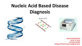
- 1. Nucleic Acid Based Disease Diagnosis Jayanta SubhashTiple B.F.Sc. 3rd year. Enll.No.F/15/061 College of Fishery Science,Udgir.dt.Latur
- 2. • Nucleic acid based diagnosis is highly specific, sensitive and rapid method widely used in health management. • The detection is mainly based on structure and sequence of bases of nucleic acid. • Nucleic acid based diagnosis is becoming common and popular in fish and shellfish health management
- 3. Commonly employed nucleic acid based diagnosis in fish two way: • A. DNA hybridization • B. Polymerase chain reaction (PCR)
- 4. Nucleic acid are made up of nitrogen base . The primary sequence of bases in DNA or RNA have region which are unique for a particular organisms or gene. The sequence of DNA is composed of series of four phosphorylated base, i.e. Adenine, thymidine, guanidine, and cytosine.
- 5. Principle : Nucleic acid probe are segments of DNA or RNA that have been labeled with enzymes, antigenic substances and chemiluminescent moieties or radioisotopes. They would bind with high specificity to complementary sequences of nucleic acid to form double stranded molecules and this process is called hybridization. DNA hybridization:
- 6. Targets and probes • DNA hybridization whether done on solid phase, in suspension or in situ. • The Target: - Target is the sequence of DNA that is to be detected. • The Probe: -A DNA sequence labeled with a radioactive element used to identify the position of a segment with the complementary sequence by binding to it. Characteristics of probe • highly radioisotopes labeling of probe with S35 , P32 , H3 and H or other radioactive elements to the target strand. • The probe length of 20-100 base pair is the most common.
- 8. Two method of DNA hybridization: 1 .DNA hybridization on solid phase . 2. In situ DNA hybridization . • 1 .DNA hybridization on solid phase: In this method the suspected fish/shellfish sample is homogenized and dotted on to a nylon membrane or nitrocellular paper and DNA is exposed from protein usually by digesting with trichloroacetic acid.
- 9. DNA in the sample is then denatured at appropriated temperature and hybridized at appropriated temperature with a probe linked to an isotope or enzyme. Probe hybridization is detected by autoradiography or with a chromogen substrate. This protocol has been adopted to detect DNA in large number of sample to developed a dot blot hybridization. The method is ideal for diagnosis and also for genotyping of pathogen.
- 11. • 2. In situ DNA hybridization: • In this technique histological section of the suspected sample are employed. • DNA of the pathogen in the tissue is exposed using tricholroacetic acid and hybridized with isotope or enzyme labeled probe. • Hybridization is detected by autoradiography or enzyme substrate reaction . • The technique is ideal for diagnosis and studying site of replication in the target organs of the host.
- 13. Polymerase chain reaction (PCR) • PCR is an in vitro technics,by which a specific sequence of a DNA molecule can amplified by repeated enzymatic thermocyclic prosses . • PCR technique was invented by kary.b mullis in 1985. • Major steps in PCR • 1. Denaturation • 2. Primer annealing • 3. Extension
- 16. General PCR protocol used for detection of viral pathogen in aquaculture • The protocol has several steps such as collection of sample, extraction of DNA amplification of DNA and detection of the amplified DNA. 1. Sampling : fresh samples should be collected and fixed immediately in alcohol. Appropriate sample size and sampling strategy are necessary to avoid false negative result. 2. DNA extraction : samples are subjected to DNA extraction by suitable method ( ex. Alkaline lysis method)
- 17. 3.DNA amplification : target DNA is amplified with a cocktail of taq polymerase oligonucleotide primer and nucleotides in a thermal cycle in 20-30 cycle. Each cycle consist of denaturation at 1000c for 1 min and extension at 700c for 2min • If required 2 step PCR carried out by using 1-5%of amplified DNA from the above 1 step . in 2 step PCR is amplification of DNA can be performed either with the same set of primer or internal primer 4.Detection of amplified DNA :the amplified DNA along with marker is subjected to electrophoresis in a 1.5% polyacrylamide gel the gel is then stained with ethidium dibromide and fluorescing DNA bands under u.v light are recorded. Positive and negative controls should yield appropriated result in each test for validation of the DNA amplification. •
- 18. THANKS FOR KIND ATTENTION.... FINFISH AND SHELLFISH DISEASES AND MANAGEMENT