
Recommended
More Related Content
What's hot
What's hot (20)
Similar to Blotting techniques
Similar to Blotting techniques (20)
More from IndrajaDoradla
More from IndrajaDoradla (20)
Recently uploaded
Recently uploaded (20)
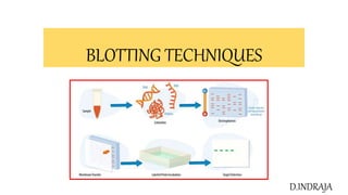
Blotting techniques
- 2. • Blotting is used in molecular biology for the identification of proteins and nucleic acids and is widely used for diagnostic purposes. • This technique immobilizes the molecule of interest on a support, which is a nitrocellulosic membrane or nylon. • It uses hybridization techniques for the identification of the specific nucleic acids and genes. • The blotting technique is a tool used in the identification of biomolecules such ad DNA, mRNA and protein during different stages of gene expression • Southern blotting was introduced by Edwin Southern in 1975 as a method to detect specific sequences of DNA in DNA samples. The other blotting techniques emerged from this method have been termed as Northern (for RNA), Western (for proteins), Eastern (for post-translational protein modifications) • TYPES OF BLOTTING TECHNIQUES • Southern blotting(DNA) • Northern blotting(RNA) • Western blotting(proteins), Far western blott(protein-protein interaction) • Eastern blotting, Far eastern blott (lipds) • Dotblot (bio molecules)
- 3. Southern Blot • Southern Blot is the analytical technique used in molecular biology, immunogenetics and other molecular methods to detect or identify DNA of interest from a mixture of DNA sample or a specific base sequence within a strand of DNA. • The technique was developed by a molecular biologist E.M. Southern in 1975 for analysing the related genes in a DNA restriction fragment and thus named as Southern blotting in his honour Principle of Southern Blot • The process involves the transfer of electrophoresis-separated DNA fragments to a carrier membrane which is usually nitrocellulose and the subsequent detection of the target DNA fragment by probe hybridization. • Hybridization Hybridization refers to the process of forming a double-stranded DNA molecule between a single-stranded DNA probe and a single-stranded target DNA. Since the probe and target DNA are complementary to each other, the reaction is specific which aids in the detection of the specific DNA fragment. • Key point If some of the DNA fragments are larger than 15 kb, then prior to blotting, the gel may be treated with an acid, such as dilute HCl. This depurinates the DNA fragments, breaking the DNA into smaller pieces, thereby allowing more efficient transfer from the gel to membrane
- 4. Steps Involved in Southern Blot 1.Extraction and purification of DNA from cells • DNA is first separated from target cells following standard methods of genomic DNA extraction and then purified. 2.Restriction Digestion or DNA Fragmentation • Restriction endonucleases are used to cut high-molecular-weight DNA strands into smaller fragments. One or more restriction enzymes can be used to achieve such fragments. 3.Separation by Electrophoresis • The separation may be done by agarose gel electrophoresis in which the negatively charged DNA fragments move towards the positively charged anode, the distance moved depending upon its size. 5.Denaturation • DNA is then denatured with a mild alkali such as an alkaline solution of NaOH. This causes the double stranded DNA to become single-stranded, making them suitable for hybridization. DNA is then neutralized with NaCl to prevent re- hybridization before addition of the probe.
- 5. 6.Blotting • The denatured fragments are then transferred onto a nylon or nitrocellulose filter membrane which is done by placing the gel on top of a buffer saturated filter paper, then laying nitrocellulose filter membrane on the top of gel. Finally some dry filter papers are placed on top of the membrane. Fragments are pulled towards the nitrocellulose filter membrane by capillary action and result in the contact print of the gel. 7.Baking • The nitrocellulose membrane is removed from the blotting stack, and the membrane with single stranded DNA bands attached on to it is baked in a vacuum or regular oven at 80 °C for 2-3 hours or exposed to ultraviolet radiation to permanently attach the transferred DNA onto the membrane. 8.Hybridization • The membrane is then exposed to a hybridization probe which is a single DNA fragment with a specific sequence whose presence in the target DNA is to be determined. The probe DNA is labeled so that it can be detected, usually by incorporating radioactivity or tagging the molecule with a fluorescent or chromogenic dye.
- 6. 9.Washing of unbound probes • After hybridization, the membrane is thoroughly washed with a buffer to remove the probe that is bound nonspecifically or any unbound probes present. 10.Autoradiograph • The hybridized regions are detected autoradiographically by placing the nitrocellulose membrane in contact with a photographic film which shows the hybridized DNA molecules. The pattern of hybridization is visualized on X-ray film by autoradiography in case of a radioactive or fluorescent probe is used or by the development of color on the membrane if a chromogenic detection method is used Excess probe is washed off
- 8. Applications of Southern Blot • Identifying specific DNA in a DNA sample. • Preparation of RFLP (Restriction Fragment Length Polymorphism) maps • Detection of mutations, deletions or gene rearrangements in DNA • For criminal identification and DNA fingerprinting (VNTR) • Detection and identification of trans gene in transgenic individual • Mapping of restriction sites • For diagnosis of infectious diseases • Prognosis of cancer and prenatal diagnosis of genetic diseases • Determination of the molecular weight of a restriction fragment and to measure relative amounts in different samples.
- 9. Northern blotting • The technique that is used in molecular biology research to study gene expression by detection of RNA or isolated mRNA in a sample is called northern blotting (RNA blotting). • It is a classical method for analysis of the size and steady state level of a specific RNA in a complex sample. • Northern blotting is a technique for detection of specific RNA sequences. Northern blotting was developed by James Alwine and George Stark at Stanford University (1979) and was named such by analogy to Southern blotting • The Northern blot involves the size separation of RNA in gels like that of DNA. Because we wish to determine the native size of the RNA transcript (and because RNA is single stranded) no restriction enzymes are ever used. • Key point Because most RNA is single stranded and can fold into various conformations through intra-molecular base pairing, the electrophoresis separation is more haphazard and the bands are often less sharp, compared to that of double stranded DNA.
- 10. • Steps Involved in Northern Blot 1. RNA isloation • RNA is isolated from several biological samples (e.g. various tissues, various developmental stages of same tissue etc.) by standard methods of islolation • The tissue or culture sample collected is first homogenized. The samples may be representative of different types of culture for comparison or it can be for the study of different stages of growth inside the culture. 2. Separation by Electrophoresis • Sample’s are loaded on gel and the RNA samples are separated according to their size on an agarose gel • The RNA samples are most commonly separated on agarose gels containing formaldehyde as a denaturing agent for the RNA to limit secondary structure • Denaturing agent (formaldehyde or glyoxal/DMSO) disrupts the secondary structure. • Separation of RNA is better in glyoxal/DMSO system as sharper band for specific RNA is detected by hybridization.
- 11. 3. Transfer of RNA to a membrane • Nitrocellulose and nylons are commonly used membranes. (Nylon membrane are more durable) • RNA are transferred via capillary or vacuum transfer. Vacuum transfer are more efficient but require special transfer apparatus. • Electrophoretic transfer method is available only for nylon membrane due to low concentration of salts required to bind the RNA. • Transferred RNA are then immobilized to membrane by baking at 80 °C or through UV crosslinking in case of nylon membrane. 4. Hybridization and Washing • Hybridization is performed using radio or fluorescently labelled probe to identify specific RNA immobilization. • Pre-hybridization blocks single stranded probe from binding on non- specific sites on the membrane. • Hybridization solution should contain 50% formamide to ensure hybridization at lower temperature and minimize RNA degradation. • The membrane is washed in the buffer containing lower concentration of salt to remove excess probes
- 12. 5. Visualization • Detection of specific transcript through autoradiography. • Membranes are place over X-ray film. • The X-ray film darkens where fragments are corresponding to the radioactive probes.
- 13. APPLICATIONS • A standard for the study of gene expression at the level of mRNA (messenger RNA transcripts) • Detection of mRNA transcript size • Study RNA degradation • Study RNA splicing • Study RNA half-life • Often used to confirm and check transgenic / knockout mice (animals) Disadvantages • Time consuming (Only one gene can be analyzed at a time). • RNA degradation risks because of RNases contamination in work environment. • Relatively expensive for large scale analysis as huge amount of RNA and reagents are required.
- 14. Western blotting • Western blot is the analytical technique used in molecular biology, immunogenetics and other molecular biology to detect specific proteins in a sample of tissue homogenate or extract. • Western blotting is called so as the procedure is similar to Southern blotting. • While Southern blotting is done to detect DNA, Western blotting is done for the detection of proteins. • Western blotting is also called protein immunoblotting because an antibody is used to specifically detect its antigen • Western blotting was first described by G.stark in 1979. It was in 1981 when W. Neal Burnette developed an improved version of the method and gave the name “western blotting” • Principle • Western blotting (protein blotting or immunoblotting) is a rapid and sensitive assay for detection and characterization of proteins. It is based on the principle of immunochromatography where proteins are separated into polyacrylamide gel according to their molecular weight. • The protein thus separated are then transferred or electrotransferred onto nitrocellulose membrane and are detected using specific primary antibody and secondary enzyme labeled antibody and substrate.
- 15. • Steps Involved in Western Blot 1. Extraction of Protein • Cell lysate is most common sample for western blotting. • Protein is extracted from cell by mechanical or chemical lysis of cell. This step is also known as tissue preparation. • To prevent denaturing of protein protease inhibitor is used. • The concentration of protein is determined by spectroscopy. • When sufficient amount of protein sample is obtained, it is diluted in loading buffer containing glycerol which helps to sink the sample in well. • Tracking dye (bromothymol blue) is also added in sample to monitor the movement of proteins. 2. Gel electrophoresis • The sample is loaded in well of SDS-PAGE Sodium dodecyl sulfate- poly- acrylamide gel electrophoresis. • The proteins are separated on the basis of electric charge, isoelectric point, molecular weight, or combination of these all. • The small size protein moves faster than large size protein. • Protein are negatively charged, so they move toward positive (anode) pole as electric current is applied.
- 16. 3. Blotting • The nitrocellulose membrane is placed on the gel. The separated protein from gel get transferred to nitrocellulose paper by capillary action. This type of blotting is time consuming and may take 1-2 days • For fast and more efficient transfer of desired protein from the gel to nitrocellulose paper electro-blotting can be used. • In electro-blotting nitrocellulose membrane is sandwich between gel and cassette of filter paper and then electric current is passed through the gel causing transfer of protein to the membrane. 4. Blocking • Blocking is very important step in western blotting. • Antibodies are also protein so they are likely to bind the nitrocellulose paper. So before adding the primary antibody the membrane is non-specifically saturated or masked by using casein or Bovine serum albumin (BSA) 5. Treatment with Primary Antibody • The primary antibody (1° Ab) is specific to desired protein so it form Ag-Ab complex 6.Treatment with secondary antibody • The secondary antibody is enzyme labelled. For eg. alkaline phosphatase or Horseradish peroxidase (HRP) is labelled with secondary antibody. • Secondary antibody (2° Ab) is antibody against primary antibody (anti-antibody) so it can bind with Ag-Ab complex.
- 17. 7. Treatment with suitable substrate • To visualize the enzyme action, the reaction mixture is incubated with specific substrate. • The enzyme convert the substrate to give visible colored product, so band of color can be visualized in the membrane. • Western blotting is also a quantitative test to determine the amount of protein in sample Detection can be done by other methods such as: •Colorimetric detection •Radioactive detection •Chemiluminescent detection •Fluorescent detection
- 19. Applications: • To determine the size and amount of protein in given sample. • Disease diagnosis: detects antibody against virus or bacteria in serum. • Western blotting technique is the confirmatory test for HIV. It detects anti HIV antibody inpatient's serum. • Useful to detect defective proteins. For eg Prions disease. • Definitive test for Creutzfeldt-Jacob disease Lyme disease Hepatitis B and Herpes
- 20. Eastern Blotting • Developed by Towbin in 1979 • The eastern blot is a biochemical technique used to analyze protein post translational modifications (PTM) such as lipids, phosphomoieties and glycoconjugates. • It is most often used to detect carbohydrate epitopes • Thus, eastern blotting can be considered an extension of the biochemical technique of western blotting. • Multiple techniques have been described by the term eastern blotting, most use proteins blotted from SDS-PAGE gel on to a nitrocellulose membrane • Transferred proteins are analyzed for post-translational modifications using probes that may detect lipids, carbohydrate, phosphorylation or any other protein modification • Eastern blotting should be used to refer to methods that detect their targets through specific interaction of the PTM and the probe, distinguishing them from a standard far-western blot. • In principle, eastern blotting is similar to lectin blotting (i.e. detection of carbohydrate epitopes on proteins or lipids).
- 21. DOT BLOT TECHNIQUE • This technique is used to detect the presence of a given sequence of DNA/RNA in the non- fractionated(not subjected to electrophoresis) DNA sample • DNA from many samples can be tested in a single test. • A Dot blot (or Slot blot) is a technique used to detect biomolecules. • It represents a simplification of the northern blot, Southern blot, or western blot methods. In a dot blot the biomolecules to be detected are not first separated • Instead, a mixture containing the molecule to be detected is applied directly on a membrane as a dot. • Then is spotted through circular templates directly onto the membrane or paper substrate. • Then followed by detection by either nucleotide probes (for a northern blot and Southern blot) or antibodies (for a western blot). • It offers no information on the size of the target biomolecule. Furthermore, if two molecules of different sizes are detected, they will still appear as a single dot. • Can only confirm the presence or absence of a biomolecule.