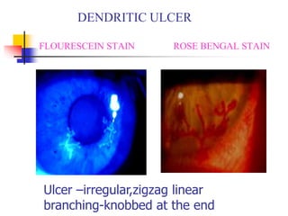
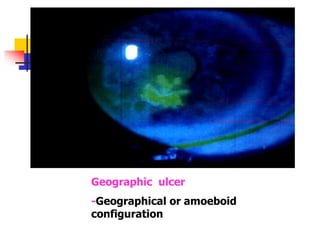
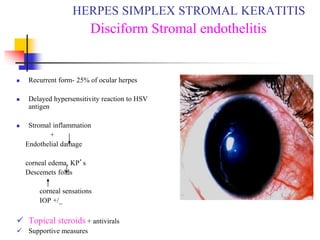
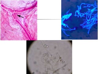
DISEASES OF THE CORNEA
Viral keratitis
 Herpes simplex keratitis
 Herpes Zoster keratitis
 Adenoviral keratitis
Herpes simplex virus
 DNA Virus
 Infection is common upto 90% of the population- most
of it is subclinical
 Humans are the only natural reservoirs
 Source of infection –
Direct contact with the lesions,
By salivary droplets
Fomites
By asymptomatic virus shedding carriers
 HSV-1 causes infection over face,lips and
eyes (mucocutaneous distribution of
trigeminal nerve)
 HSV-2 causes genital herpes
 Infection can be
- Primary
- Recurrent
HSV infection
 Congenital ocular herpes
- skin vesicles
- eye ds
- microencephalopathy
 Neonatal HSV keratitis
- 2 days to 2 weeks
 Primary ocular herpes
 Recurrent ocular herpes
Primary infection
 Occurs in early childhood
 Uncommon during first 6 months
 Transmission through droplet infection or
direct innoculation
 May be subclinical or fever,malaise and
URTI
VIRAL KERATITIS
HERPES SIMPLEX KERATITIS
Primary

Vesicular blepharitis
 Follicular conjunctivitis
 Keratitis Punctate
keratitis
Dendritic ulcer
Recurrent infection
 36% at 5 yrs
 63% at 20 years
 Activated by various trigger factors
- fever
- surgery
- systemic illness
- immunosupression etc
RECURRENT
HERPES SIMPLEX KERATITIS
PUNCTATE EPITHELIAL KERATITIS
 Fine or coarse
superficial punctate
lesions
DENDRITIC ULCER
FLOURESCEIN STAIN ROSE BENGAL STAIN
Ulcer –irregular,zigzag linear
branching-knobbed at the end
Geographic ulcer
-Geographical or amoeboid
configuration
 A,B-PEK
 C,D-Dendritic ulcer
 E,F-Geographical ulcer
 G,H-Disciform keratitis
TREATMENT
 Topical antivirals Acyclovir eye oint. 3%
Ganciclovir gel 0.15%
Triflurothymidine drops 1%
Vidarabine oint. 3%
 Cycloplegics
 Debridement of ulcer edges
 Systemic Antivirals Acyclovir 400mg tid/bd
Valacyclovir 500 mg bd
 Supportive measures
 Topical steroids ×××
HERPES SIMPLEX
EPITHELIAL KERATITIS
 Recurrent form- 25% of ocular herpes
 Delayed hypersensitivity reaction to HSV
antigen
 Stromal inflammation
+
Endothelial damage
corneal edema, KP’s
Descemets folds
corneal sensations
IOP +/_
 Topical steroids + antivirals
 Supportive measures
HERPES SIMPLEX STROMAL KERATITIS
Disciform Stromal endothelitis
DISCIFORM ENDOTHELITIS
 Active viral invasion
+
Tissue destruction
 Necrotic cheesy white infiltrates
 Mild iritis,KP’s(herpetic keratouveitis)
 Stromal vascularization
 Treatment topical steroids
antivirals
cycloplegics
HERPES SIMPLEX STROMAL KERATITIS
STROMAL NECROTIC KERATITIS
Metaherpetic keratitis /
Neurotrophic epithelial keratitis
 Occurs in patient with previous HSV
epithelial disease
 Due to impared corneal innervation
 Nonhealing sterile ulceration
 Round ,oval ulcers with grey thickenned
rolled up margins
 Complication – scarring , neovascularization,
necrosis , perforation, 2̊ bacterial infection
Metaherpetic keratitis / Neurotrophic
epithelial keratitis
Treatment
 Bandage soft contact lens
 Topical antibiotics
 Tear substitutes
 PK
 Lid closure (tarsorrhaphy)
HERPES ZOSTER OPHTHALMICUS
 Varicella-zoster virus
 Latency - Gasserian ganglion
 Ophthalmic div. of 5th nerve
 50% have ocular lesions
 Unilateral
 Systemic features
 Cutaneous lesions
 Ocular lesions
 Seen more in older age group - >75yrs
 Systemic – fever , malaise and eruptions
preceded by sever neuralgic pain
 Ocular manifestations are uncommon
 Ocular lesions-Acute phase
Lids Keratitis
Conjunctivitis SPK
Episcleritis & Scleritis Dendritic
Iridocyclitis Nummular
Secondary glaucoma Disciform
Acute retinal necrosis
Motor nerve palsies
Optic neuritis
Encephalitis
 A,B-PEK
 C,D-
microdendritic
epithelial ulcer
 E,F-nummular
keratitis
 G,H-disciform
keratitis
NUMMULAR KERATITIS
Ocular lesions-chronic
 Neurotrophic keratitis
 Scleritis-scleral atrophy
 Mucous plaque keratitis
 Lipid degeneration
 Lipid granulomata
 Eyelid scarring-entropion/extropion
TREATMENT OF HZO
SYSTEMIC THERAPY
 Oral antivirals Acyclovir -
800mg x10days
Valciclovir
 Analgesics
 Systemic steroids
 Amitryptyline
 Cimetidine
LOCAL THERAPY
Ocular lesions
Topical acyclovir
Topical steroids
Cycloplegics
Anti glaucoma drugs
Artificial tears
Tarsorrhaphy
Keratoplasty
Protozoal kerititis
 Acanthamoeba
 Microsporidia
ACANTHAMOEBA KERATITIS
Free lying amoeba soil, sewage, air
fresh, well, sea water
Ocular infection CL wearers using home made saline
Swimming in contaminated water
Mild trauma
Opportunistic infection
Orthokeratology
Life cycle
 2 stages
- Trophozoit stage
- Cyst stage - dormant
 Symptoms:
- foreign body sensation
- photophobia
- severe pain
ACANTHAMOEBA KERATITIS
SEVERE PAIN disproportional to signs
SIGNS
Epithelial stippling with
microcystic edema
Dendritiform appearance of
epithelium
Radial Keratoneuritis
Ring infiltrate
Stromal infection – overlying epithelium may
be intact
Stromal neovascularization is never seen even
in sever and longstanding cases
Scleritis
ACANTHAMOEBA
CYSTS KERATITIS
ACANTHAMOEBA KERATITIS
Diagnosis Treatment
 High index of suspicion
 KOH wet mount
 Calcofluor white stain
 Culture on E.coli enriched
non nutrient agar
 Confocal microscopy
 PCR of corneal biopsy
specimens
 Neomycin drops 1%
 Propamidine isethionate0.1%
drops
 Polyhexamethyl biguanide
0.02% drops
 Chlorhexidine 0.02%
 Penetrating keratoplasty
Fungal keratitis
 Most common organism is Aspergillus
 Infections are more common when there is
high humidity
 Incidence increased in recent years d/t
injudicious use of antibiotics & steroids
Classification
 Filamentous
1. Septate
- Nonpigmented – Fusarium
Aspergillus
Penicillium
- Pigmented - Curvularia
Alternaria
2. Nonseptate
Rhizopus
 Yeast
Candida
FUNGAL (MYCOTIC)
CORNEAL ULCERS
 Etiology Trauma with organic matter
Injury with animal tail
Systemic/ local immune suppresion
 Causative agent Aspergillus , Fusarium
Candida , Cryptococcus
Curvularia, Alternaria
 Indolent course
 Symptom – foreign body sensation , photophobia , blurred
vision and discharge
SIGNS MORE THAN
SYMPTOMS
SIGNS
Soft creamy raised exudates
Dry looking
Feathery margins
Satellite lesions
Sterile Immune ring of
Wesseley
Hypopyon +/-
Endothelial plaque
Posterior abscess
DIAGNOSIS TREATMENT
 History Organic
matter
 Typical clinical picture
 Corneal scrapings
KOH wet mount
Gram, Giemsa staining
Calcoflour white
Culture on SDA
 Topical antifungals
Natamycin 5%
Itraconazole 1%
Fluconazole 0.2%
Amphotericin B 0.1-0.2%
 Intracameral &
intracorneal/intrastromal
voriconazole
 Systemic antifungals
 Cycloplegics
 Anti inflammatory drugs
 Therapeutic PK in unresponsive
cases
THANK YOU!

Lect._II_Cornea copy.pptx

  • 1.
  • 2.
    Viral keratitis  Herpessimplex keratitis  Herpes Zoster keratitis  Adenoviral keratitis
  • 3.
    Herpes simplex virus DNA Virus  Infection is common upto 90% of the population- most of it is subclinical  Humans are the only natural reservoirs  Source of infection – Direct contact with the lesions, By salivary droplets Fomites By asymptomatic virus shedding carriers
  • 4.
     HSV-1 causesinfection over face,lips and eyes (mucocutaneous distribution of trigeminal nerve)  HSV-2 causes genital herpes  Infection can be - Primary - Recurrent
  • 5.
    HSV infection  Congenitalocular herpes - skin vesicles - eye ds - microencephalopathy  Neonatal HSV keratitis - 2 days to 2 weeks  Primary ocular herpes  Recurrent ocular herpes
  • 6.
    Primary infection  Occursin early childhood  Uncommon during first 6 months  Transmission through droplet infection or direct innoculation  May be subclinical or fever,malaise and URTI
  • 7.
    VIRAL KERATITIS HERPES SIMPLEXKERATITIS Primary  Vesicular blepharitis  Follicular conjunctivitis  Keratitis Punctate keratitis Dendritic ulcer
  • 8.
    Recurrent infection  36%at 5 yrs  63% at 20 years  Activated by various trigger factors - fever - surgery - systemic illness - immunosupression etc
  • 9.
  • 10.
    PUNCTATE EPITHELIAL KERATITIS Fine or coarse superficial punctate lesions
  • 11.
    DENDRITIC ULCER FLOURESCEIN STAINROSE BENGAL STAIN Ulcer –irregular,zigzag linear branching-knobbed at the end
  • 12.
    Geographic ulcer -Geographical oramoeboid configuration
  • 13.
     A,B-PEK  C,D-Dendriticulcer  E,F-Geographical ulcer  G,H-Disciform keratitis
  • 14.
    TREATMENT  Topical antiviralsAcyclovir eye oint. 3% Ganciclovir gel 0.15% Triflurothymidine drops 1% Vidarabine oint. 3%  Cycloplegics  Debridement of ulcer edges  Systemic Antivirals Acyclovir 400mg tid/bd Valacyclovir 500 mg bd  Supportive measures  Topical steroids ××× HERPES SIMPLEX EPITHELIAL KERATITIS
  • 15.
     Recurrent form-25% of ocular herpes  Delayed hypersensitivity reaction to HSV antigen  Stromal inflammation + Endothelial damage corneal edema, KP’s Descemets folds corneal sensations IOP +/_  Topical steroids + antivirals  Supportive measures HERPES SIMPLEX STROMAL KERATITIS Disciform Stromal endothelitis
  • 16.
  • 17.
     Active viralinvasion + Tissue destruction  Necrotic cheesy white infiltrates  Mild iritis,KP’s(herpetic keratouveitis)  Stromal vascularization  Treatment topical steroids antivirals cycloplegics HERPES SIMPLEX STROMAL KERATITIS STROMAL NECROTIC KERATITIS
  • 18.
    Metaherpetic keratitis / Neurotrophicepithelial keratitis  Occurs in patient with previous HSV epithelial disease  Due to impared corneal innervation  Nonhealing sterile ulceration  Round ,oval ulcers with grey thickenned rolled up margins  Complication – scarring , neovascularization, necrosis , perforation, 2̊ bacterial infection
  • 19.
    Metaherpetic keratitis /Neurotrophic epithelial keratitis
  • 20.
    Treatment  Bandage softcontact lens  Topical antibiotics  Tear substitutes  PK  Lid closure (tarsorrhaphy)
  • 21.
    HERPES ZOSTER OPHTHALMICUS Varicella-zoster virus  Latency - Gasserian ganglion  Ophthalmic div. of 5th nerve  50% have ocular lesions  Unilateral  Systemic features  Cutaneous lesions  Ocular lesions
  • 22.
     Seen morein older age group - >75yrs  Systemic – fever , malaise and eruptions preceded by sever neuralgic pain  Ocular manifestations are uncommon
  • 23.
     Ocular lesions-Acutephase Lids Keratitis Conjunctivitis SPK Episcleritis & Scleritis Dendritic Iridocyclitis Nummular Secondary glaucoma Disciform Acute retinal necrosis Motor nerve palsies Optic neuritis Encephalitis
  • 24.
     A,B-PEK  C,D- microdendritic epithelialulcer  E,F-nummular keratitis  G,H-disciform keratitis
  • 25.
  • 26.
    Ocular lesions-chronic  Neurotrophickeratitis  Scleritis-scleral atrophy  Mucous plaque keratitis  Lipid degeneration  Lipid granulomata  Eyelid scarring-entropion/extropion
  • 27.
    TREATMENT OF HZO SYSTEMICTHERAPY  Oral antivirals Acyclovir - 800mg x10days Valciclovir  Analgesics  Systemic steroids  Amitryptyline  Cimetidine LOCAL THERAPY Ocular lesions Topical acyclovir Topical steroids Cycloplegics Anti glaucoma drugs Artificial tears Tarsorrhaphy Keratoplasty
  • 28.
  • 29.
    ACANTHAMOEBA KERATITIS Free lyingamoeba soil, sewage, air fresh, well, sea water Ocular infection CL wearers using home made saline Swimming in contaminated water Mild trauma Opportunistic infection Orthokeratology
  • 30.
    Life cycle  2stages - Trophozoit stage - Cyst stage - dormant
  • 31.
     Symptoms: - foreignbody sensation - photophobia - severe pain
  • 32.
    ACANTHAMOEBA KERATITIS SEVERE PAINdisproportional to signs SIGNS Epithelial stippling with microcystic edema Dendritiform appearance of epithelium Radial Keratoneuritis Ring infiltrate
  • 33.
    Stromal infection –overlying epithelium may be intact Stromal neovascularization is never seen even in sever and longstanding cases Scleritis
  • 34.
  • 35.
    ACANTHAMOEBA KERATITIS Diagnosis Treatment High index of suspicion  KOH wet mount  Calcofluor white stain  Culture on E.coli enriched non nutrient agar  Confocal microscopy  PCR of corneal biopsy specimens  Neomycin drops 1%  Propamidine isethionate0.1% drops  Polyhexamethyl biguanide 0.02% drops  Chlorhexidine 0.02%  Penetrating keratoplasty
  • 36.
    Fungal keratitis  Mostcommon organism is Aspergillus  Infections are more common when there is high humidity  Incidence increased in recent years d/t injudicious use of antibiotics & steroids
  • 37.
    Classification  Filamentous 1. Septate -Nonpigmented – Fusarium Aspergillus Penicillium - Pigmented - Curvularia Alternaria 2. Nonseptate Rhizopus  Yeast Candida
  • 38.
    FUNGAL (MYCOTIC) CORNEAL ULCERS Etiology Trauma with organic matter Injury with animal tail Systemic/ local immune suppresion  Causative agent Aspergillus , Fusarium Candida , Cryptococcus Curvularia, Alternaria  Indolent course  Symptom – foreign body sensation , photophobia , blurred vision and discharge
  • 39.
    SIGNS MORE THAN SYMPTOMS SIGNS Softcreamy raised exudates Dry looking Feathery margins Satellite lesions Sterile Immune ring of Wesseley Hypopyon +/- Endothelial plaque Posterior abscess
  • 44.
    DIAGNOSIS TREATMENT  HistoryOrganic matter  Typical clinical picture  Corneal scrapings KOH wet mount Gram, Giemsa staining Calcoflour white Culture on SDA  Topical antifungals Natamycin 5% Itraconazole 1% Fluconazole 0.2% Amphotericin B 0.1-0.2%  Intracameral & intracorneal/intrastromal voriconazole  Systemic antifungals  Cycloplegics  Anti inflammatory drugs  Therapeutic PK in unresponsive cases
  • 45.