Respiratory system
•Download as PPTX, PDF•
9 likes•2,274 views

Its about repiratory system of living thing, Hope usefull for you.

Recommended
More Related Content
What's hot
What's hot (20)
Similar to Respiratory system
Similar to Respiratory system (20)
Recently uploaded
Recently uploaded (20)
Respiratory system
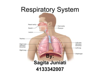
- 2. Introduction • Respiratory System Controls Gases exchange • The organs of the respiratory system, there are: Nose, pharynx, larynx, trachea, bronchi and their smaller branches, and the lungs, which contain the alveoli.
- 3. The Nose • External portion that contain: Nostrils Two nasal cavities( separated by septum; composed cartilage and bone) conchae Paranasal sinuses
- 4. The Pharynx Three parts: • The nasopharynx, where the nasal cavities open posterior to the soft palate; • the oropharynx, where the oral cavity joins the pharynx; and • the Hypopharynx, which opens into the larynx. Is the continouslly nose track that make air pass the larynx
- 5. The Larynx • Consists of cartilage plate • The movement of inner walls by muscle; glottis is the gap that connected the pharynx-Trachea • Consists of vocal cords, when air pass, vibration, speak. • Consists of valve=ephyglotis; always open, closed if food enter to the pharynx.
- 7. The Trachea • Called the windpipe, is a tube connecting the larynx to the primary bronchi. • Arranged by cartilage rings • Placed front of esophagus. • Covered by mucous membrane; Slippery • Pipe form • Layer of Pseudostratified ciliated columnar epithelium to keep the lungs from dust/little particle on air
- 8. The Bronchial Tree • Connected lungs and Trachea • Found on left and right of heart • Cartilage plate • The walls arranged by smooth muscle • The bronchi branch called bronchiolus
- 9. The Lungs and alveolus
- 10. The Lungs • Cone-shaped organs • Central area - the primary bronchi, the heart, and other organs. • The right lung has three lobes; left lung has two lobes; allowing room for the heart whose apex points left • Lobe – lobules – bronchiole - many alveoli • enclosed by a double layer of serous membrane called the pleura. • Surface tension-the tendency for water molecules to cling to each other and to form a droplet. • Surface tension holds the two pleural layers together when the lungs recoil during expiration.
- 11. The Alveoli • inhalation, air passes by way of the bronchial tree to the alveoli. • alveolar sac is made up of simple squamous epithelium; surrounded by blood capillaries • Gas exchange occurs between the air in the alveoli and the blood in the capillaries. • Oxygen diffuses - alveolar and capillary walls- bloodstream - carbon dioxide diffuses from the blood - the alveoli.
- 12. Mechanism of Breathing • Conscious and insensbly • Expiration and Inspiration • Based on placed and the ways of breath on Expiration and Inspiration, divided into: 1. Thorax respiration 2. Abdominal respiration
- 13. Thorax respiration • Inspiration : muskulus interkostalis constractionLiftpad of Margin boneThorax cavity will be dilate, the lung also expand pressure of Air cavity of lungs ↓ at outer ↑The air from the outer entered to the lungs • Expiration : muskulus interkostalis relaxatationThe thorax bone will be downThorax cavity become narrow, the lungs become small pressure of Air cavity of lungs ↑ at outer↓Air released from the lungs
- 15. Abdominal respiration • Inspiration: Diaphgram Muscle ConstractionDiaphgram has flatThorax cavity and the lungs will be expandAir Pressure of Lungs cavity↓Air from the outer enter to the lungs. • Expiration: Diaphragm muscle relaxationcurved Diaphragm Thorax cavity and the lungs will be decline Air Pressure of Lungs cavity ↑ The air out of the lungs
- 17. Volume and capacity of the Lungs • Every people is different • Depend of the lungs size, The power of breathing and the ways of breathing • The lungs volume of Adult; 5-6 L; there are: – Volume tidal/ tidal volume(VT) – Volume cadangan inspirasi/volume reseve inspiration (VCI) – Volume cadangan ekspirasi/volume reseve expiration(VCE) – Volume residu/residual volume (VR)
- 18. Volume and capacity of the Lungs • Volume tidal (VT): The air volume-Result of Inspiration/expiration-nomal breathing, ± 500cc/ml for the young adult • Volume reseve inspiration (VCI): Extra air volume—from the inspiration after tidal volume, ± 3000cc/ml
- 19. Volume and capacity of the Lungs • Volume reseve expiration (VCE): the volume of air that can still be in a strong expiration at the end of a normal expiration; ± 1100cc/ml • Residual Volume (VR):volume of air that still remains in the lungs after expiration strong , ± 1200cc / ml
- 20. The frequently of breathing • Slow or fast breathing is affected by: –Age –Gender –Temperature of environmental –Position of body
- 21. Exchanges mechanism of oxygen and carbondioxide
- 23. Repiratory system of Pisces
- 24. Repiratory system of Pisces Internal Gills The gills of bony fishes are located between the buccal (mouth) cavity and the opercular cavities The buccal cavity can be opened and closed by opening and closing the mouth, and the opercular cavity can be opened and closed by movements of the operculum, or gill cover.
- 25. Respiratory of Amphibian • Breathing organ of amphibian is Oral cavity, coane/Nose cavity and lungs also skin Skin is Thin, moist and many blood capillary; Doing when sleep and diffussion ways → Oxygen though skin → pulmonary vein cutanea → Heart → all body
- 27. Respiratory of Amphibian • Inspiration Sternohioideus muscle contraction → oral cavity enlarges → O2 entry through koane ( slit nose ) → koane shut → submandibular muscles and muscle geniohioideus are contraction → oral cavity become small → O2 pushed into the lungs through the cracks → gas exchange occurs in the lungs (O2 bound by the walls of the blood in the capillaries of the lungs, the CO2 released into the environment).
- 28. Respiratory of Amphibian • Expiration gas exchange in the lungs → submandibular muscle relaxation → and sternohioideus stomach muscles to contract → lungs shrink → pressured air out and into the oral cavity → koane open → slit throat closes → and geniohioideus submandibular muscles to contract → the oral cavity decreases → CO2 driven exit through koane .
- 30. Repiratory of Reptile • Phase of inspiration : the rib muscles to contract so that the chest cavity enlarges followed lungs expand, air in through the nostrils , trachea , bronchi, and lungs . O2 gas intake by nose → mouth cavity → long trachea → bronchioles in the lungs → O2 from the lungs to the blood is transported throughout the body tissues. • Phase of expiration : rib muscle relaxation so that the chest cavity and lungs shrink, air from the lungs out through the lungs, bronchi, trachea, and nostrils. From the body tissue in the transport of CO2 → blood to the heart → Artery to the lungs→bronkiolus → the long tracheal → uvula → oral cavity → the nostrils
- 32. Respiratory of Aves The flow of breathing ( when perch ) : • Inspiration; The ribs move down - enlarged chest cavity - lungs inflate - Air enters the lungs - Purse rear Eve - Lungs - Purse weather ahead • Expiration; The ribs move up - thorax deflate - Lung shrink - Air out of the coffers of the weather - Lungs ( diffusion ) - Exit
- 33. Respiratory of Aves The flow of breathing ( when flying ) : • Inspiration; Wing lift - coffers swell korakoid desires - desires abdominal Air enters coffers - Lungs ( partially ) and the coffers of the air ( mostly ) . • Expiration; Wing down - weather coffers korakoid pinched - purses weather thorax expands - Air pushed out
- 34. Disease of Human breathing • Faringitis • Pneumonia; The alveolus consist fluid and the amount of eritrosit are excessive • Asma • Dipteri • Asfiksi • Tuberkulosis • Hipoksia • Asidosis • Sianosis
- 35. Reference • Elaine,M.Marieb.2015. Essential of human anatomy and physiology.USA: Library of Congress Cataloging-in-Publication Data • http://www.bimbie.com/frekuensi-volume- pernafasan-manusia.htm • Ananymous. Respiratory.pdf • Helen,Colbourne.2007. Inquiry into BIOLOGY. Canada: McGraw-Hill Ryerson Inquiry Into Biology • https://extraordinarnee.wordpress.com/2013/10 /04/pernapasan-amfibi/