Head and neck anatomy 3 meningese & the brain
•Download as PPTX, PDF•
0 likes•111 views

anatomy

Recommended
More Related Content
What's hot
What's hot (20)
Similar to Head and neck anatomy 3 meningese & the brain
Similar to Head and neck anatomy 3 meningese & the brain (20)
Recently uploaded
Recently uploaded (20)
Head and neck anatomy 3 meningese & the brain
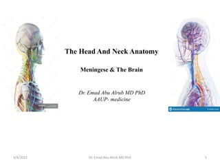
- 1. The Head And Neck Anatomy Meningese & The Brain 3/4/2022 1 Dr. Emad Abu Alrub MD PhD Dr. Emad Abu Alrub MD PhD AAUP- medicine
- 2. INTERIOR OF THE SKULL Meninges Three membranes (meninges) surround and protect the brain in the skull. From superficial to deep, these are: The dura mater, Arachnoid mater, and Pia mater. They are mostly continuous with the spinal meninges that surround the spinal cord in the vertebral column 3/4/2022 2 Dr. Emad Abu Alrub MD PhD
- 3. 3/4/2022 3 Dr. Emad Abu Alrub MD PhD
- 4. 3/4/2022 4 Dr. Emad Abu Alrub MD PhD
- 5. Dura Mater The dura mater is conventionally described as two layers: The endosteal layer The meningeal layer The dural folds/ Four septa 1. Falx cerebri 2. Falx cerebelli 3. Tentorium cerebelli 4. Diaphragma sellae 3/4/2022 5 Dr. Emad Abu Alrub MD PhD
- 6. 3/4/2022 Dr. Emad Abu Alrub MD PhD 6 Dural venous sinuses The dural venous sinuses are formed in following two ways: (a) by separation of the two layers of cerebral dura, and (b) by reduplication of the meningeal layer of dura (Fig. 16.2). The dural venous sinuses are lined by endothelium which becomes continuous with the endothelial lining of the veins.
- 7. 3/4/2022 7 Dr. Emad Abu Alrub MD PhD
- 8. 3/4/2022 Dr. Emad Abu Alrub MD PhD 8 Classification The dural venous sinuses are classified into two types, unpaired and paired
- 9. Dural Nerve Supply Branches of the trigeminal, vagus, and first three cervical nerves and branches from the sympathetic system pass to the dura. Dural Arterial Supply Numerous arteries from the internal carotid, maxillary, ascending pharyngeal, occipital, and vertebral arteries supply the dura mater. From a clinical standpoint, the most important is the middle meningeal artery, which is commonly damaged in head injuries. Dural Venous Drainage The meningeal veins lie in the endosteal layer of dura. The middle meningeal vein 3/4/2022 9 Dr. Emad Abu Alrub MD PhD
- 10. 3/4/2022 10 Arachnoid mater Arachnoid mater is a thin, transparent membrane lying between the pia mater internally and dura mater externally. • It invests the brain loosely and continues as spinal arachnoid at the foramen magnum, which ends at the level of second sacral vertebra. • Sub arachnoid space contains CSF • Arachnoid villi • Arachnoid granulations
- 11. 11 Dr. Emad Abu Alrub MD PhD 3/4/2022 Pia mater Pia mater is thin transparent vascular membrane which closely invests the surface of the brain. It follows the surface irregularities of the cerebral hemisphere, and dips in every sulcus forming folds. All the blood vessels to brain run on it before entering the brain.
- 12. THE BRAIN 3/4/2022 12 Dr. Emad Abu Alrub MD PhD
- 13. Brain development • The telencephalon develops into the cerebrum and lateral ventricles. • The diencephalon forms the thalamus, hypothalamus, epithalamus, and third ventricle. • The metencephalon becomes the Pons, cerebellum, and upper part of the fourth ventricle. • The myelencephalon forms the medulla oblongata and lower part of the fourth ventricle. • The mesencephalon (mes-en-SEF-a-lon), or midbrain, gives rise to the midbrain and aqueduct of the midbrain (cerebral aqueduct). 3/4/2022 13 Dr. Emad Abu Alrub MD PhD
- 14. Brainstem The brainstem is the stalk-like part of the brain which connects the spinal cord with the forebrain. From below upwards it consists of three parts: medulla oblongata, pons, and midbrain. It contains important autonomic reflex centres (vital centres) associated with the control of respiration heart rate and blood pressure. 3/4/2022 14 Dr. Emad Abu Alrub MD PhD
- 15. 15 Dr. Emad Abu Alrub MD PhD 3/4/2022 Diencephalon The diencephalon is the part of brain between the cerebrum and the brainstem. The cavity within it is termed third ventricle. components • Pars dorsalis lies above (dorsal) to the hypothalamic sulcus and consists of: 1. Thalamus, 2. Metathalamus 3. Epithalamus / pineal body (gland), • Pars ventralis lies below (ventral) to the hypothalamic sulcus and consists of: 1. Subthalamus, and 2. Hypothalamus. Functionally, the thalamus is generally considered as the great sensory gateway to the cerebral cortex.
- 16. 3/4/2022 16 Dr. Emad Abu Alrub MD PhD
- 17. 3/4/2022 Dr. Emad Abu Alrub MD PhD 17 The cerebrum • The cerebrum is the largest part of the human brain that fills most of the cranial cavity. • The cerebrum is a heavily, convoluted bilobed structure. • The two lateral halves are called cerebral hemispheres. • A deep median cleft, the longitudinal cerebral fissure, incompletely separates the two cerebral hemispheres. • The corpus callosum which is a large mass of white fibres joins the two cerebral hemispheres across the median plane. • Lateral ventricles.
- 18. 3/4/2022 Dr. Emad Abu Alrub MD PhD 18 Superior view of the cerebrum.
- 19. Lobes of Cerebral Hemisphere • The frontal lobe • The parietal lobe • The temporal lobe • The occipital lobe Insula 3/4/2022 19 Dr. Emad Abu Alrub MD PhD
- 20. • Central sulcus • Precentral gyrus- motor • Post central gyrus- somatosensory Wernicke's area Broca's area Aphasia 3/4/2022 20 Dr. Emad Abu Alrub MD PhD
- 21. 3/4/2022 21 Dr. Emad Abu Alrub MD PhD
- 22. The cerebellum Lobes 3/4/2022 22 Dr. Emad Abu Alrub MD PhD
- 23. Cerebellum- regulates equilibrium, muscle tone, postural control, fine movement and coordination of voluntary muscle movement. Locati on 3/4/2022 23 Dr. Emad Abu Alrub MD PhD
- 24. Brain Blood flow Internal carotid Vertebral arteries The dural venous sinuses drain into the internal jugular veins to return blood from the head to the Heart. 3/4/2022 24 Dr. Emad Abu Alrub MD PhD
- 25. 3/4/2022 25 Dr. Emad Abu Alrub MD PhD
- 26. 3/4/2022 26 Dr. Emad Abu Alrub MD PhD
- 27. Cranial Nerves
- 28. 3/4/2022 28 Dr. Emad Abu Alrub MD PhD
- 29. 3/4/2022 Dr. Emad Abu Alrub MD PhD 29 Thank you!