2. and the Peripheral Nervous System (PNS).
brain (located in the cranial cavity) and the
spinal cord (located in the vertebral cavity), which serve as the main
control centers for all body activities.
nerves derived from the brain and spinal cord (12
pairs of cranial nerves and 31 pairs of spinal nerves) which serve as
linkage between the CNS and the body.
3.
4.
5.
6.
7.
8.
9.
10.
11.
12.
13.
14.
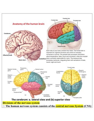
15. Organization of the brain: The human brain consists of 4 major parts:
1. Brain stem: is continuous with the spinal cord and consists of: medulla oblongata, pons, and
midbrain.
2. cerebellum: (cerebellum=little brain) it is posterior to the brain stem
3. diencephalon: (di=through; encephalon=brain) it is superior to the brain stem, it consists of
thalamus, hypothalamus, and epithalamus.
4. Cerebrum: (cerebrum=brain) the cerebral cortex is a region of gray matter that forms the
outer rim of the cerebrum, it is found on the diencephalon and brain stem.
Lobes of the cerebrum: Each cerebral hemisphere can be further divided into 4 lobes. The lobes
are named after the bones that cover them: frontal lobe, parietal lobe, temporal lobe and
occipital lobe.
Cerebral cortex: (cortex=rind or park) is a region of gray matter that forms the outer rim of the
cerebrum with thickness of about only 2 – 4 mm, and contains billions of neurons.
The folds are called gyri (jiri=circular) or convolutions. The deepest grooves between folds are
known as fissures; the shallower groves between folds are called as sulci (sulsi=grooves).
Longitudinal fissure separates the cerbrum into right and left halves called cerebral
hemispheres. The cerebral hemispheres are connected internally by the corpus callosum
26. Cerebrospinal Fluid (CSF)
CSF is a clear, colorless liquid that nourishes and protects the CNS from chemical and physical injury
It circulates through the subarachnoid space around the brain and spinal cord and through the four ventricles
(cavities) within the brain
It contains glucose, proteins, lactic acid urea, ions, some lymphocytes.
27. - Formed by selective transport across ependymal cells
- Volume 125-150 ml and is replaced > 3 times/day, flow maintained by 10 mmHg pressure gradient
- Path: ventricles ® subarachnoid space, reabsorbed into blood in dural sinuses through arachnoid villi
Functions of CSF:
- Shock-absorbing medium
- Provides a optimum and stable environment for generating nerve impulses
- Provides a medium for the exchange of nutrients and wastes between blood and nervous tissue.
Production of CSF
The choroid plexus is a network of blood capillaries in the walls of the ventricles
The capillaries are covered by ependymal cells that produces the CSF.
The ependymal cells provide a fluid tight barrier around the capillaries called the blood-brain barrie
28. Blood-Brain Barrier
Formed by capillary endothelial cells formed with tight junctions
The tight junctions restrict transport between cells
Astrocytes induce formation of tight junctions and help control transcellular transport such as K+
Some materials pass the blood-brain barrier
o - Materials that diffuse through the lipid bilayer cannot be restricted –O2, CO2, alcohol, steroids, H2O
- Materials that require carrier transport are restricted – glucose, amino acids, and ions
- Some materials cannot pass the barrier – potentially harmful substances, some hormones/drugs
Functions of the blood-brain barrier:
o - Protects CNS from chemical fluctuations
- Prevents entry of harmful substances
- Prevents entry of molecules that could act as neurotransmitters
There are only 3 places in the brain that do not have a blood brain barrier:
1. Choroid plexuses (because they make CSF)
2. Hypothalamus (needs to let hormones into bloodstream)
3. Pineal gland (also needs to let hormones into the bloodstream
42. Protective covering of the brain: the cranial meninges are continuous with the spinal meninges.
The outer dura mater, the middle arachnoid mater, and the inner pia mater.
43. The ventricular system of the human brain:
There are 4 ventricles (little cavities) filled with cerebrospinal fluid (CSF) within the brain. A
lateral ventricle is located in each hemisphere of the cerebrum. Anteriorly, the lateral ventricles
are separated by a thin membrane, the septum pellucidum (pellucidum=transparent). The third
ventricle is superior to the hypothalamus and between the right and left halves of the thalamus.
The fourth ventricle lies between the brain stem and the cerebellum. CSF escapes into the
subarachnoid space via small apertures near the base of the cerebellum. In the subarachnoid
space, CSF is absorbed into the blood. The total volume of CSF is 125-150 ml. Normal resting
pressure of the CSF is between 150-180 mm H2O. Total production of CSF is about 400-500
ml/day (about .36 ml/min)
44.
45.
46. Cerebellum:
Differentiation of the midbrain: The midbrain differenciates into the tectum (means: roof) and
the tegmentum (the floor of the midbrain). The CSF-filled space at the core of the midbrain in
the cerebral aqueduct.
The tectum differenciates into two structures: superior colliculus (control eye movement ) and
47. inferior colliculus (receives sensory information from ear and send it to thalamus). Colliculus
means mound
The tegmentum contains two colorful regions (substantia nigra and red nucleus) both regions
control voluntary movement
**Differentiation of the rostral hindbrain: The rostral hindbrain differentiates into the
cerebellum and pons. The cerebellum is formed by the growth and fusion of the rhombic lips.
The CSF-filled space at the core of the hindbrain is the fourth ventricle
**The pons serves as a massive switchboard connecting the cerebral cortex to the cerebellum.
(pons means bridge)
**Differentiation of the caudal hindbrain: The caudal hindbrain differentiates into the medulla.
The medullary pyramids are bundles of axons coursing caudally toward the spinal cord. The
CSF-filled space at the core of the medulla is the fourth ventricle
**The pyramidal decussation: the corticospinal tract crosses from one side to the other in the
medulla
**Differentiation of the spinal cord: The butterfly-shaped core of the spinal cord is gray matter,
divisible into dorsal and ventral horns, and intermediated zone. Surrounding the gray matter
are white matter columns running rostrocaudally, up and down the cord
Dorsal columns: are bundles of axons running along the dorsal surface of the cord
Lateral columns: are bundles of axons lateral to the spinal gray matter on each side
Ventral columns: are bundles of axons on the ventral surface of the cord
48. **The surface area of the human cerebral cortex measures about 1100cm2
The Hippocampus
49. The hippocampus is located deep in the brain and plays a huge role in fear conditioning. Studies have
suggested that damage to the hippocampus could contribute to avoidance learning, which is a
considerable component of anxiety.
The Hippocampus turns threatening events into memories. People who are victims of child abuse or
who have been involved in combat have shown in studies to have a smaller hippocampus. Not
everyone gets PTSD from traumatic events, but researchers believe that a dysfunction hippocampus
plays a role in developing the condition after a traumatic event.
A team of researchers has also recently discovered anxiety cells in the hippocampus. The team
found that when mice went into an open field or in an elevated part of a maze, these anxiety
neurons responded strongly. When the research team was able to suppress these anxiety neurons in
the mice, it made the mice more comfortable in unfamiliar places. And when they excited these
neurons, it made the mice have anxiety in safe and enclosed spaces.
How CBD Can Help With Anxiety
Chronic stress can impair signaling in the endocannabinoid system with the amygdala and the
hippocampus which can lead to anxiety disorders. Studies suggest that CBD oil can help reduce
activity in the amygdala giving anxiolytic effects. Studies have also shown that CBD may be able
to help with the disturbed signaling in the endocannabinoid system involving the hippocampus.
Possibly even aiding in cell proliferation, which is increasing the number of cells. More findings are
requested to be conclusive, but scientists are on a hot track to discovering the ways that CBD hemp
oil can help with anxiety symptoms and it causes.
50. The hippocampus (from the Greek "seahorse" from hippos, "horse" and kampos, "sea-
monster") is a major component of the brain of humans and other vertebrates. Humans
and other mammals have two hippocampi, one in each side of the brain. The
hippocampus is part of the limbic system, and plays important roles in
the consolidation of information from short-term memory to long-term memory, and
in spatial memory that enables navigation. The hippocampus is located under
the cerebral cortex in the allocortex,and in primates it is in the medial temporal lobe. It
contains two main interlocking parts: the hippocampus proper (also called Ammon's
horn)and the dentate gyrus.
In Alzheimer's disease (and other forms of dementia), the hippocampus is one of the first regions of
the brain to suffer damage; short-term memory loss and disorientation are included among the early
symptoms. Damage to the hippocampus can also result from oxygen starvation (hypoxia), encephalitis,
or medial temporal lobe epilepsy. People with extensive, bilateral hippocampal damage may
experience anterograde amnesia: the inability to form and retain new memories.
Since different neuronal cell types are neatly organized into layers in the hippocampus, it has
frequently been used as a model system for studying neurophysiology. The form of neural
plasticity known as long-term potentiation (LTP) was initially discovered to occur in the hippocampus
and has often been studied in this structure. LTP is widely believed to be one of the main neural
mechanisms by which memories are stored in the brain.
51. In rodents as model organisms, the hippocampus has been studied extensively as part of a brain system
responsible for spatial memory and navigation. Many neurons in the rat and mouse hippocampus
respond as place cells: that is, they fire bursts of action potentials when the animal passes through a
specific part of its environment. Hippocampal place cells interact extensively with head direction cells,
whose activity acts as an inertial compass, and conjecturally with grid cells in the
neighboring entorhinal cortex.
Mammalian brains