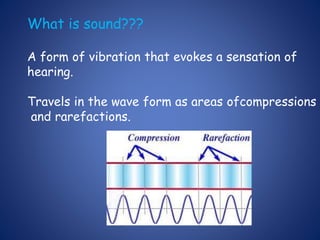
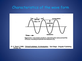
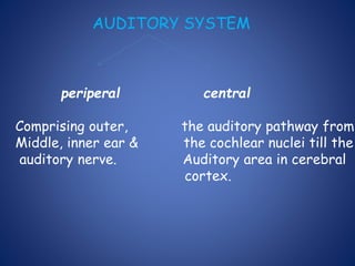
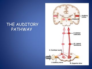
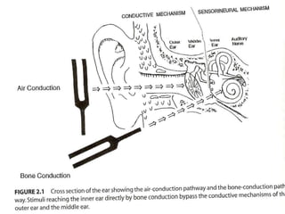
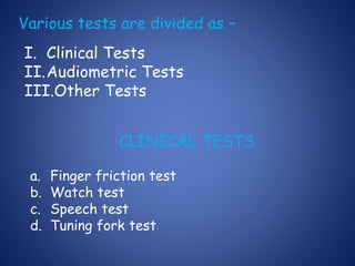
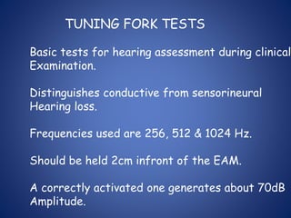
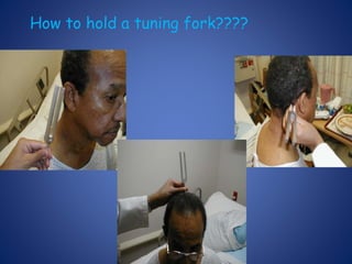
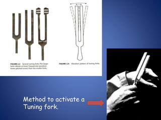
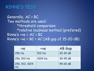
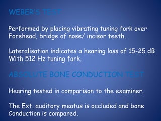
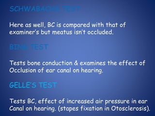
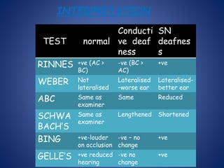
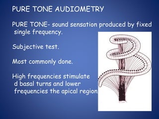
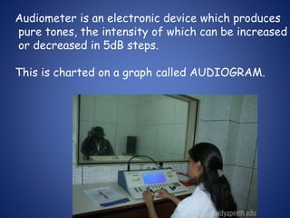
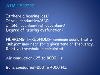
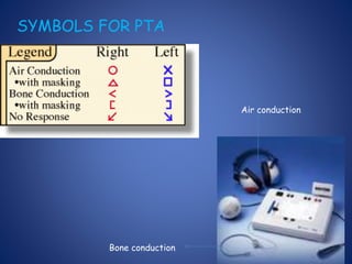
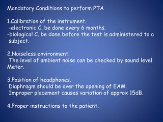
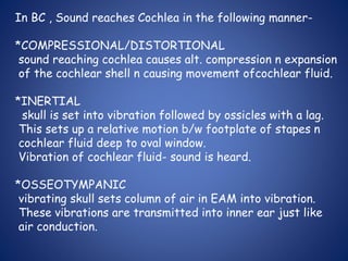
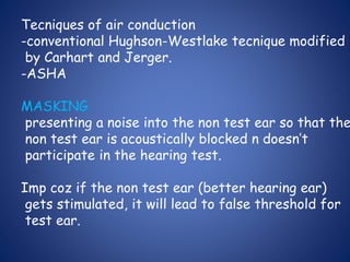
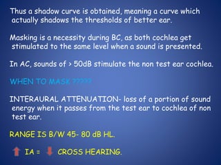
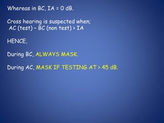
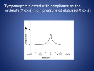
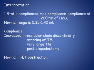
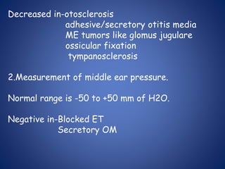
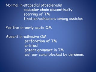
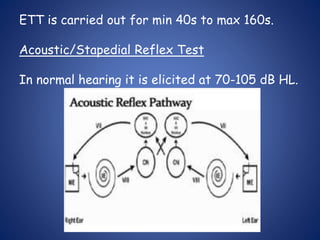
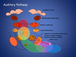
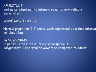
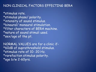
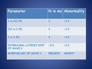
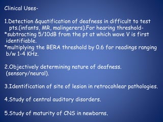
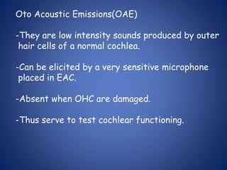
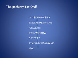
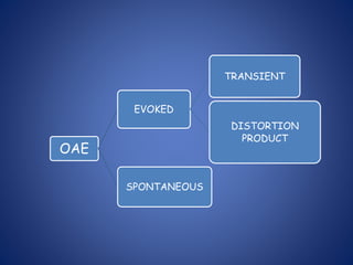
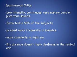
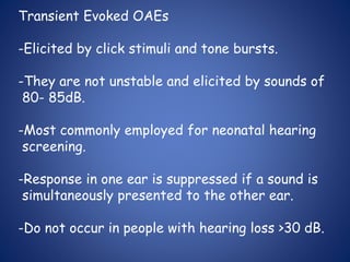
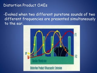
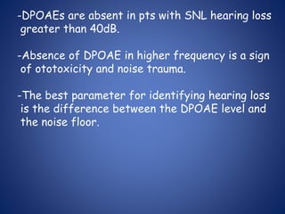
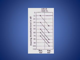
4. WHAT IS DECIBEL???
Decibel (dB) = 1/10 Bel.
Bel is a base10 logarithmic ratio of intensity
of given sound to threshold of hearing in
normal subjects at 1000Hz.
Named after Alexander Graham Bel – the
inventor of telephone.
8. Various tests are divided as –
I. Clinical Tests
II.Audiometric Tests
III.Other Tests
CLINICAL TESTS
a. Finger friction test
b. Watch test
c. Speech test
d. Tuning fork test
9. TUNING FORK TESTS
Basic tests for hearing assessment during clinical
Examination.
Distinguishes conductive from sensorineural
Hearing loss.
Frequencies used are 256, 512 & 1024 Hz.
Should be held 2cm infront of the EAM.
A correctly activated one generates about 70dB
Amplitude.
12. RINNE’S TEST
Generally, AC > BC.
Two methods are used-
*threshold comparison
*relative loudness method (prefered)
Rinne’s +ve = AC > BC
Rinne’s –ve = BC > AC (AB gap of 15-20 dB)
-ve +ve AB Gap
256 Hz 512 Hz 20-30 dB
256, 512 Hz 1024 Hz 30-45 dB
256, 512, 1024
Hz
45-60 dB
13. WEBER’S TEST
Performed by placing vibrating tuning fork over
Forehead, bridge of nose/ incisor teeth.
Lateralisation indicates a hearing loss of 15-25 dB
With 512 Hz tuning fork.
ABSOLUTE BONE CONDUCTION TEST
Hearing tested in comparison to the examiner.
The Ext. auditory meatus is occluded and bone
Conduction is compared.
14. SCHWABACHS TEST
Here as well, BC is compared with that of
examiner’s but meatus isn’t occluded.
BING TEST
Tests bone conduction & examines the effect of
Occlusion of ear canal on hearing.
GELLE’S TEST
Tests BC, effect of increased air pressure in ear
Canal on hearing. (stapes fixation in Otosclerosis).
15. INTERPRETATION
TEST normal
Conducti
ve deaf
ness
SN
deafnes
s
RINNES +ve (AC >
BC)
-ve (BC >
AC)
+ve
WEBER Not
lateralised
Lateralised
–worse ear
Lateralised-
better ear
ABC Same as
examiner
Same Reduced
SCHWA
BACH’S
Same as
examiner
Lengthened Shortened
BING +ve-louder
on occlusion
-ve – no
change
+ve
GELLE’S +ve reduced
hearing
-ve no
change
+ve
16. PURE TONE AUDIOMETRY
PURE TONE- sound sensation produced by fixed
single frequency.
Subjective test.
Most commonly done.
High frequencies stimulate
d basal turns and lower
frequencies the apical region.
17. Audiometer is an electronic device which produces
pure tones, the intensity of which can be increased
or decreased in 5dB steps.
This is charted on a graph called AUDIOGRAM.
18. AIM IS?????
Is there a hearing loss?
If yes, conductive/SN?
If SN, cochlear/retrocochlear?
Degree of hearing dysfunction?
HEARING THRESHOLD- minimum sound that a
subject may hear for a given tone or frequency.
Relative threshold is calculated.
Air conduction-125 to 8000 Hz
Bone conduction-250 to 4000 Hz.
20. Mandatory Conditions to perform PTA
1.Calibration of the instrument.
-electronic C. be done every 6 months.
-biological C. be done before the test is administered to a
subject.
2.Noiseless environment.
The level of ambient noise can be checked by sound level
Meter.
3.Position of headphones.
Diaphragm should be over the opening of EAM.
Improper placement causes variation of approx 15dB.
4.Proper instructions to the patient.
21. In BC , Sound reaches Cochlea in the following manner-
*COMPRESSIONAL/DISTORTIONAL
sound reaching cochlea causes alt. compression n expansion
of the cochlear shell n causing movement ofcochlear fluid.
*INERTIAL
skull is set into vibration followed by ossicles with a lag.
This sets up a relative motion b/w footplate of stapes n
cochlear fluid deep to oval window.
Vibration of cochlear fluid- sound is heard.
*OSSEOTYMPANIC
vibrating skull sets column of air in EAM into vibration.
These vibrations are transmitted into inner ear just like
air conduction.
22. Tecniques of air conduction
-conventional Hughson-Westlake tecnique modified
by Carhart and Jerger.
-ASHA
MASKING
presenting a noise into the non test ear so that the
non test ear is acoustically blocked n doesn’t
participate in the hearing test.
Imp coz if the non test ear (better hearing ear)
gets stimulated, it will lead to false threshold for
test ear.
23. Thus a shadow curve is obtained, meaning a curve which
actually shadows the thresholds of better ear.
Masking is a necessity during BC, as both cochlea get
stimulated to the same level when a sound is presented.
In AC, sounds of > 50dB stimulate the non test ear cochlea.
WHEN TO MASK ?????
INTERAURAL ATTENUATION- loss of a portion of sound
energy when it passes from the test ear to cochlea of non
test ear.
RANGE IS B/W 45- 80 dB HL.
IA = CROSS HEARING.
24. Whereas in BC, IA = 0 dB.
Cross hearing is suspected when;
AC (test) – BC (non test) > IA
HENCE,
During BC, ALWAYS MASK.
During AC, MASK IF TESTING AT > 45 dB.
26. HOOD’S Plateau Method of Masking (used clinically)
Unmasked AC thresholds found for both ears
Non test ear masked ( AC + 15 dB)
Test ear presented with sound
Not heard, raise by 5dB heard, this is actual threshold
Heard tone not heard
Raise masking by 5dB raise tone by 5dB till pt
starts to hear
hears tone
Raise masking by 5 dB, if still
heard it is the actual threshold.
27. For BC, min. masking
Bt + ( Am – Bm )
For AC, min. masking
At – 45 + (Am – Bm)
What sounds to use for masking??
*white noise- equal amount of sound of all frequencies.
*narrow band noise (most effective)- has sound 100- 200 Hz above n
below the test frequency.
*complex noise- made of low frequency plus multiples of test frequency
upto 4000 Hz.
28. MASKING DILEMMA
*when min masking is more than the max masking.
*seen in pts with large AB gap.
*solution is using insert ear phones to present the
masking sound.
29. INTERPRETATION
Range of Hearing Loss
10 – 25 dB HL = Normal range
26 – 40 dB HL = Mild hearing
loss
41 – 55 dB HL = Moderate
56 – 70 dB HL = Moderately
Severe
71 – 90 dB HL= Severe
Greater than 90 dB HL =
Profound
Pure tone average is calculated for
500, 1000 and 2000 Hz.
30. What we interpret???
AC threshold.
BC threshold.
AB gap.
Requisite masking done?
If it matches the clinical findings and tuning fork
tests.
34. SHAPES OF AUDIOGRAMS FOR INTERPRETATION
*for CONDUCTIVE lesion
-left sloping- inc stiffness; eg otosclerosis
-right sloping- secretory; eg secretory OM
-large AB gap- ossicular discontinuity
-notch at BC HL at 2000 Hz- otosclerosis
*for SNL lesion
-flat audiogram- atrophy of stria vascularis
-descending- lesions in organ of corti.
-notch at 4000 Hz- acoustic trauma
-ascending curve (slope to left) – early endolymphatic hydrops
-trough shaped- congenital SNL lesion.
36. USES
-Measures threshold of hearing by air&bone conduction;
thus degre&type of hearing loss.
-Record for future reference.
-Essential for prescription of hearing aid.
-Find degree of handicap for medicolegal purpose.
-Predict speech receprion threshold.
37. SPEECH AUDIOMETRY
Subjective test which measures hearing sensitivity.
Measures two variables-
Speech Reception Threshold(SRT).
Speech Discrimination Score(SDS).
SRT- lowest level in dB HL at which 50% of spondee
Words are correctly identified.
Interepretation
in normal hearing, SRT usually 2dB lesser than avg
Of pure tone hearing thresholds at 500&1000 Hz.
38. In neural pathology, several dB higher than the pure
tone average.
SDS-%age of correctly identified words,when words
from a specially prepared list called PB word list is
presented to te subject.
Interpretation
Normal= 90-100%
Neural lesions= considerably low
Roll over ratio=(PBmax-PBmin)/ PB max
Cochlear lesions = < 0.40
Neural lesions = > 0.45
39. SD Score Ability to understand
speech
90-100% Normal
76-88% Slight difficulty
60-74% Moderate difficulty
40-58% Poor
< 40% Very poor
DRAWBACK
in high frequency deafness, SDS becomes poor
irrespective of whether it is cochlear or neural.
40. BEKESY AUDIOMETRY
-Self recording audiometry, not performed now.
-Two tracings- continuous and pulsed tone are
obtained.
Interpretation
Type 1-cont n pulsed tones superimposed.
normal and conductive deafness.
Type 2-cont n pulsed tones superimposed upto 1000
Hz, falls behind pulsed tones after that.
cochlear lesions.
41. Type 3-continuous falls behind pulsed tones from
the very beginning.
neural lesions.
Type 4- pattern between type 2 and 3.
neural lesions.
Type 5- tracing for continuous tone above that of
pulsed tone.
malingerers.
42. IMPEDANCE AUDIOMETRY
Objective test, widely used.
Comprises 3 separate but related tests-
*tympanometry
*eustachian tube function test
*acoustic/stapedial reflex test
USES-
-objective differentiation b/w conductive & SNHL.
-DD in cases of conductive deafness.
-measurement of middle ear pressure & evaluation
of ET function.
-DD of SNHL
-Identification of lesion in facial paralysis,brain
43. Tympanometry
Definition- Measurement of change of impedance of
middle ear at the plane of the TM as a result of
changes in air pressure in ext. auditory meatus.
Principle- When sound strikes the TM, some of it is
absorbed and rest reflected.
Middle ear acts as an impedance matching device.
Equipment has a probe with 3 channels-
*deliver tone of 220Hz.
*pick up reflected sound via microphone.
*bring changes in air pressure in ear canal.
46. Interpretation
1.Static compliance= max compliance-compliance at
+200mm of H2O.
Normal range is 0.35-1.40 ml.
Compliance
Increased in-ossicular chain discontinuity
scarring of TM
very large TM
post stapedectomy
Normal in-ET obstruction
47. Decreased in-otosclerosis
adhesive/secretory otitis media
ME tumors like glomus jugulare
ossicular fixation
tympanosclerosis
2.Measurement of middle ear pressure.
Normal range is -50 to +50 mm of H2O.
Negative in-Blocked ET
Secretory OM
48. Normal in-stapedial otosclerosis
ossicular chain discontinuity
scarring of TM
fixation/adhesions among ossicles
Positive in-early acute OM
Absent in-adhesive OM
perforation of TM
artifact
patent grommet in TM
ext ear canal blocked by cerumen.
49. 3. Shape of tympanogram
Jerger classified them as follows-
Type A- N ears, otosclerosis
Type As-N pressure, dec compliance
otosclerosis, thickened TM
50. Type Ad- N pressure, inc compliance
ossicular chain discontinuty/scarrinf of TM
Type B-little/no compliance n no sharp peak.
OM with effusion/adhesive OM/perforation
in TM.
52. Feldman used 3 parameters for interpretation-
*peak pressure
*compliance/amplitude
*type/configuration
FALLACY
When two types of ME pathology are present togethe
static compliance is representative of the more
lateral pathology.
53. Eustachian Tube Function Test
Physiological functions assessed i.e.
ventilatory function
mucociliary clearance function
*WILLIAM’S TEST
measures 3 pressures
P1-ambient pressure
P2-pressure after swallowing
P3-pressure after valsalva manouvere
P1 is normal air pressure, during swallowing pressure
dec n during valsalva inc. Any variation is considered
abnormal.
54. *TOYNBEE’S TEST
-Impedance audiometer is programmed to artificially
inc or dec the air pressure in middle ear n record
change of air pressure in middle ear, each time the
pt swallows.
-in perforated drum, as change of pressure during
swallowing n recorded as step ladder type graph.
-it should be completely neutralized by 3 to 4
swallows or considered abnormal.
55. ETT is carried out for min 40s to max 160s.
Acoustic/Stapedial Reflex Test
In normal hearing it is elicited at 70-105 dB HL.
56. ON AFFERENT SIDE
sound enters ear- passes through middle ear- reaches cochlea- passes
through 8th CN- reaches cochlear nucleus on same side- passes to
superior olivary complex in the brain stem.
CROSSING OF PATHWAYS OCCUR AND EFFERENT PATHWAY IS
BILATERAL.
ON EFFERENT SIDE
Superior olivary complex- impulse to 7th CN nucleus of both side- passes
through facial N.- passes through N. to stapedius- stapedius muscle
contracts on both sides and pulls the stapes outwards and upwards.
CHANGE IN IMPEDANCE OCCURS IN BOTH I/L N C/L EAR.
57. IMP- # of both stapedial crura is probably the only
middle ear pathology which does not abolish the
reflex.
Interpretation
-sensitive for middle ear pathology- absent in an ear with
slighest of middle ear or tympanic membrane pathology.
-cochlear lesions ,are present at comparitively lesser
sound intensity levels due to loudness recruitment
.
-absence/presence in SNL depends on severity as
well as the type.
58. SPAR
It is based on the diff b/w acoustic reflex threshol
ds to pure tone vs broadband noise(BBN) &on change
in BBN thresholds bt not pure tone thresholds as a
result of SNL loss.
If a patient’s SPAR value is < 15, high probability of
SNL hearing loss.
Imp- A correction factor is applied to yield a SPAR
value of 20 in normal hearing subjects.
59. METZ RECRUITMENT TEST
-normal Acoustic Reflex hearing threshold is 70-105 dB above Pure Tone
hearing threshold.
-In cochlear lesions, gap is reduced due to loudness recruitment.
-When range < 60 dB , cochlear lesion be suspected.
ACOUSTIC REFLEX DECAY TEST
-Change in intensity of acoustic reflex during first 10s of contraction
of stapedial muscle, when sustained sound stimulus is presented.
-If amplitude falls to less than 50 %- ABNORMAL DECAY- NEURAL/
RETROCOCHLEAR PATHOLOGY.
60. III- Other Tests.
these include Auditory Evoked Potential Tests i.e.
ECoG
BERA
MLR
LLR
OAE
Tone Decay Tests
SISI
ABLB
62. *A non invasive procedure.
*More standardized parameters.
*No effect of sleep/anaesthesia.
-Random electrical activity EEG continuously occurs
in brain but sound evoked electrical activity is
time specific & occurs at a fixed point of time
after sound stimulation.
-PTA readings are required to find out the hearing
threshold.
63. Auditory Pathway
Medial geniculate body
Inferior colliculus
Lateral lemniscus
Superior & accessory olive area
Dorsal cochlear nucleus
Ventral cochlear nucleus
Auditory cortex
VIIIth nerve
I
II
IV
III
VI
V
64. Principle- Sound reaches the cochlea, converts into
electric impulse and passes to auditory cortex.
BERA evaluates the integrity of this pathway upto
the level of lateral leminiscus.
Evoked response can be recoreded upto 500ms.
Upto 5ms – ECoG
Upto 10ms- BERA
10-250ms – MLR
50-250ms- LLR (CERA)
65. Types of stimulus-
*CLICKS- mixture of all frequencies but more amplification
of middle and higher freq sounds with greatest energy at
3 KHz.
*TONE PIPS- pure tone sounds of a specific frequency.
PREREQUISITES- AC room with good earthing, pt lying
down, eyes closed and preferably asleep.
A total of 2000- 4000 recordings are done and then
averaged and superimposed.
66. Method
Elicited by click stimulus, 50-60 dB above average
pure tone hearing level for 0.1ms.
Masking is done with white noise.
3 electrodes are used-
67. Active electrode(red)- over the vertex.
Reference electrode(white/black)- on mastoid/ear
lobe of I/L ear.
Ground electrode(green)- over forehead just above
nasion /over C/L mastoid.
Interpretation
Recorded on a graph with amplitude in uV(ordinate)
and time (ms) as abscissa.
68. WAVE Site Of Neural Generator.
I COCHLEAR N. (DISTAL END)
II COCHLEAR N. (PROXIMAL END)
III COCHLEAR NUCLEUS
IV SUPERIOR OLIVARY COMPLEX
V LATERAL LEMINISCUS & INFERIOR COLLICULUS
VI & VII NOT DEFINITELY KNOWN.
69. Wave V- largest & most robust.
most reliable & easily identifiable.
hallmark-sharp negative deflection immediately
following the peak.
Last wave to disappear & persists till intensity of
stimulus is just about 10dB above avg pure tone
hearing threshold.
Wave IV- peak just preceding wave V.
separately identifiable, present in 50-60% of
subjects.
If it gets superimposed with wave V, C/L ear is
used as reference.
70. Wave III- preceding wave IV. upward peak b/w
II & IV just beyond 3ms mark on graph.
Amplitude= 0.2-0.25uV
Present around 3.8ms mark
Wave II- immediately preceding wave III.
Wave I- sharp peak beyond 1ms mark.
Imp to recognize as it gives us an idea –whether
stimulus has crossed over from cochlea & distal
end of auditory N.
Amplitude is approx.= 1.8uV.
Latency value = 1.65- 1.75 ms.
71. INTERPRETATION
The parameters studied-
-latency of the wave- absolute, interwave, interaural
-amplitude- absolute & relative.
-wave form morphology.
-latency intensity functions of wave V.
Absolute latency of wave V is most important.
72. LATENCY
Latency of wave V is dependant on the intensity of sound
stimulus.
intensity of latency of wave V.
Sound stimulus
Interwave latency b/w wave I & V in
Adults = 4ms
Children = 5ms
Interaural latency of wave V, provided same suprathreshold
stimulus has been presented = 0.3s. (correction factor
is to be applied if hearing loss at 4000Hz is more than 50dB)
High freq hearing loss inc the latency of wave V irrespective
of whether a tumor is present/not.
73. AMPLITUDE
not as constant as the latency, so not a very reliable
parameter.
WAVE MORPHOLOGY
Normal graph has 5-7 peaks, each separated by a time interval
of about 1ms.
In NEWBORNS
3 peaks ; waves III & IV are inconspicuous.
larger wave I and smaller wave V as compared to adults.
74. NON CLINICAL FACTORS EFFECTING BERA
*stimulus rate.
*stimulus phase/ polarity.
*intensity of sound stimulus.
*binaural/ monaural stimulation.
*filter characters of BERA machine.
*nature of sound stimuli used.
*sex/age of the pt.
NORMAL VALUES are for a clinic if-
*60dB of suprathreshold stimulus.
*stimulus rate of 10- 20 Hz.
*rarefaction stimulus polarity.
*age b/w 2-60yrs.
75. Parameter N in ms Abnormality
I to III IPL 2 > 2.4
III to V IPL 2 > 2.4
I to V IPL 4 > 4.4
INTERAURAL LATENCY DIFF
OF WAVE V
< 0.3 > 0.3
MORPHOLOGY OF WAVE V PRESENT ABSENT
76. Clinical Uses-
1.Detection &quatification of deafness in difficult to test
pts.(infants, MR, malingerers).For hearing threshold-
*subtracting 5/10dB from the pt at which wave V is first
identifiable.
*multiplying the BERA threshold by 0.6 for readings ranging
b/w 1-4 KHz.
2.Objectively determining nature of deafness.
(sensory/neural).
3.Identification of site of lesion in retrocohlear pathologies.
4.Study of central auditory disorders.
5.Study of maturity of CNS in newborns.
77. FEATURES for a correct BERA tracing-
-prominent waves I, III & V.
-long negativity following wave V.
-wave V be having the largest peak.
-approx. 2ms gap b/w waves I & III and between waves III
& V.
78. DERIVED BAND BERA
-objective is to have a response from several discrete
segments of the cochlea using click stimulus.
-Cochlea arbitrarily divided into 5 discrete segments-
1= above 8000 Hz (extreme basal).
2= 4000- 8000 Hz.
3= 2000- 4000 Hz.
4= 1000- 2000 Hz.
5= 500- 1000 Hz (apical)
Click sound and masking noise is given simultaneously to test
ear and results obtained.
79. ADVANTAGE
precise idea of neural activity from all different portions
of cochlea is obtained.
STACKED BERA
is derived by stacking the derived band BERA.
Hence, the sensitivity and specificity of the test are very
high.
80. MLR ( Middle Latency Response)
Occurs within first 50ms.
Two successive positive peaks- Pa at 25- 35ms
Pb at 40- 60ms
Detects sound in low freq range.
The peaks have some myogenic contribution. Esp
from neck- reduces on relaxation of neck muscles.
Affected by sleep and anaesthesia.
81. MAIN USES are audiological (not neurological) as-
*waves are unstable.
*contamination by myogenic potential.
*neurogenic generators of wave peaks are controversial.
ABNORMALITY suggests disorder in thalamo cortical
region.
82. LLR (Late Latency Response) / CERA
Elicited by tone pips of 1000/ 2000 Hz.
About 50 stimuli are presented & response averaged
by the computer.
Within first 250ms of signal onset.
It has a negative peak (N1) at 90ms and positive
peak (P1) at 180ms.
Best recorded when pt is awake & carefully respond
ing as wave peaks depend on psychomatic factors
like concentration & intelligence.
It originates from the cerebral cortex.
86. Parameters Measured
-Cochlear microphonics( outer hair cell response)
-Summating potential
-Compound action potential (auditory N. activity)
These parameters are measured within the first
5ms of stimulus.
87. USES
1. Diagnosis of Meniere’s Disease
ratio of SP/AP > 0.45
whereas in normal it is < 0.4.
88. 2. Ascertaining hearing thresholds.
the level of sound in dB HL at which compound
action potential is just recorded.
Average pure tone hearing level lies within few dB
(5-10) of ECochG thresholds.
3. Monitoring integrity of cochlea and cochlear N.
during neuro otological surgical procedures.
4. Helping in interpretation of BERA in cases when
wave I isn’t identifiable.
#Adv – C/L masking isn’t required.
89. Oto Acoustic Emissions(OAE)
-They are low intensity sounds produced by outer
hair cells of a normal cochlea.
-Can be elicited by a very sensitive microphone
placed in EAC.
-Absent when OHC are damaged.
-Thus serve to test cochlear functioning.
90. The pathway for OAE
OUTER HAIR CELLS
BASILAR MEMBRANE
PERILYMPH
OVAL WINDOW
OSSICLES
TYMPANIC MEMBRANE
EAC
92. Spontaneous OAEs
-Low intensity, continuous, very narrow band or
pure tone sounds.
-Detected in 50% of the subjects.
-present more frequently in females.
-more commonly in right ear.
-Its absence doesn’t imply deafness in the tested
ear.
93. Transient Evoked OAEs
-Elicited by click stimuli and tone bursts.
-They are not unstable and elicited by sounds of
80- 85dB.
-Most commonly employed for neonatal hearing
screening.
-Response in one ear is suppressed if a sound is
simultaneously presented to the other ear.
-Do not occur in people with hearing loss >30 dB.
94. Distortion Product OAEs
-Evoked when two different puretone sounds of two
different frequencies are presented simultaneously
to the ear.
95. f2 =f1 1.22
Frequency of evoked DPOAE is 2f1- f2.
The response will represent the cochlear function
of the f2 frequency region of cochlea.
96. -DPOAEs are absent in pts with SNL hearing loss
greater than 40dB.
-Absence of DPOAE in higher frequency is a sign
of ototoxicity and noise trauma.
-The best parameter for identifying hearing loss
is the difference between the DPOAE level and
the noise floor.
98. Hence,
TEOAE more sensitive for low v hearing loss.
DPOAE for high v hearing loss.
TEOAEs usually done using click sounds and DPOAE
using pure tone sounds, so more frequency specific.
99. Tone Decay Test
-to be performed in all cases of SNL deafness esp
if U/L.
Principle- pathology in auditory nerve causes an
abnormally rapid detioration in threshold of hearing
of a tone, if tone presented continuously to the ear.
Various methods- Carhart’s method
- Green’s modified method
- Olsen & Noffsinger Test
- Rosenberg’s Method
- STAT
101. ABLB TEST
-Can be preformed if there is significant difference
in hearing thresholds i.e. 25dB.
-The deafer ear is made the reference ear & better
ear the variable one.
Plotting of Results
It is plotted as a laddergram.
104. SISI
This test determines the capacity of a pt to detect
a brief 1 dB increment in a 20dB suprathreshold
tone in various frequencies.
20 as 1dB increments are given.
The number detected multiplied by 5 gives the
SISI score.
105. Interpretation
70-100%= cochlear
0-20% = retrocochlear
Limitations
pt with very severe deafness (> 85dB) cannot be
tested as test done at 20dB suprathreshold level.