SlideShare a Scribd company logo
1 of 45
Special Senses - Vision
Dr. Aniket A. Shilwant
Assistant Professor,
Dept. of Kriya Sharir
GJP-IASR, CVM University
Human Eyeball
• Bulbus oculi
• Diameter 24 mm
• 2segments
• Anterior – 1/6th portion – Cornea
• Posterior – 5/6th portion – Sclera
• Centre of anterior curvature – anterior pole
• Centre of posterior curvature – posterior pole
• Line joining both poles – Optical axis.
• Line joining to point on cornea to slight medial to anterior pole to fovea centralis (lateral to posterior
pole) – Visual axis (Light rays pass through visual axis)
Dr. Aniket Shilwant, GJPIASR
2
Layers of Human Eyeball
• Outer layer (Fibrous) – Cornea & Sclera
• Middle layer (Vascular) – Choroid, Ciliary
Body & Iris
• Inner layer (Nervous) – Retina
Dr. Aniket Shilwant, GJPIASR
3
Fibrous Coat – Cornea
• Transparent, colorless, thin layer covers 1/6th anterior part of eyeball
• Covers mainly iris & pupil thus Reflects color of pupil
• No blood supply, but abundant free nerve endings thus sensitive to pain
• 5layers-
• Stratified epithelium
• Bowmann’s membrane / anterior elastic lamina
• Substantia proper
• Descement layer / posterior elastic lamina
• Endothelial layer
Dr. Aniket Shilwant, GJPIASR
4
Fibrous Coat – Sclera
• Outer fibrous layer - White and elastic
fibers
• Covers posterior 5/6th part of eyeball
• Perforated part posteriorly where
optic nerve leaves – Lamina Cribrosa
Dr. Aniket Shilwant, GJPIASR
5
Vascular Coat – Choroid
• Thin layer
• Highly vascular as rich in capillary plexus, small arteries and veins
• Location- Between sclera and retina
• Forms posterior 5/6th part
• Choroid extended anteriorly by insertion of ciliary muscle
(junction – Ora serrata)
Dr. Aniket Shilwant, GJPIASR
6
Vascular Coat – Ciliary Body
• Thick anterior part of middle layer of eyeball.
• Location-in front of Ora Serrate
• Attaches to lens with - Suspensory ligaments
• 3parts
• Orbicularis ciliaris – Forms posterior 2/3rd of Ciliary
body
• Ciliary body proper – Ciliary muscles (Outer
Longitudinal & Inner Circular) – PNS
• Ciliary processes – Finger like process from inner
surface of Ciliary body toward central axis of eye.
Dr. Aniket Shilwant, GJPIASR
7
Dr. Aniket Shilwant, GJPIASR
8
Vascular Coat – Iris
• Thin curtain like structure of eyeball forms circular Diaphragm.
• Location – In front of lens
• A central opening – Pupil
• Separates space between cornea and lens in 2chambers – Anterior &
Posterior
• Aqueous humor
Dr. Aniket Shilwant, GJPIASR
9
Shades of Iris & Pupil
Dr. Aniket Shilwant, GJPIASR
10
Nervous Coat – Retina
• Thin, delicate photo sensitive membrane
• Covers the area from optic disc to Ciliary body
• Ends at dentate border at Ora Serrata
• 10 layers
Dr. Aniket Shilwant, GJPIASR
11
Layers of Retina
Dr. Aniket Shilwant, GJPIASR
12
Layers of retina from outside in:
1. Layer of pigment epithelium
2. Layer of rods and cones
3. External limiting membrane
4. Outer nuclear layer
5. Outer plexiform layer
6. Inner nuclear layer
7. Inner plexiform layer
8. Ganglion cell layer
9. Layer of nerve fibers
10. Internal limiting membrane.
Layers of Retina
Dr. Aniket Shilwant, GJPIASR
13
Layers of retina from outside in:
1. Layer of pigment epithelium –
Pigment granules – Melanin (Fuscin)
Epithelial cells – Storage (Vit. A –
Retinol & Phagocytic action – removal of
cellular debris)
2. Layer of Rods and Cones –
Photosensitive receptors
Layers of Retina
Dr. Aniket Shilwant, GJPIASR
14
3. External limiting membrane –
Supporting Muller fibers
4. Outer nuclear layer – Granular layer
of Rods & Cones having Nucleus
5. Outer plexiform layer – Reticular
meshwork – Synaptic Terminal fibers &
Dendrites of Bipolar cells
Layers of Retina
Dr. Aniket Shilwant, GJPIASR
15
6. Inner nuclear layer – Bipolar cells, Muller supporting
fibers, Synapses
(a. With ganglionic cell layer & outer plexiform layer)
7. Inner plexiform layer – Synapses (Ganglionic cells
bipolar cells)
8. Ganglion cell layer – Multipolar cell layer (Giant
ganglion cells & Midget Ganglion cells), Axons of these
cells leave orbit as – Optic nerve
Layers of Retina
Dr. Aniket Shilwant, GJPIASR
16
9. Layer of nerve fibers – Layer of
non-myelinated axons converges at
Optic disc & leave as – Optic Nerve
10. Internal limiting membrane –
Hyaline membrane separates Retina
from inner Vitreous body
Dr. Aniket Shilwant, GJPIASR
17
Retinal 10 layers
Dr. Aniket Shilwant, GJPIASR
18
Dr. Aniket Shilwant, GJPIASR
19
Dr. Aniket Shilwant, GJPIASR
20
Fundus Oculi - Macula Lutea / Fovea centralis 21
• Small, yellowish area, also called Yellow Spot
• Location – Lateral to optic disc in retina
• It is yellow due to presence of pigment which is
yellow in color
• It has depression in the centre where all retinal
layers become thin – Fovea centralis
• It is the area for precise vision as it contains only –
Cones
Dr. Aniket Shilwant, GJPIASR
Fundus Oculi – Optic Disc 22
• Pale disc
• Location – Centre of posterior wall of eyeball
• Formed by convergence of axons from ganglion
cells
• Contains all layers of retina except Rods & Cones
• Thus it is insensitive to light and hence called as –
Blind spot
Dr. Aniket Shilwant, GJPIASR
Physiology Of Vision
Retinal image formation
Photo sensitive receptors
Photo transduction
Visual pathway
Dr. Aniket Shilwant, GJPIASR
23
Retinal image formation
Dr. Aniket Shilwant, GJPIASR
24
• Light rays pass through 4medias to reach retina
• Cornea – Aqueous Humor - Lens – Vitreous Humor
• Retinal image formation
Refraction of light rays
Accommodation of lens
Constriction of pupil
Convergence of eyeball
Dr. Aniket Shilwant, GJPIASR
25
Refraction of light rays
• Light rays pass through 4refractory Medias
Lighter air – denser cornea
Denser cornea to less denser aqueous humor
Less dense aqueous humor to denser lens
Denser lens to less denser vitreous humor
Retinal image formation –Refraction of Light rays
Dr. Aniket Shilwant, GJPIASR
26
Accommodation of lens
• Ability of lens to gets adjust by giving
curvature to itself when an eye is focusing an
object
• It is done so because light rays must any how
fall on fovea centralis
• In nearby vision – ciliary muscle contracts –
thickened, shorten and bulging lens
• In distant vision – ciliary muscle relaxed –
flattened lens
Retinal image formation –Accommodation of Lens
Retinal image formation – Diameter of Pupil
Constriction of pupil
• Adjusting the diameter
of hole through which
light enters the eyeball
• Constricts – Bright
light
• Dilates – Dim light
Dr. Aniket Shilwant, GJPIASR
27
Convergence of eyeball
• Human eye – Single binocular vision
• As object moves closer to eyes, eyeball start moving
medially so that light rays must hit on identical spots on
retina.
• Medial rotation of eyeball for proper adjustment of
vision – Convergence of eyeball.
Dr. Aniket Shilwant, GJPIASR
28
Retinal image formation– Convergence of Eyeball
Photosensitive Receptors
• Present in 2nd Layer of Retina from outside
• Layer of Rods & Cones
• Rods – Rhodopsin
Chromophore (Vit. A – Retinal) & Opsin (Scotopsin)
• Cones – Photopsin
Chromophore (Vit. A – Retinal) & Opsin (Porphyropsin, Idopsin &
Cyanopsin)
Dr. Aniket Shilwant, GJPIASR
29
Photosensitive Receptors – Rods
• Cylindrical in shape
• Length – 40 µ to 60 µ Diameter – 2 µ
• Outer segment –
• Contains about 1000 floating flat membranous disks
• These disks contains photosensitive pigment – Rhodopsin
• Newly formed disks at the inner portion of outer segment and older
disks are pushed towards the tip.
• Rate of formation of membranous disks – 3 or 4 per hour
Dr. Aniket Shilwant, GJPIASR
30
• Inner segment –
• It is connected to outer segment by Cilium
• Contains large number of mitochondria
• Cell body –
• It is the portion containing nucleus of the cell.
• Synaptic terminal –
• It synapses with dendrites of bipolar cell and horizontal cell
• Involved neurotransmitter – Glutamate
Dr. Aniket Shilwant, GJPIASR
31
Photosensitive Receptors – Rods
FUNCTIONS OF RODS
• These cells have a very low threshold to light
• Very sensitive towards light
• Rod cell are responsible for Dim Light Vision or Night Vision or
Scotopic Vision
• Vision by Rod cell is black and white or rather grey
Dr. Aniket Shilwant, GJPIASR
32
Photosensitive Receptors – Rods
Dr. Aniket Shilwant, GJPIASR
33
Photosensitive Receptors – Cones
• Cone or Flask shaped
• Length - 35 µ to 40 µ Diameter – 5 µ
• Outer segment –
• Small, conical
• Does not contain separate membranous disks
• The inward folding of cell membrane called as – Saccules
• Photosensitive pigment synthesized in inner segment and
incorporated into folding forming saccule
• Inner segment –
• It is connected to outer segment by Cilium
• Contains large number of mitochondria
Dr. Aniket Shilwant, GJPIASR
34
• Cell body –
• It is the portion containing nucleus of the cell.
• Synaptic terminal –
• It synapses with dendrites of bipolar cell and horizontal cell
• Involved neurotransmitter – Glutamate
Dr. Aniket Shilwant, GJPIASR
35
Photosensitive Receptors – Cones
FUNCTIONS OF CONE CELL:-
• These cells have high threshold to light
• Cones are sensitive only to bright light
• Cone cell are responsible for Bright Light Vision or Daylight
Vision or Photopic Vision
• Cone cells are also responsible for Acuity of Vision and Color
Vision.
Dr. Aniket Shilwant, GJPIASR
36
Photosensitive Receptors – Cones
Dark & Light Adaptation
Dr. Aniket Shilwant, GJPIASR
37
Causes for Dark Adaptation
 Increased sensitivity of rods as a result of resynthesis of rhodopsin
 Dilatation of pupil
Causes for Light Adaptation
 Decreased sensitivity of rods as a result of resynthesis of rhodopsin
 Constriction of pupil
Photochemical changes in Synthesis of Rhodopsin
Dr. Aniket Shilwant, GJPIASR
38
Rhodopsin
Batho-Rhodopsin
Lumi-Rhodopsin
Meta-Rhodopsin I
Meta-Rhodopsin II
Scotopsin & Trans Retinal
Trans Retinal – Cis Retinal
Trans Retinol – Cis Retinol
Cis Retinal + Scotopsin
Photon absorbed
11 cis retinal gets decomposed to Metarhodopsin II (active from)by
Rhodopsin of Rod cell in retina
Metarhodopsin II activates G protein – Transducin
Transducin activates cGMP phosphodiesterase
Reduction of cGMP to 5’-GMP
Closure of sodium channels in membrane of visual receptors
Blocked entry of sodium ions leading to more negativity –Hyperpolarization
Development of Receptor Potential
Dr. Aniket Shilwant, GJPIASR
39
PHOTO-TRANSDUCTION
Dr. Aniket Shilwant, GJPIASR
40
Dr. Aniket Shilwant, GJPIASR
41
Dark Current
Normally resting membrane potential = -70 to -90mV
RMP in visual receptors = -40mV
Sodiums leaked from inner segment
Re enters cells in outer segment via sodium channels
Na channels remain open in presence of cGMP
Entry of Na again back in cells gives mild depolarization
RMP remains constant = -40mV
This is dark current.
Photo-Transduction
• Light rays striking on retina
• Photon absorption
• Rhodopsin dissociation – Meta-
Rhodopsin & Retinene
• Meta-Rhodopsin stimulates Transducin
(G-protein)
• Transducin – cGMP
phosphodiesterase
• cGMP is hydrolyzed into 5’cGMP
• Decreased concentration of cGMP
• Closure of sodium channels
• Addition to this – Na+K+ pump
• Extra negativity inside cells
• Hyperpolarization
• Development of Receptor potential
Dr. Aniket Shilwant, GJPIASR
42
Visual Pathway
Dr. Aniket Shilwant, GJPIASR
43
• Axon fibers from retina leave eyeball as – Optic N.
• Crossing of fibers at – Optic Chiasma (Only Nasal Fibers crosses)
• Fibers thereafter terminate at –
 Lateral geniculate body (Thalamus)
 Superior colliculi (Midbrain)
• From both the optic radiations terminate at – Primary visual center
(Occipital lobe – Area no. 17)
• Primary visual centre is associated with –
 Visual Association Area (Area No. 18) &
 Occipital Eyefield Area (Area No. 19)
Dr. Aniket Shilwant, GJPIASR
44
Thank You All !!!
Dr. Aniket A. Shilwant
Assistant Professor
Department of Kriya Sharir
GJP-IASR, CVM University
Email –
ayuraniket18@gmail.com
http://ayugjac.edu.in/Staff_CV.aspx?dl=dn3Mja19480dn3Mja19
http://scholar.google.co.in/citations?user=636K2sMAAAAJ&hl=en
https://www.researchgate.net/profile/Aniket_Shilwant

More Related Content

What's hot

HNS PART XI SPECIAL SENSES GUSTATORY & OLFACTORY
HNS PART XI SPECIAL SENSES GUSTATORY & OLFACTORY HNS PART XI SPECIAL SENSES GUSTATORY & OLFACTORY
HNS PART XI SPECIAL SENSES GUSTATORY & OLFACTORY Dr. Aniket Shilwant
 
HNS PART VII - AUTONOMIC NERVOUS SYSTEM
HNS PART VII - AUTONOMIC NERVOUS SYSTEM HNS PART VII - AUTONOMIC NERVOUS SYSTEM
HNS PART VII - AUTONOMIC NERVOUS SYSTEM Dr. Aniket Shilwant
 
HNS PART VII - SOMATOMOTOR & SOMATOSENSORY SYSTEM
HNS PART VII - SOMATOMOTOR & SOMATOSENSORY SYSTEM HNS PART VII - SOMATOMOTOR & SOMATOSENSORY SYSTEM
HNS PART VII - SOMATOMOTOR & SOMATOSENSORY SYSTEM Dr. Aniket Shilwant
 
STUDY OF BASIC FUNCTIONAL ANATOMY OF NEPHRON
STUDY OF BASIC FUNCTIONAL ANATOMY OF NEPHRONSTUDY OF BASIC FUNCTIONAL ANATOMY OF NEPHRON
STUDY OF BASIC FUNCTIONAL ANATOMY OF NEPHRONDr. Aniket Shilwant
 
MICTURITION AND DEFECATION REFLEXES.pptx
MICTURITION AND DEFECATION REFLEXES.pptxMICTURITION AND DEFECATION REFLEXES.pptx
MICTURITION AND DEFECATION REFLEXES.pptxDr. Aniket Shilwant
 
PHYSIOLOGY OF CONCENTRATION OF URINE.pptx
PHYSIOLOGY OF CONCENTRATION OF URINE.pptxPHYSIOLOGY OF CONCENTRATION OF URINE.pptx
PHYSIOLOGY OF CONCENTRATION OF URINE.pptxDr. Aniket Shilwant
 
PHYSIOLOGY OF MUSCLE CONTRACTION.pptx
PHYSIOLOGY OF MUSCLE CONTRACTION.pptxPHYSIOLOGY OF MUSCLE CONTRACTION.pptx
PHYSIOLOGY OF MUSCLE CONTRACTION.pptxDr. Aniket Shilwant
 
PHYSIOLOGY OF CELL TRANSPORT.pptx
PHYSIOLOGY OF CELL TRANSPORT.pptxPHYSIOLOGY OF CELL TRANSPORT.pptx
PHYSIOLOGY OF CELL TRANSPORT.pptxDr. Aniket Shilwant
 
PHYSIOLOGY OF SPEECH & ARTICULATION
PHYSIOLOGY OF SPEECH & ARTICULATION PHYSIOLOGY OF SPEECH & ARTICULATION
PHYSIOLOGY OF SPEECH & ARTICULATION Dr. Aniket Shilwant
 
GENERAL HISTORY TAKING & PHYSICAL EXAMINATION
GENERAL HISTORY TAKING & PHYSICAL EXAMINATION GENERAL HISTORY TAKING & PHYSICAL EXAMINATION
GENERAL HISTORY TAKING & PHYSICAL EXAMINATION Dr. Aniket Shilwant
 
HISTORY TAKING AND EXAMINATION PROTOCOL
HISTORY TAKING AND EXAMINATION PROTOCOLHISTORY TAKING AND EXAMINATION PROTOCOL
HISTORY TAKING AND EXAMINATION PROTOCOLDr. Aniket Shilwant
 
PHYSIOLOGY OF TEMPERATURE REGULATION
PHYSIOLOGY OF TEMPERATURE REGULATION PHYSIOLOGY OF TEMPERATURE REGULATION
PHYSIOLOGY OF TEMPERATURE REGULATION Dr. Aniket Shilwant
 
CARDIOVASCULAR SYSTEMIC EXAMINATION
CARDIOVASCULAR SYSTEMIC EXAMINATION CARDIOVASCULAR SYSTEMIC EXAMINATION
CARDIOVASCULAR SYSTEMIC EXAMINATION Dr. Aniket Shilwant
 
Meninges the_spinal_cord
Meninges  the_spinal_cordMeninges  the_spinal_cord
Meninges the_spinal_cordPersonal
 

What's hot (20)

RESPIRATORY SYSTEM-2.ppt
RESPIRATORY SYSTEM-2.pptRESPIRATORY SYSTEM-2.ppt
RESPIRATORY SYSTEM-2.ppt
 
HNS PART XI SPECIAL SENSES GUSTATORY & OLFACTORY
HNS PART XI SPECIAL SENSES GUSTATORY & OLFACTORY HNS PART XI SPECIAL SENSES GUSTATORY & OLFACTORY
HNS PART XI SPECIAL SENSES GUSTATORY & OLFACTORY
 
HNS PART VII - AUTONOMIC NERVOUS SYSTEM
HNS PART VII - AUTONOMIC NERVOUS SYSTEM HNS PART VII - AUTONOMIC NERVOUS SYSTEM
HNS PART VII - AUTONOMIC NERVOUS SYSTEM
 
HNS PART VII - SOMATOMOTOR & SOMATOSENSORY SYSTEM
HNS PART VII - SOMATOMOTOR & SOMATOSENSORY SYSTEM HNS PART VII - SOMATOMOTOR & SOMATOSENSORY SYSTEM
HNS PART VII - SOMATOMOTOR & SOMATOSENSORY SYSTEM
 
STUDY OF BASIC FUNCTIONAL ANATOMY OF NEPHRON
STUDY OF BASIC FUNCTIONAL ANATOMY OF NEPHRONSTUDY OF BASIC FUNCTIONAL ANATOMY OF NEPHRON
STUDY OF BASIC FUNCTIONAL ANATOMY OF NEPHRON
 
MICTURITION AND DEFECATION REFLEXES.pptx
MICTURITION AND DEFECATION REFLEXES.pptxMICTURITION AND DEFECATION REFLEXES.pptx
MICTURITION AND DEFECATION REFLEXES.pptx
 
ECG BASICS
ECG BASICS ECG BASICS
ECG BASICS
 
CEREBROSPINAL FLUID (CSF)
CEREBROSPINAL FLUID (CSF)CEREBROSPINAL FLUID (CSF)
CEREBROSPINAL FLUID (CSF)
 
PHYSIOLOGY OF CONCENTRATION OF URINE.pptx
PHYSIOLOGY OF CONCENTRATION OF URINE.pptxPHYSIOLOGY OF CONCENTRATION OF URINE.pptx
PHYSIOLOGY OF CONCENTRATION OF URINE.pptx
 
PHYSIOLOGY OF MUSCLE CONTRACTION.pptx
PHYSIOLOGY OF MUSCLE CONTRACTION.pptxPHYSIOLOGY OF MUSCLE CONTRACTION.pptx
PHYSIOLOGY OF MUSCLE CONTRACTION.pptx
 
INSTRUMENTS IMAGES.pptx
INSTRUMENTS IMAGES.pptxINSTRUMENTS IMAGES.pptx
INSTRUMENTS IMAGES.pptx
 
PHYSIOLOGY OF SLEEP & DREAMS
PHYSIOLOGY OF SLEEP & DREAMS PHYSIOLOGY OF SLEEP & DREAMS
PHYSIOLOGY OF SLEEP & DREAMS
 
PHYSIOLOGY OF CELL TRANSPORT.pptx
PHYSIOLOGY OF CELL TRANSPORT.pptxPHYSIOLOGY OF CELL TRANSPORT.pptx
PHYSIOLOGY OF CELL TRANSPORT.pptx
 
PHYSIOLOGY OF SPEECH & ARTICULATION
PHYSIOLOGY OF SPEECH & ARTICULATION PHYSIOLOGY OF SPEECH & ARTICULATION
PHYSIOLOGY OF SPEECH & ARTICULATION
 
GENERAL HISTORY TAKING & PHYSICAL EXAMINATION
GENERAL HISTORY TAKING & PHYSICAL EXAMINATION GENERAL HISTORY TAKING & PHYSICAL EXAMINATION
GENERAL HISTORY TAKING & PHYSICAL EXAMINATION
 
HISTORY TAKING AND EXAMINATION PROTOCOL
HISTORY TAKING AND EXAMINATION PROTOCOLHISTORY TAKING AND EXAMINATION PROTOCOL
HISTORY TAKING AND EXAMINATION PROTOCOL
 
PHYSIOLOGY OF TEMPERATURE REGULATION
PHYSIOLOGY OF TEMPERATURE REGULATION PHYSIOLOGY OF TEMPERATURE REGULATION
PHYSIOLOGY OF TEMPERATURE REGULATION
 
CARDIOVASCULAR SYSTEMIC EXAMINATION
CARDIOVASCULAR SYSTEMIC EXAMINATION CARDIOVASCULAR SYSTEMIC EXAMINATION
CARDIOVASCULAR SYSTEMIC EXAMINATION
 
V.appratus
V.appratusV.appratus
V.appratus
 
Meninges the_spinal_cord
Meninges  the_spinal_cordMeninges  the_spinal_cord
Meninges the_spinal_cord
 

Similar to HNS PART IX SPECIAL SENSES - VISION

ocular anatomy of eye for optometry and optalomolgy
ocular anatomy of eye for optometry and optalomolgyocular anatomy of eye for optometry and optalomolgy
ocular anatomy of eye for optometry and optalomolgydevaramgehlot
 
The physiology of vision part 1
The physiology of vision   part 1The physiology of vision   part 1
The physiology of vision part 1Santosh Deo
 
Ocular anatomy DR TEK
Ocular anatomy DR TEKOcular anatomy DR TEK
Ocular anatomy DR TEKdrtek
 
Neuro-Ophthalmology_Dr. Bastola.pptx
Neuro-Ophthalmology_Dr. Bastola.pptxNeuro-Ophthalmology_Dr. Bastola.pptx
Neuro-Ophthalmology_Dr. Bastola.pptxDr. Pradeep Bastola
 
Anatomy and physiology of retina
Anatomy and physiology of retinaAnatomy and physiology of retina
Anatomy and physiology of retinamohitgoyal179
 
RETINA - anatomy & physiology
RETINA - anatomy & physiologyRETINA - anatomy & physiology
RETINA - anatomy & physiologymonika pallan
 
Anatomy of optic nerve and its clinical significance
Anatomy of optic nerve and its clinical significanceAnatomy of optic nerve and its clinical significance
Anatomy of optic nerve and its clinical significancePabita Dhungel
 
Anatomy and physilogy of eye,nose and throat
Anatomy and physilogy of eye,nose and throatAnatomy and physilogy of eye,nose and throat
Anatomy and physilogy of eye,nose and throatDeeps Gupta
 
Introduction to the Eye.pptx
Introduction to the Eye.pptxIntroduction to the Eye.pptx
Introduction to the Eye.pptxZaid Azhar
 
The human eye. group 1
The human eye. group 1The human eye. group 1
The human eye. group 1Domlan Mathias
 
Anatomy of Uvea track
Anatomy of Uvea trackAnatomy of Uvea track
Anatomy of Uvea trackLhacha
 

Similar to HNS PART IX SPECIAL SENSES - VISION (20)

ocular_anatomy.pptx
ocular_anatomy.pptxocular_anatomy.pptx
ocular_anatomy.pptx
 
ocular anatomy of eye for optometry and optalomolgy
ocular anatomy of eye for optometry and optalomolgyocular anatomy of eye for optometry and optalomolgy
ocular anatomy of eye for optometry and optalomolgy
 
The physiology of vision part 1
The physiology of vision   part 1The physiology of vision   part 1
The physiology of vision part 1
 
Eye
EyeEye
Eye
 
Ocular anatomy DR TEK
Ocular anatomy DR TEKOcular anatomy DR TEK
Ocular anatomy DR TEK
 
Vision1
Vision1Vision1
Vision1
 
Neuro-Ophthalmology_Dr. Bastola.pptx
Neuro-Ophthalmology_Dr. Bastola.pptxNeuro-Ophthalmology_Dr. Bastola.pptx
Neuro-Ophthalmology_Dr. Bastola.pptx
 
Retina & the vision
Retina & the visionRetina & the vision
Retina & the vision
 
Vision.ppt
Vision.pptVision.ppt
Vision.ppt
 
Anatomy and physiology of retina
Anatomy and physiology of retinaAnatomy and physiology of retina
Anatomy and physiology of retina
 
Special sense organs
Special sense organsSpecial sense organs
Special sense organs
 
RETINA - anatomy & physiology
RETINA - anatomy & physiologyRETINA - anatomy & physiology
RETINA - anatomy & physiology
 
Lens
LensLens
Lens
 
Anatomy of optic nerve and its clinical significance
Anatomy of optic nerve and its clinical significanceAnatomy of optic nerve and its clinical significance
Anatomy of optic nerve and its clinical significance
 
Anatomy and physilogy of eye,nose and throat
Anatomy and physilogy of eye,nose and throatAnatomy and physilogy of eye,nose and throat
Anatomy and physilogy of eye,nose and throat
 
11.eye
11.eye11.eye
11.eye
 
Presentation.pdf
Presentation.pdfPresentation.pdf
Presentation.pdf
 
Introduction to the Eye.pptx
Introduction to the Eye.pptxIntroduction to the Eye.pptx
Introduction to the Eye.pptx
 
The human eye. group 1
The human eye. group 1The human eye. group 1
The human eye. group 1
 
Anatomy of Uvea track
Anatomy of Uvea trackAnatomy of Uvea track
Anatomy of Uvea track
 

More from Dr. Aniket Shilwant

More from Dr. Aniket Shilwant (9)

INTRODUCTION TO BASICS OF HUMAN CELL PHYSIOLOGY
INTRODUCTION TO BASICS OF HUMAN CELL PHYSIOLOGYINTRODUCTION TO BASICS OF HUMAN CELL PHYSIOLOGY
INTRODUCTION TO BASICS OF HUMAN CELL PHYSIOLOGY
 
BLOOD COLLECTION & PRACTICAL HEMATOLOGY.pptx
BLOOD COLLECTION & PRACTICAL HEMATOLOGY.pptxBLOOD COLLECTION & PRACTICAL HEMATOLOGY.pptx
BLOOD COLLECTION & PRACTICAL HEMATOLOGY.pptx
 
ANTICOAGULANTS.pptx
ANTICOAGULANTS.pptxANTICOAGULANTS.pptx
ANTICOAGULANTS.pptx
 
KAPHA DOSHA BHEDA.pptx
KAPHA DOSHA BHEDA.pptxKAPHA DOSHA BHEDA.pptx
KAPHA DOSHA BHEDA.pptx
 
PITTA DOSHA BHEDA.pptx
PITTA DOSHA BHEDA.pptxPITTA DOSHA BHEDA.pptx
PITTA DOSHA BHEDA.pptx
 
VATA DOSHA BHEDA.pptx
VATA DOSHA BHEDA.pptxVATA DOSHA BHEDA.pptx
VATA DOSHA BHEDA.pptx
 
PHYSIO-BIOCHEMISTRY OF ACID BASE BALANCE.pptx
PHYSIO-BIOCHEMISTRY OF ACID BASE BALANCE.pptxPHYSIO-BIOCHEMISTRY OF ACID BASE BALANCE.pptx
PHYSIO-BIOCHEMISTRY OF ACID BASE BALANCE.pptx
 
DIGESTIVE JUICES.pptx
DIGESTIVE JUICES.pptxDIGESTIVE JUICES.pptx
DIGESTIVE JUICES.pptx
 
RESPIRATORY SYSTEM-1.ppt
RESPIRATORY SYSTEM-1.pptRESPIRATORY SYSTEM-1.ppt
RESPIRATORY SYSTEM-1.ppt
 

Recently uploaded

Accessible design: Minimum effort, maximum impact
Accessible design: Minimum effort, maximum impactAccessible design: Minimum effort, maximum impact
Accessible design: Minimum effort, maximum impactdawncurless
 
Web & Social Media Analytics Previous Year Question Paper.pdf
Web & Social Media Analytics Previous Year Question Paper.pdfWeb & Social Media Analytics Previous Year Question Paper.pdf
Web & Social Media Analytics Previous Year Question Paper.pdfJayanti Pande
 
Measures of Central Tendency: Mean, Median and Mode
Measures of Central Tendency: Mean, Median and ModeMeasures of Central Tendency: Mean, Median and Mode
Measures of Central Tendency: Mean, Median and ModeThiyagu K
 
1029 - Danh muc Sach Giao Khoa 10 . pdf
1029 -  Danh muc Sach Giao Khoa 10 . pdf1029 -  Danh muc Sach Giao Khoa 10 . pdf
1029 - Danh muc Sach Giao Khoa 10 . pdfQucHHunhnh
 
“Oh GOSH! Reflecting on Hackteria's Collaborative Practices in a Global Do-It...
“Oh GOSH! Reflecting on Hackteria's Collaborative Practices in a Global Do-It...“Oh GOSH! Reflecting on Hackteria's Collaborative Practices in a Global Do-It...
“Oh GOSH! Reflecting on Hackteria's Collaborative Practices in a Global Do-It...Marc Dusseiller Dusjagr
 
18-04-UA_REPORT_MEDIALITERAСY_INDEX-DM_23-1-final-eng.pdf
18-04-UA_REPORT_MEDIALITERAСY_INDEX-DM_23-1-final-eng.pdf18-04-UA_REPORT_MEDIALITERAСY_INDEX-DM_23-1-final-eng.pdf
18-04-UA_REPORT_MEDIALITERAСY_INDEX-DM_23-1-final-eng.pdfssuser54595a
 
Call Girls in Dwarka Mor Delhi Contact Us 9654467111
Call Girls in Dwarka Mor Delhi Contact Us 9654467111Call Girls in Dwarka Mor Delhi Contact Us 9654467111
Call Girls in Dwarka Mor Delhi Contact Us 9654467111Sapana Sha
 
Advanced Views - Calendar View in Odoo 17
Advanced Views - Calendar View in Odoo 17Advanced Views - Calendar View in Odoo 17
Advanced Views - Calendar View in Odoo 17Celine George
 
Separation of Lanthanides/ Lanthanides and Actinides
Separation of Lanthanides/ Lanthanides and ActinidesSeparation of Lanthanides/ Lanthanides and Actinides
Separation of Lanthanides/ Lanthanides and ActinidesFatimaKhan178732
 
A Critique of the Proposed National Education Policy Reform
A Critique of the Proposed National Education Policy ReformA Critique of the Proposed National Education Policy Reform
A Critique of the Proposed National Education Policy ReformChameera Dedduwage
 
How to Make a Pirate ship Primary Education.pptx
How to Make a Pirate ship Primary Education.pptxHow to Make a Pirate ship Primary Education.pptx
How to Make a Pirate ship Primary Education.pptxmanuelaromero2013
 
SOCIAL AND HISTORICAL CONTEXT - LFTVD.pptx
SOCIAL AND HISTORICAL CONTEXT - LFTVD.pptxSOCIAL AND HISTORICAL CONTEXT - LFTVD.pptx
SOCIAL AND HISTORICAL CONTEXT - LFTVD.pptxiammrhaywood
 
mini mental status format.docx
mini    mental       status     format.docxmini    mental       status     format.docx
mini mental status format.docxPoojaSen20
 
Contemporary philippine arts from the regions_PPT_Module_12 [Autosaved] (1).pptx
Contemporary philippine arts from the regions_PPT_Module_12 [Autosaved] (1).pptxContemporary philippine arts from the regions_PPT_Module_12 [Autosaved] (1).pptx
Contemporary philippine arts from the regions_PPT_Module_12 [Autosaved] (1).pptxRoyAbrique
 
Hybridoma Technology ( Production , Purification , and Application )
Hybridoma Technology  ( Production , Purification , and Application  ) Hybridoma Technology  ( Production , Purification , and Application  )
Hybridoma Technology ( Production , Purification , and Application ) Sakshi Ghasle
 
Software Engineering Methodologies (overview)
Software Engineering Methodologies (overview)Software Engineering Methodologies (overview)
Software Engineering Methodologies (overview)eniolaolutunde
 
Introduction to ArtificiaI Intelligence in Higher Education
Introduction to ArtificiaI Intelligence in Higher EducationIntroduction to ArtificiaI Intelligence in Higher Education
Introduction to ArtificiaI Intelligence in Higher Educationpboyjonauth
 
Industrial Policy - 1948, 1956, 1973, 1977, 1980, 1991
Industrial Policy - 1948, 1956, 1973, 1977, 1980, 1991Industrial Policy - 1948, 1956, 1973, 1977, 1980, 1991
Industrial Policy - 1948, 1956, 1973, 1977, 1980, 1991RKavithamani
 

Recently uploaded (20)

Accessible design: Minimum effort, maximum impact
Accessible design: Minimum effort, maximum impactAccessible design: Minimum effort, maximum impact
Accessible design: Minimum effort, maximum impact
 
Web & Social Media Analytics Previous Year Question Paper.pdf
Web & Social Media Analytics Previous Year Question Paper.pdfWeb & Social Media Analytics Previous Year Question Paper.pdf
Web & Social Media Analytics Previous Year Question Paper.pdf
 
Measures of Central Tendency: Mean, Median and Mode
Measures of Central Tendency: Mean, Median and ModeMeasures of Central Tendency: Mean, Median and Mode
Measures of Central Tendency: Mean, Median and Mode
 
1029 - Danh muc Sach Giao Khoa 10 . pdf
1029 -  Danh muc Sach Giao Khoa 10 . pdf1029 -  Danh muc Sach Giao Khoa 10 . pdf
1029 - Danh muc Sach Giao Khoa 10 . pdf
 
“Oh GOSH! Reflecting on Hackteria's Collaborative Practices in a Global Do-It...
“Oh GOSH! Reflecting on Hackteria's Collaborative Practices in a Global Do-It...“Oh GOSH! Reflecting on Hackteria's Collaborative Practices in a Global Do-It...
“Oh GOSH! Reflecting on Hackteria's Collaborative Practices in a Global Do-It...
 
Mattingly "AI & Prompt Design: The Basics of Prompt Design"
Mattingly "AI & Prompt Design: The Basics of Prompt Design"Mattingly "AI & Prompt Design: The Basics of Prompt Design"
Mattingly "AI & Prompt Design: The Basics of Prompt Design"
 
18-04-UA_REPORT_MEDIALITERAСY_INDEX-DM_23-1-final-eng.pdf
18-04-UA_REPORT_MEDIALITERAСY_INDEX-DM_23-1-final-eng.pdf18-04-UA_REPORT_MEDIALITERAСY_INDEX-DM_23-1-final-eng.pdf
18-04-UA_REPORT_MEDIALITERAСY_INDEX-DM_23-1-final-eng.pdf
 
Call Girls in Dwarka Mor Delhi Contact Us 9654467111
Call Girls in Dwarka Mor Delhi Contact Us 9654467111Call Girls in Dwarka Mor Delhi Contact Us 9654467111
Call Girls in Dwarka Mor Delhi Contact Us 9654467111
 
Advanced Views - Calendar View in Odoo 17
Advanced Views - Calendar View in Odoo 17Advanced Views - Calendar View in Odoo 17
Advanced Views - Calendar View in Odoo 17
 
Separation of Lanthanides/ Lanthanides and Actinides
Separation of Lanthanides/ Lanthanides and ActinidesSeparation of Lanthanides/ Lanthanides and Actinides
Separation of Lanthanides/ Lanthanides and Actinides
 
A Critique of the Proposed National Education Policy Reform
A Critique of the Proposed National Education Policy ReformA Critique of the Proposed National Education Policy Reform
A Critique of the Proposed National Education Policy Reform
 
How to Make a Pirate ship Primary Education.pptx
How to Make a Pirate ship Primary Education.pptxHow to Make a Pirate ship Primary Education.pptx
How to Make a Pirate ship Primary Education.pptx
 
SOCIAL AND HISTORICAL CONTEXT - LFTVD.pptx
SOCIAL AND HISTORICAL CONTEXT - LFTVD.pptxSOCIAL AND HISTORICAL CONTEXT - LFTVD.pptx
SOCIAL AND HISTORICAL CONTEXT - LFTVD.pptx
 
mini mental status format.docx
mini    mental       status     format.docxmini    mental       status     format.docx
mini mental status format.docx
 
Mattingly "AI & Prompt Design: Structured Data, Assistants, & RAG"
Mattingly "AI & Prompt Design: Structured Data, Assistants, & RAG"Mattingly "AI & Prompt Design: Structured Data, Assistants, & RAG"
Mattingly "AI & Prompt Design: Structured Data, Assistants, & RAG"
 
Contemporary philippine arts from the regions_PPT_Module_12 [Autosaved] (1).pptx
Contemporary philippine arts from the regions_PPT_Module_12 [Autosaved] (1).pptxContemporary philippine arts from the regions_PPT_Module_12 [Autosaved] (1).pptx
Contemporary philippine arts from the regions_PPT_Module_12 [Autosaved] (1).pptx
 
Hybridoma Technology ( Production , Purification , and Application )
Hybridoma Technology  ( Production , Purification , and Application  ) Hybridoma Technology  ( Production , Purification , and Application  )
Hybridoma Technology ( Production , Purification , and Application )
 
Software Engineering Methodologies (overview)
Software Engineering Methodologies (overview)Software Engineering Methodologies (overview)
Software Engineering Methodologies (overview)
 
Introduction to ArtificiaI Intelligence in Higher Education
Introduction to ArtificiaI Intelligence in Higher EducationIntroduction to ArtificiaI Intelligence in Higher Education
Introduction to ArtificiaI Intelligence in Higher Education
 
Industrial Policy - 1948, 1956, 1973, 1977, 1980, 1991
Industrial Policy - 1948, 1956, 1973, 1977, 1980, 1991Industrial Policy - 1948, 1956, 1973, 1977, 1980, 1991
Industrial Policy - 1948, 1956, 1973, 1977, 1980, 1991
 

HNS PART IX SPECIAL SENSES - VISION

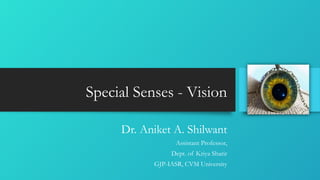
  • 1. Special Senses - Vision Dr. Aniket A. Shilwant Assistant Professor, Dept. of Kriya Sharir GJP-IASR, CVM University
  • 2. Human Eyeball • Bulbus oculi • Diameter 24 mm • 2segments • Anterior – 1/6th portion – Cornea • Posterior – 5/6th portion – Sclera • Centre of anterior curvature – anterior pole • Centre of posterior curvature – posterior pole • Line joining both poles – Optical axis. • Line joining to point on cornea to slight medial to anterior pole to fovea centralis (lateral to posterior pole) – Visual axis (Light rays pass through visual axis) Dr. Aniket Shilwant, GJPIASR 2
  • 3. Layers of Human Eyeball • Outer layer (Fibrous) – Cornea & Sclera • Middle layer (Vascular) – Choroid, Ciliary Body & Iris • Inner layer (Nervous) – Retina Dr. Aniket Shilwant, GJPIASR 3
  • 4. Fibrous Coat – Cornea • Transparent, colorless, thin layer covers 1/6th anterior part of eyeball • Covers mainly iris & pupil thus Reflects color of pupil • No blood supply, but abundant free nerve endings thus sensitive to pain • 5layers- • Stratified epithelium • Bowmann’s membrane / anterior elastic lamina • Substantia proper • Descement layer / posterior elastic lamina • Endothelial layer Dr. Aniket Shilwant, GJPIASR 4
  • 5. Fibrous Coat – Sclera • Outer fibrous layer - White and elastic fibers • Covers posterior 5/6th part of eyeball • Perforated part posteriorly where optic nerve leaves – Lamina Cribrosa Dr. Aniket Shilwant, GJPIASR 5
  • 6. Vascular Coat – Choroid • Thin layer • Highly vascular as rich in capillary plexus, small arteries and veins • Location- Between sclera and retina • Forms posterior 5/6th part • Choroid extended anteriorly by insertion of ciliary muscle (junction – Ora serrata) Dr. Aniket Shilwant, GJPIASR 6
  • 7. Vascular Coat – Ciliary Body • Thick anterior part of middle layer of eyeball. • Location-in front of Ora Serrate • Attaches to lens with - Suspensory ligaments • 3parts • Orbicularis ciliaris – Forms posterior 2/3rd of Ciliary body • Ciliary body proper – Ciliary muscles (Outer Longitudinal & Inner Circular) – PNS • Ciliary processes – Finger like process from inner surface of Ciliary body toward central axis of eye. Dr. Aniket Shilwant, GJPIASR 7
  • 9. Vascular Coat – Iris • Thin curtain like structure of eyeball forms circular Diaphragm. • Location – In front of lens • A central opening – Pupil • Separates space between cornea and lens in 2chambers – Anterior & Posterior • Aqueous humor Dr. Aniket Shilwant, GJPIASR 9
  • 10. Shades of Iris & Pupil Dr. Aniket Shilwant, GJPIASR 10
  • 11. Nervous Coat – Retina • Thin, delicate photo sensitive membrane • Covers the area from optic disc to Ciliary body • Ends at dentate border at Ora Serrata • 10 layers Dr. Aniket Shilwant, GJPIASR 11
  • 12. Layers of Retina Dr. Aniket Shilwant, GJPIASR 12 Layers of retina from outside in: 1. Layer of pigment epithelium 2. Layer of rods and cones 3. External limiting membrane 4. Outer nuclear layer 5. Outer plexiform layer 6. Inner nuclear layer 7. Inner plexiform layer 8. Ganglion cell layer 9. Layer of nerve fibers 10. Internal limiting membrane.
  • 13. Layers of Retina Dr. Aniket Shilwant, GJPIASR 13 Layers of retina from outside in: 1. Layer of pigment epithelium – Pigment granules – Melanin (Fuscin) Epithelial cells – Storage (Vit. A – Retinol & Phagocytic action – removal of cellular debris) 2. Layer of Rods and Cones – Photosensitive receptors
  • 14. Layers of Retina Dr. Aniket Shilwant, GJPIASR 14 3. External limiting membrane – Supporting Muller fibers 4. Outer nuclear layer – Granular layer of Rods & Cones having Nucleus 5. Outer plexiform layer – Reticular meshwork – Synaptic Terminal fibers & Dendrites of Bipolar cells
  • 15. Layers of Retina Dr. Aniket Shilwant, GJPIASR 15 6. Inner nuclear layer – Bipolar cells, Muller supporting fibers, Synapses (a. With ganglionic cell layer & outer plexiform layer) 7. Inner plexiform layer – Synapses (Ganglionic cells bipolar cells) 8. Ganglion cell layer – Multipolar cell layer (Giant ganglion cells & Midget Ganglion cells), Axons of these cells leave orbit as – Optic nerve
  • 16. Layers of Retina Dr. Aniket Shilwant, GJPIASR 16 9. Layer of nerve fibers – Layer of non-myelinated axons converges at Optic disc & leave as – Optic Nerve 10. Internal limiting membrane – Hyaline membrane separates Retina from inner Vitreous body
  • 17. Dr. Aniket Shilwant, GJPIASR 17
  • 18. Retinal 10 layers Dr. Aniket Shilwant, GJPIASR 18
  • 19. Dr. Aniket Shilwant, GJPIASR 19
  • 20. Dr. Aniket Shilwant, GJPIASR 20
  • 21. Fundus Oculi - Macula Lutea / Fovea centralis 21 • Small, yellowish area, also called Yellow Spot • Location – Lateral to optic disc in retina • It is yellow due to presence of pigment which is yellow in color • It has depression in the centre where all retinal layers become thin – Fovea centralis • It is the area for precise vision as it contains only – Cones Dr. Aniket Shilwant, GJPIASR
  • 22. Fundus Oculi – Optic Disc 22 • Pale disc • Location – Centre of posterior wall of eyeball • Formed by convergence of axons from ganglion cells • Contains all layers of retina except Rods & Cones • Thus it is insensitive to light and hence called as – Blind spot Dr. Aniket Shilwant, GJPIASR
  • 23. Physiology Of Vision Retinal image formation Photo sensitive receptors Photo transduction Visual pathway Dr. Aniket Shilwant, GJPIASR 23
  • 24. Retinal image formation Dr. Aniket Shilwant, GJPIASR 24 • Light rays pass through 4medias to reach retina • Cornea – Aqueous Humor - Lens – Vitreous Humor • Retinal image formation Refraction of light rays Accommodation of lens Constriction of pupil Convergence of eyeball
  • 25. Dr. Aniket Shilwant, GJPIASR 25 Refraction of light rays • Light rays pass through 4refractory Medias Lighter air – denser cornea Denser cornea to less denser aqueous humor Less dense aqueous humor to denser lens Denser lens to less denser vitreous humor Retinal image formation –Refraction of Light rays
  • 26. Dr. Aniket Shilwant, GJPIASR 26 Accommodation of lens • Ability of lens to gets adjust by giving curvature to itself when an eye is focusing an object • It is done so because light rays must any how fall on fovea centralis • In nearby vision – ciliary muscle contracts – thickened, shorten and bulging lens • In distant vision – ciliary muscle relaxed – flattened lens Retinal image formation –Accommodation of Lens
  • 27. Retinal image formation – Diameter of Pupil Constriction of pupil • Adjusting the diameter of hole through which light enters the eyeball • Constricts – Bright light • Dilates – Dim light Dr. Aniket Shilwant, GJPIASR 27
  • 28. Convergence of eyeball • Human eye – Single binocular vision • As object moves closer to eyes, eyeball start moving medially so that light rays must hit on identical spots on retina. • Medial rotation of eyeball for proper adjustment of vision – Convergence of eyeball. Dr. Aniket Shilwant, GJPIASR 28 Retinal image formation– Convergence of Eyeball
  • 29. Photosensitive Receptors • Present in 2nd Layer of Retina from outside • Layer of Rods & Cones • Rods – Rhodopsin Chromophore (Vit. A – Retinal) & Opsin (Scotopsin) • Cones – Photopsin Chromophore (Vit. A – Retinal) & Opsin (Porphyropsin, Idopsin & Cyanopsin) Dr. Aniket Shilwant, GJPIASR 29
  • 30. Photosensitive Receptors – Rods • Cylindrical in shape • Length – 40 µ to 60 µ Diameter – 2 µ • Outer segment – • Contains about 1000 floating flat membranous disks • These disks contains photosensitive pigment – Rhodopsin • Newly formed disks at the inner portion of outer segment and older disks are pushed towards the tip. • Rate of formation of membranous disks – 3 or 4 per hour Dr. Aniket Shilwant, GJPIASR 30
  • 31. • Inner segment – • It is connected to outer segment by Cilium • Contains large number of mitochondria • Cell body – • It is the portion containing nucleus of the cell. • Synaptic terminal – • It synapses with dendrites of bipolar cell and horizontal cell • Involved neurotransmitter – Glutamate Dr. Aniket Shilwant, GJPIASR 31 Photosensitive Receptors – Rods
  • 32. FUNCTIONS OF RODS • These cells have a very low threshold to light • Very sensitive towards light • Rod cell are responsible for Dim Light Vision or Night Vision or Scotopic Vision • Vision by Rod cell is black and white or rather grey Dr. Aniket Shilwant, GJPIASR 32 Photosensitive Receptors – Rods
  • 33. Dr. Aniket Shilwant, GJPIASR 33
  • 34. Photosensitive Receptors – Cones • Cone or Flask shaped • Length - 35 µ to 40 µ Diameter – 5 µ • Outer segment – • Small, conical • Does not contain separate membranous disks • The inward folding of cell membrane called as – Saccules • Photosensitive pigment synthesized in inner segment and incorporated into folding forming saccule • Inner segment – • It is connected to outer segment by Cilium • Contains large number of mitochondria Dr. Aniket Shilwant, GJPIASR 34
  • 35. • Cell body – • It is the portion containing nucleus of the cell. • Synaptic terminal – • It synapses with dendrites of bipolar cell and horizontal cell • Involved neurotransmitter – Glutamate Dr. Aniket Shilwant, GJPIASR 35 Photosensitive Receptors – Cones
  • 36. FUNCTIONS OF CONE CELL:- • These cells have high threshold to light • Cones are sensitive only to bright light • Cone cell are responsible for Bright Light Vision or Daylight Vision or Photopic Vision • Cone cells are also responsible for Acuity of Vision and Color Vision. Dr. Aniket Shilwant, GJPIASR 36 Photosensitive Receptors – Cones
  • 37. Dark & Light Adaptation Dr. Aniket Shilwant, GJPIASR 37 Causes for Dark Adaptation  Increased sensitivity of rods as a result of resynthesis of rhodopsin  Dilatation of pupil Causes for Light Adaptation  Decreased sensitivity of rods as a result of resynthesis of rhodopsin  Constriction of pupil
  • 38. Photochemical changes in Synthesis of Rhodopsin Dr. Aniket Shilwant, GJPIASR 38 Rhodopsin Batho-Rhodopsin Lumi-Rhodopsin Meta-Rhodopsin I Meta-Rhodopsin II Scotopsin & Trans Retinal Trans Retinal – Cis Retinal Trans Retinol – Cis Retinol Cis Retinal + Scotopsin
  • 39. Photon absorbed 11 cis retinal gets decomposed to Metarhodopsin II (active from)by Rhodopsin of Rod cell in retina Metarhodopsin II activates G protein – Transducin Transducin activates cGMP phosphodiesterase Reduction of cGMP to 5’-GMP Closure of sodium channels in membrane of visual receptors Blocked entry of sodium ions leading to more negativity –Hyperpolarization Development of Receptor Potential Dr. Aniket Shilwant, GJPIASR 39
  • 41. Dr. Aniket Shilwant, GJPIASR 41 Dark Current Normally resting membrane potential = -70 to -90mV RMP in visual receptors = -40mV Sodiums leaked from inner segment Re enters cells in outer segment via sodium channels Na channels remain open in presence of cGMP Entry of Na again back in cells gives mild depolarization RMP remains constant = -40mV This is dark current.
  • 42. Photo-Transduction • Light rays striking on retina • Photon absorption • Rhodopsin dissociation – Meta- Rhodopsin & Retinene • Meta-Rhodopsin stimulates Transducin (G-protein) • Transducin – cGMP phosphodiesterase • cGMP is hydrolyzed into 5’cGMP • Decreased concentration of cGMP • Closure of sodium channels • Addition to this – Na+K+ pump • Extra negativity inside cells • Hyperpolarization • Development of Receptor potential Dr. Aniket Shilwant, GJPIASR 42
  • 43. Visual Pathway Dr. Aniket Shilwant, GJPIASR 43 • Axon fibers from retina leave eyeball as – Optic N. • Crossing of fibers at – Optic Chiasma (Only Nasal Fibers crosses) • Fibers thereafter terminate at –  Lateral geniculate body (Thalamus)  Superior colliculi (Midbrain) • From both the optic radiations terminate at – Primary visual center (Occipital lobe – Area no. 17) • Primary visual centre is associated with –  Visual Association Area (Area No. 18) &  Occipital Eyefield Area (Area No. 19)
  • 44. Dr. Aniket Shilwant, GJPIASR 44
  • 45. Thank You All !!! Dr. Aniket A. Shilwant Assistant Professor Department of Kriya Sharir GJP-IASR, CVM University Email – ayuraniket18@gmail.com http://ayugjac.edu.in/Staff_CV.aspx?dl=dn3Mja19480dn3Mja19 http://scholar.google.co.in/citations?user=636K2sMAAAAJ&hl=en https://www.researchgate.net/profile/Aniket_Shilwant