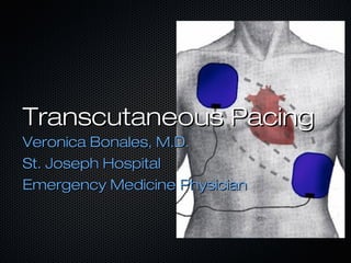
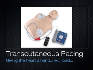
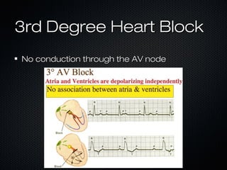
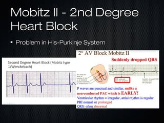
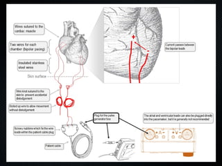
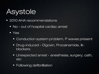
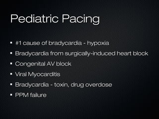
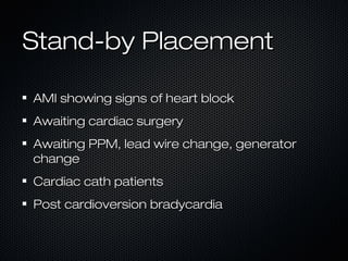
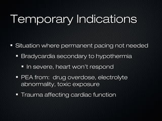
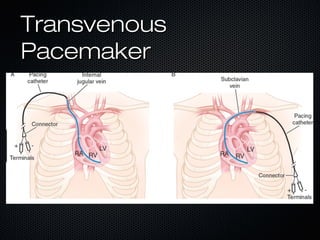
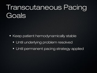
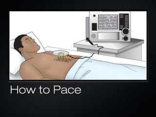
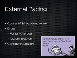
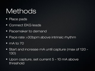
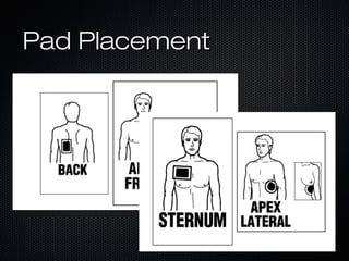
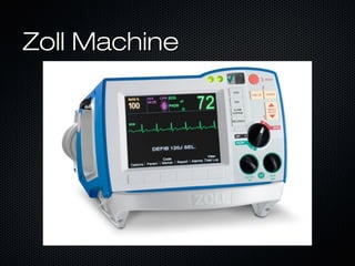
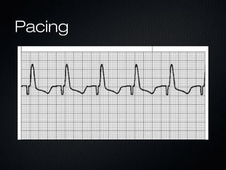
Transcutaneous PacingTranscutaneous Pacing
Veronica Bonales, M.D.Veronica Bonales, M.D.
St. Joseph HospitalSt. Joseph Hospital
Emergency Medicine PhysicianEmergency Medicine Physician
Why do we care...?Why do we care...?
AHA 2016AHA 2016
OOH - >350K - 12% survivalOOH - >350K - 12% survival
IH - 209K - 24.8% survivalIH - 209K - 24.8% survival
#1 Cause of death - heart disease#1 Cause of death - heart disease
VF, VT, high degree AV BlockVF, VT, high degree AV Block
Back toBack to
the Basicsthe Basics
Coronary muscleCoronary muscle
cells got the beatcells got the beat
Cardiac MyocytesCardiac Myocytes
Intrinsic rhythmIntrinsic rhythm
Not coordinated effortNot coordinated effort
Back toBack to
the Basicsthe Basics
Heart’s internalHeart’s internal
pacemakerpacemaker
Heart ConductionHeart Conduction
SA nodeSA node
AV nodeAV node
BOHBOH
Purkinje fibersPurkinje fibers
Heart ConductionHeart Conduction
Back toBack to
the Basicsthe Basics
Coronary arteryCoronary artery
anatomyanatomy
Coronary arteryCoronary artery
anatomyanatomy
Left MainLeft Main
““Widowmaker”Widowmaker”
lesionlesion
Right MainRight Main
Loss of intrinsicLoss of intrinsic
pacemakerpacemaker
Right Coronary Artery MIRight Coronary Artery MI
HypotensionHypotension
after NTGafter NTG
BradycardiaBradycardia
Nausea/vomitingNausea/vomiting
HiccoughingHiccoughing
Urge to defecateUrge to defecate
Transcutaneous PacingTranscutaneous Pacing
Giving the heart a hand... er... pad...Giving the heart a hand... er... pad...
IndicationsIndications
BradycardiaBradycardia
3rd degree heart block3rd degree heart block
Mobitz type II 2nd degree heart blockMobitz type II 2nd degree heart block
When hemodynamically unstableWhen hemodynamically unstable
Overdrive PacingOverdrive Pacing
AsystoleAsystole
PediatricPediatric
StandbyStandby
Bradycardia (HR < 50)Bradycardia (HR < 50)
Hemodynamic InstabilityHemodynamic Instability
Acute AMSAcute AMS
Severe Chest PainSevere Chest Pain
IschemicIschemic
CHFCHF
SyncopeSyncope
3rd Degree Heart Block3rd Degree Heart Block
No conduction through the AV nodeNo conduction through the AV node
Mobitz II - 2nd DegreeMobitz II - 2nd Degree
Heart BlockHeart Block
Problem in His-Purkinje SystemProblem in His-Purkinje System
Overdrive PacingOverdrive Pacing
Tachydysrhythmia not responsive to drugTachydysrhythmia not responsive to drug
therapy or electrocardioversiontherapy or electrocardioversion
Usually only good HR <180Usually only good HR <180
Goal to break re-entry circuitGoal to break re-entry circuit
AV node able to take controlAV node able to take control
Can cause VFibCan cause VFib
AsystoleAsystole
2010 AHA recommendations2010 AHA recommendations
No - out of hospital cardiac arrestNo - out of hospital cardiac arrest
YesYes
Conduction system problem, P waves presentConduction system problem, P waves present
Drug-induced - Digoxin, Procainamide,Drug-induced - Digoxin, Procainamide, ΒΒ--
blockersblockers
Unexpected arrest - anesthesia, surgery, cath,Unexpected arrest - anesthesia, surgery, cath,
etcetc
Following defibrilliationFollowing defibrilliation
Pediatric PacingPediatric Pacing
#1 cause of bradycardia - hypoxia#1 cause of bradycardia - hypoxia
Bradycardia from surgically-induced heart blockBradycardia from surgically-induced heart block
Congenital AV blockCongenital AV block
Viral MyocarditisViral Myocarditis
Bradycardia - toxin, drug overdoseBradycardia - toxin, drug overdose
PPM failurePPM failure
Stand-by PlacementStand-by Placement
AMI showing signs of heart blockAMI showing signs of heart block
Awaiting cardiac surgeryAwaiting cardiac surgery
Awaiting PPM, lead wire change, generatorAwaiting PPM, lead wire change, generator
changechange
Cardiac cath patientsCardiac cath patients
Post cardioversion bradycardiaPost cardioversion bradycardia
Temporary IndicationsTemporary Indications
Situation where permanent pacing not neededSituation where permanent pacing not needed
Bradycardia secondary to hypothermiaBradycardia secondary to hypothermia
In severe, heart won’t respondIn severe, heart won’t respond
PEA from: drug overdose, electrolytePEA from: drug overdose, electrolyte
abnormality, toxic exposureabnormality, toxic exposure
Trauma affecting cardiac functionTrauma affecting cardiac function
TransvenousTransvenous
PacemakerPacemaker
Temporary IndicationsTemporary Indications
When unable to place transvenous pacerWhen unable to place transvenous pacer
Non-native Tricuspid valveNon-native Tricuspid valve
Potential for bleeding - anticoagulantsPotential for bleeding - anticoagulants
Potential for infection - immunocompromisedPotential for infection - immunocompromised
Transcutaneous PacingTranscutaneous Pacing
GoalsGoals
Keep patient hemodynamically stableKeep patient hemodynamically stable
Until underlying problem resolvedUntil underlying problem resolved
Until permanent pacing strategy appliedUntil permanent pacing strategy applied
How to PaceHow to Pace
External PacingExternal Pacing
Consent/Make patient awareConsent/Make patient aware
DrugsDrugs
Fentanyl/versedFentanyl/versed
Morphine/ativanMorphine/ativan
Consider intubationConsider intubation
MethodsMethods
Place padsPlace pads
Connect EKG leadsConnect EKG leads
Pacemaker to demandPacemaker to demand
Pace rate >30bpm above intrinsic rhythmPace rate >30bpm above intrinsic rhythm
mA to 70mA to 70
Start and increase mA until capture (max of 120 -Start and increase mA until capture (max of 120 -
130)130)
Upon capture, set current 5 - 10 mA aboveUpon capture, set current 5 - 10 mA above
thresholdthreshold
Pad PlacementPad Placement
MethodsMethods
Place padsPlace pads
Connect EKG leadsConnect EKG leads
Pacemaker to demandPacemaker to demand
Pace rate >30bpm above intrinsic rhythmPace rate >30bpm above intrinsic rhythm
mA to 70mA to 70
Start and increase mA until capture (max of 120 -Start and increase mA until capture (max of 120 -
130)130)
Upon capture, set current 5 - 10 mA aboveUpon capture, set current 5 - 10 mA above
thresholdthreshold
Zoll MachineZoll Machine
PacingPacing
ResultsResults
Associated with reduced L ventricular systolicAssociated with reduced L ventricular systolic
pressurepressure
Lower stroke index 2nd to AV dyssynchronyLower stroke index 2nd to AV dyssynchrony
Because of muscle contractions, can provideBecause of muscle contractions, can provide
greater COgreater CO
ComplicationsComplications
Failure to paceFailure to pace
Failure to captureFailure to capture
DiscomfortDiscomfort
Skin burns (check q 30 minutes)Skin burns (check q 30 minutes)
Questions...?Questions...?

Using External Pacemaker

  • 1.
    Transcutaneous PacingTranscutaneous Pacing VeronicaBonales, M.D.Veronica Bonales, M.D. St. Joseph HospitalSt. Joseph Hospital Emergency Medicine PhysicianEmergency Medicine Physician
  • 2.
    Why do wecare...?Why do we care...? AHA 2016AHA 2016 OOH - >350K - 12% survivalOOH - >350K - 12% survival IH - 209K - 24.8% survivalIH - 209K - 24.8% survival #1 Cause of death - heart disease#1 Cause of death - heart disease VF, VT, high degree AV BlockVF, VT, high degree AV Block
  • 3.
    Back toBack to theBasicsthe Basics Coronary muscleCoronary muscle cells got the beatcells got the beat
  • 4.
    Cardiac MyocytesCardiac Myocytes IntrinsicrhythmIntrinsic rhythm Not coordinated effortNot coordinated effort
  • 5.
    Back toBack to theBasicsthe Basics Heart’s internalHeart’s internal pacemakerpacemaker
  • 6.
    Heart ConductionHeart Conduction SAnodeSA node AV nodeAV node BOHBOH Purkinje fibersPurkinje fibers
  • 7.
  • 8.
  • 9.
    Back toBack to theBasicsthe Basics Coronary arteryCoronary artery anatomyanatomy
  • 10.
  • 11.
  • 12.
    Right MainRight Main Lossof intrinsicLoss of intrinsic pacemakerpacemaker
  • 13.
    Right Coronary ArteryMIRight Coronary Artery MI HypotensionHypotension after NTGafter NTG BradycardiaBradycardia Nausea/vomitingNausea/vomiting HiccoughingHiccoughing Urge to defecateUrge to defecate
  • 14.
    Transcutaneous PacingTranscutaneous Pacing Givingthe heart a hand... er... pad...Giving the heart a hand... er... pad...
  • 15.
    IndicationsIndications BradycardiaBradycardia 3rd degree heartblock3rd degree heart block Mobitz type II 2nd degree heart blockMobitz type II 2nd degree heart block When hemodynamically unstableWhen hemodynamically unstable Overdrive PacingOverdrive Pacing AsystoleAsystole PediatricPediatric StandbyStandby
  • 16.
    Bradycardia (HR <50)Bradycardia (HR < 50) Hemodynamic InstabilityHemodynamic Instability Acute AMSAcute AMS Severe Chest PainSevere Chest Pain IschemicIschemic CHFCHF SyncopeSyncope
  • 17.
    3rd Degree HeartBlock3rd Degree Heart Block No conduction through the AV nodeNo conduction through the AV node
  • 18.
    Mobitz II -2nd DegreeMobitz II - 2nd Degree Heart BlockHeart Block Problem in His-Purkinje SystemProblem in His-Purkinje System
  • 19.
    Overdrive PacingOverdrive Pacing Tachydysrhythmianot responsive to drugTachydysrhythmia not responsive to drug therapy or electrocardioversiontherapy or electrocardioversion Usually only good HR <180Usually only good HR <180 Goal to break re-entry circuitGoal to break re-entry circuit AV node able to take controlAV node able to take control Can cause VFibCan cause VFib
  • 21.
    AsystoleAsystole 2010 AHA recommendations2010AHA recommendations No - out of hospital cardiac arrestNo - out of hospital cardiac arrest YesYes Conduction system problem, P waves presentConduction system problem, P waves present Drug-induced - Digoxin, Procainamide,Drug-induced - Digoxin, Procainamide, ΒΒ-- blockersblockers Unexpected arrest - anesthesia, surgery, cath,Unexpected arrest - anesthesia, surgery, cath, etcetc Following defibrilliationFollowing defibrilliation
  • 22.
    Pediatric PacingPediatric Pacing #1cause of bradycardia - hypoxia#1 cause of bradycardia - hypoxia Bradycardia from surgically-induced heart blockBradycardia from surgically-induced heart block Congenital AV blockCongenital AV block Viral MyocarditisViral Myocarditis Bradycardia - toxin, drug overdoseBradycardia - toxin, drug overdose PPM failurePPM failure
  • 23.
    Stand-by PlacementStand-by Placement AMIshowing signs of heart blockAMI showing signs of heart block Awaiting cardiac surgeryAwaiting cardiac surgery Awaiting PPM, lead wire change, generatorAwaiting PPM, lead wire change, generator changechange Cardiac cath patientsCardiac cath patients Post cardioversion bradycardiaPost cardioversion bradycardia
  • 24.
    Temporary IndicationsTemporary Indications Situationwhere permanent pacing not neededSituation where permanent pacing not needed Bradycardia secondary to hypothermiaBradycardia secondary to hypothermia In severe, heart won’t respondIn severe, heart won’t respond PEA from: drug overdose, electrolytePEA from: drug overdose, electrolyte abnormality, toxic exposureabnormality, toxic exposure Trauma affecting cardiac functionTrauma affecting cardiac function
  • 25.
  • 26.
    Temporary IndicationsTemporary Indications Whenunable to place transvenous pacerWhen unable to place transvenous pacer Non-native Tricuspid valveNon-native Tricuspid valve Potential for bleeding - anticoagulantsPotential for bleeding - anticoagulants Potential for infection - immunocompromisedPotential for infection - immunocompromised
  • 27.
    Transcutaneous PacingTranscutaneous Pacing GoalsGoals Keeppatient hemodynamically stableKeep patient hemodynamically stable Until underlying problem resolvedUntil underlying problem resolved Until permanent pacing strategy appliedUntil permanent pacing strategy applied
  • 28.
  • 29.
    External PacingExternal Pacing Consent/Makepatient awareConsent/Make patient aware DrugsDrugs Fentanyl/versedFentanyl/versed Morphine/ativanMorphine/ativan Consider intubationConsider intubation
  • 30.
    MethodsMethods Place padsPlace pads ConnectEKG leadsConnect EKG leads Pacemaker to demandPacemaker to demand Pace rate >30bpm above intrinsic rhythmPace rate >30bpm above intrinsic rhythm mA to 70mA to 70 Start and increase mA until capture (max of 120 -Start and increase mA until capture (max of 120 - 130)130) Upon capture, set current 5 - 10 mA aboveUpon capture, set current 5 - 10 mA above thresholdthreshold
  • 31.
  • 32.
    MethodsMethods Place padsPlace pads ConnectEKG leadsConnect EKG leads Pacemaker to demandPacemaker to demand Pace rate >30bpm above intrinsic rhythmPace rate >30bpm above intrinsic rhythm mA to 70mA to 70 Start and increase mA until capture (max of 120 -Start and increase mA until capture (max of 120 - 130)130) Upon capture, set current 5 - 10 mA aboveUpon capture, set current 5 - 10 mA above thresholdthreshold
  • 33.
  • 34.
  • 35.
    ResultsResults Associated with reducedL ventricular systolicAssociated with reduced L ventricular systolic pressurepressure Lower stroke index 2nd to AV dyssynchronyLower stroke index 2nd to AV dyssynchrony Because of muscle contractions, can provideBecause of muscle contractions, can provide greater COgreater CO
  • 36.
    ComplicationsComplications Failure to paceFailureto pace Failure to captureFailure to capture DiscomfortDiscomfort Skin burns (check q 30 minutes)Skin burns (check q 30 minutes)
  • 37.