
Recommended
More Related Content
What's hot
What's hot (20)
Similar to orbital cellulitis.pptx
Similar to orbital cellulitis.pptx (20)
Recently uploaded
Recently uploaded (20)
orbital cellulitis.pptx
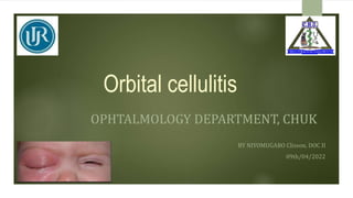
- 1. Orbital cellulitis OPHTALMOLOGY DEPARTMENT, CHUK BY NIYOMUGABO Clisson, DOC II 09th/04/2022
- 2. Outline Introduction Clinical presentation Etiology Management complications
- 3. Introduction Orbital cellulitis is infection of tissues (orbital fats and extraocular muscles) behind the orbital septum. The orbital septum is the major anatomical landmark separating anterior part(eyelids) from posterior part (orbit) Infection to tissues anterior to orbital septum is known preseptal cellulitis and infection to tissues posterior to it is known as orbital cellulitis Life threatening, Serious orbit infection, Most commonly caused by : S. pneumonia, S. aureus, S. pyogenes and H. influenza. Highly associated with sinusal Infection especially ethmoid sinus infection. More commonly seen in children . Incidence 1.6 per 100,000 compared to adults 0.1 per 100,000.
- 4. Clinical presentation Proptosis ophthalomoplegia Edema Pain Afferent pupillary defect Proptosis Optic nerve swelling Other symptoms: fever, headache, malaise, rhinorrhea.
- 5. Pathogenesis Occurs in 3 ways: Extension of an infection from the paranasal sinuses or other periorbital structures such as the face, lacrimal sac, or globe. Ethymoid sinus account for 90% of all cases. direct inoculation of the orbit from trauma or surgery (orbital decompression). Hematogenous spread from bacteremia
- 6. investigations Labs: CBC ESR, CRP Urea and creatinine Blood cultures Imaging: CT scan with contrast for better visualization of soft tissues MRI if available is a better option US to rule out orbital myositis
- 7. Management Patient with orbital cellulitis should be hospitalized for treatment until the patient is afebrile and improved clinically. Medication: broad spectrum iv antibiotics until culture results. IV Atbs for 2 weeks, then oral Atbs for 2-3 weeks. Fungal infection require iv antifungal therapy along with surgical debridement. Surgery: Canthotomy and cantholysis if compartment syndrome is diagnosed. Surgical drainage if: Decrease in vision Proptosis progresses Size of abscess not decrease on CT Scan after 48 hrs.
- 8. complications Orbital abcess Subperiosteal abcess Cavernous sinus thrombosis Optic neuropathy Raised IOP Occlusion of central retinal artery Parotid/temporal abcess Meningitis Brain abcess Septicemia
- 9. Thank you