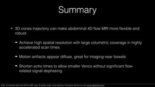
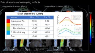
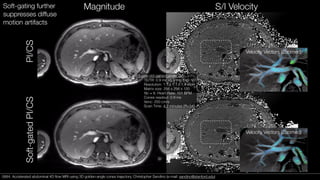
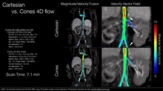
0684. Accelerated abdominal 4D flow MRI
using 3D golden-angle cones trajectory
Christopher M. Sandino1, Joseph Y. Cheng2, Marcus T. Alley2, Michael Carl3, Shreyas S. Vasanawala2
1Department of Electrical Engineering, Stanford University, Stanford, CA. 2Department of Radiology, Stanford
University, Stanford, CA. 3Applied Sciences Laboratory, GE Healthcare, San Diego, CA.
Contact: sandino@stanford.edu
Speaker Name: Christopher Sandino
I have the following financial interest or relationship to disclose with regard to the subject
matter of this presentation:
Company Name: GE Healthcare
Type of Relationship: Research Agreement
Declaration of
Financial Interests or Relationships
Cartesian 4D flow1,2 in the abdomen
Many challenges…
• Low Venc
• Respiratory & bowel motion
• High spatiotemporal resolution
required for simultaneous
DCE+4D Flow (XD Flow3)
• Long scan times
11-year-old female patient w/ ferumoxytol
TE/TR: 2.16 ms / 6.16 ms; Flip: 20°
Resolution: 1.0 x 1.4 x 1.0 mm3
Nc = 10, Heart Rate: 100 BPM
Acceleration: 4.0 x 2.5
Venc: 100 cm/s
Scan Time: 9.4 minutes
[1] M Markl, et al. Time-resolved 3DPC MRI. JMRI, 2003.
[2] JY Cheng, et al. Comprehensive 4D flow MRI. JMRI, 2016.
[3] JY Cheng, et al. XD Flow. Scientific reports, 2017.
0684. Accelerated abdominal 4D flow MRI using 3D golden-angle cones trajectory. Christopher Sandino (e-mail: sandino@stanford.edu)
kz
kx
Non-Cartesian 4D Flow
• Previous work: 3D radial4,5 and
stack-of-spirals6,7
• 3D cones trajectory8,9 combines
advantages of both:
• Short echo times
• Motion robust
• High sampling efficiency
[4] T Gu, et al. PC-VIPR. Am J Neuroradiology, 2005.
[5] KM Johnson, et al. Renal PC-VIPR. JCMR, 2008.
[6] A Sigfridsson, et al. Spiral 4D flow. MRM, 2012.
[7] H Dyvorne, et al. Abdominal spiral 4D flow. Radiology, 2014.
[8] PT Gurney, et al. 3D cones. MRM, 2006.
[9] EJ Zucker, et al. Pediatric chest MRI w/ cones. JMRI, 2017.
0684. Accelerated abdominal 4D flow MRI using 3D golden-angle cones trajectory. Christopher Sandino (e-mail: sandino@stanford.edu)
0.6 0.7 0.8 0.9 1 1.1 1.2 1.3 1.4 1.5 1.6
Readout duration (ms)
5
10
15
20
25
30
35Totalscantime(min)
Venc = 300 cm/s
Venc = 150 cm/s
Venc = 75 cm/s
Venc = 25 cm/s
Sampling Efficiency of Radial vs. Cones
Stretch = 0
-1
0
1
-1
0
1
0
2
4
0
1
2
1
Stretch = 1
4
0 0
-1 -1
0
1
2
1
Stretch = 1.5
4
0 0
-1 -1
0
1
2
1
Stretch = 2
4
0 0
-1 -1
0
1
2
1
Stretch = 2.5
4
0 0
-1 -1
0
1
2
1
Stretch = 3
4
0 0
-1 -1
Stretch = 0
-1
0
1
-1
0
1
0
2
4
0
1
2
1
Stretch = 1
4
0 0
-1 -1
0
1
2
1
Stretch = 1.5
4
0 0
-1 -1
0
1
2
1
Stretch = 2
4
0 0
-1 -1
0
1
2
1
Stretch = 2.5
4
0 0
-1 -1
0
1
2
1
Stretch = 3
4
0 0
-1 -1
Stretch = 0
-1
0
1
-1
0
1
0
2
4
0
1
2
1
Stretch = 1
4
0 0
-1 -1
0
1
2
1
Stretch = 1.5
4
0 0
-1 -1
0
1
2
1
Stretch = 2
4
0 0
-1 -1
0
1
2
1
Stretch = 2.5
4
0 0
-1 -1
0
1
2
1
Stretch = 3
4
0 0
-1 -1
Stretch = 0
-1
0
1
-1
0
1
0
2
4
0
1
2
1
Stretch = 1
4
0 0
-1 -1
0
1
2
1
Stretch = 1.5
4
0 0
-1 -1
0
1
2
1
Stretch = 2
4
0 0
-1 -1
0
1
2
1
Stretch = 2.5
4
0 0
-1 -1
0
1
2
1
Stretch = 3
4
0 0
-1 -1
(Radial) Simulation Parameters
Resolution: 1.0 x 1.0 x 2.0 mm3
Field of view: 25.6 x 25.6 x 24.0 cm
Single cardiac phase
Fully-sampled
0684. Accelerated abdominal 4D flow MRI using 3D golden-angle cones trajectory. Christopher Sandino (e-mail: sandino@stanford.edu)
Sequence Implementation
TR
Golden-angle re-ordering scheme
0684. Accelerated abdominal 4D flow MRI using 3D golden-angle cones trajectory. Christopher Sandino (e-mail: sandino@stanford.edu)
Estimation of respiratory motion
[10] T Zhang, et al. Robust body MRI with dense coil arrays. MRM, 2016.
Multi-channel DC navigators Respiratory signal
filtering,
clustering10,
etc.
0684. Accelerated abdominal 4D flow MRI using 3D golden-angle cones trajectory. Christopher Sandino (e-mail: sandino@stanford.edu)
PI/CS Reconstruction
• CPU reconstruction time: 4-5 hours (implemented using BART12)
• Initially intractable due to memory required for oversampling in all 3 spatial dimensions
➡ PCA coil compression13 (32->16 channels)
➡ Tradeoffs with temporal resolution
m = argminm
1
2
kW(Am y)k2
2 +
X
i
i|Ri(m)|1
Forward model A includes:
• Coil sensitivities (ESPIRiT)
• Non-uniform FFT
Weighting matrix W includes:
• Density compensation
• Respiratory motion state
consistency (soft-gating11)
Sparsity promoters Ri include:
• 3D spatial Wavelet
• Temporal finite differences
m = reconstructed images
y = acquired k-space data
x = spatial domain
c = cardiac phase
r = respiratory phase
t = temporal phase
[11] KM Johnson, et al. Improved least squares recon. MRM, 2012.
[12] M Uecker, et al. BART. ISMRM, 2015.
[13] F Huang, et al. Coil compression. MRM, 2007.
0684. Accelerated abdominal 4D flow MRI using 3D golden-angle cones trajectory. Christopher Sandino (e-mail: sandino@stanford.edu)
Preliminary Studies
• Cones 4D flow acquired in three pediatric patients referred for contrast-
enhanced (ferumoxytol) abdominal MRI
• Three experiments:
1. Robustness to high rates of acceleration with respect to vessel delineation
and flow quantification
2. Utility of soft-gating reconstruction
3. Comparison of image quality between Cartesian and cones 4D flow
0684. Accelerated abdominal 4D flow MRI using 3D golden-angle cones trajectory. Christopher Sandino (e-mail: sandino@stanford.edu)0684. Accelerated abdominal 4D flow MRI using 3D golden-angle cones trajectory. Christopher Sandino (e-mail: sandino@stanford.edu)
Cones 4D flow (4:56 min, R=17.5)
5-month-old male patient under GA:
TE/TR: 1.0 ms / 3.8 ms; Flip: 15°
Resolution: 1.0 x 1.4 x 1.0 mm3
Matrix size: 256 x 120 x 256
Nc = 8, Heart Rate: 121 BPM
Cones readout: 0.9 ms
Venc: 150 cm/s
AbsoluteVelocity(cm/s)
100
0
Robustness to undersampling artifacts
Cones 4D flow (2:59 min, R=30)
0684. Accelerated abdominal 4D flow MRI using 3D golden-angle cones trajectory. Christopher Sandino (e-mail: sandino@stanford.edu)0684. Accelerated abdominal 4D flow MRI using 3D golden-angle cones trajectory. Christopher Sandino (e-mail: sandino@stanford.edu)
Velocity Vectors (Zoomed)
3-year-old patient under GA:
TE/TR: 0.9 ms / 3.6 ms; Flip: 15°
Resolution: 1.1 x 1.1 x 1.4 mm3
Matrix size: 256 x 256 x 120
Nc = 8, Heart Rate: 101 BPM
Cones readout: 0.9 ms
Venc: 250 cm/s
Scan Time: 4.2 minutes (R=34)
Magnitude S/I Velocity
PI/CSSoft-gatedPI/CS
Soft-gating further
suppresses diffuse
motion artifacts
Velocity Vectors (Zoomed)
0684. Accelerated abdominal 4D flow MRI using 3D golden-angle cones trajectory. Christopher Sandino (e-mail: sandino@stanford.edu)0684. Accelerated abdominal 4D flow MRI using 3D golden-angle cones trajectory. Christopher Sandino (e-mail: sandino@stanford.edu)
Cartesian
vs. Cones 4D flow
9-year-old male patient w/o GA:
Cartesian 4D flow (Coronal)
TE/TR: 2.2 ms / 6.3 ms; Flip: 15°
Resolution: 1.1 x 2.6 x 0.8 mm3
Matrix Size: 204 x 140 x 320
Nc = 8, Heart Rate: 159 BPM
Acceleration: 10
Venc: 150 cm/s
Cones 4D flow (Axial)
TE/TR: 1.1 ms / 4.9 ms; Flip: 15°
Resolution: 0.8 x 0.8 x 2.0 mm3
Matrix Size: 256 x 256 x 120
Nc = 8, Heart Rate: 159 BPM
Cones readout: 1.0 ms
Acceleration: 16
Venc: 150 cm/s
Scan Time: 7.1 min
Magnitude/Velocity Fusion Velocity Vector Field
CartesianCones
AbsoluteVelocity(cm/s)
75
0
0684. Accelerated abdominal 4D flow MRI using 3D golden-angle cones trajectory. Christopher Sandino (e-mail: sandino@stanford.edu)0684. Accelerated abdominal 4D flow MRI using 3D golden-angle cones trajectory. Christopher Sandino (e-mail: sandino@stanford.edu)
Limitations
• Spatiotemporal resolution currently limited by reconstruction
• Eddy current-related phase errors appeared to be worse in cones before
phase correction14
• Off-resonance effects not observed in short readout reconstructions, but
will need to be corrected for extended readouts
[14] PG Walker, et al. Semi-automatic phase error correction. JMRI, 1993.
0684. Accelerated abdominal 4D flow MRI using 3D golden-angle cones trajectory. Christopher Sandino (e-mail: sandino@stanford.edu)0684. Accelerated abdominal 4D flow MRI using 3D golden-angle cones trajectory. Christopher Sandino (e-mail: sandino@stanford.edu)
Summary
• 3D cones trajectory can make abdominal 4D flow MRI more flexible and
robust:
➡ Achieve high spatial resolution with large volumetric coverage in highly
accelerated scan times
➡ Motion artifacts appear diffuse, great for imaging near bowels
➡ Shorten echo times to allow smaller Vencs without significant flow-
related signal dephasing
0684. Accelerated abdominal 4D flow MRI using 3D golden-angle cones trajectory. Christopher Sandino (e-mail: sandino@stanford.edu)0684. Accelerated abdominal 4D flow MRI using 3D golden-angle cones trajectory. Christopher Sandino (e-mail: sandino@stanford.edu)
Acknowledgements
Stanford University
Pediatric MR Group
Shreyas Vasanawala
Brian Hargreaves
Joseph Cheng
Marcus Alley
Morteza Mardani
Jianmin Yuan
Adam Bush
Feiyu Chen
Magnetic Resonance Systems
Research Laboratory
Dwight Nishimura
John Pauly
David Zeng
Srivathsan Koundinyan
Mario Malavé
Kwang Eun Jang
UC Berkeley
Miki Lustig
Frank Ong
Jonathan Tamir
GE Healthcare
Michael Carl
Peng Lai
Valentina Taviani
Anne Menini
Funding Sources
NIH R01 EB0096900
NSF Graduate Research
Fellowship
GE Healthcare
0684. Accelerated abdominal 4D flow MRI using 3D golden-angle cones trajectory. Christopher Sandino (e-mail: sandino@stanford.edu)0684. Accelerated abdominal 4D flow MRI using 3D golden-angle cones trajectory. Christopher Sandino (e-mail: sandino@stanford.edu)

Accelerated abdominal 4D flow MRI using 3D golden-angle cones trajectory

  • 1.
    0684. Accelerated abdominal4D flow MRI using 3D golden-angle cones trajectory Christopher M. Sandino1, Joseph Y. Cheng2, Marcus T. Alley2, Michael Carl3, Shreyas S. Vasanawala2 1Department of Electrical Engineering, Stanford University, Stanford, CA. 2Department of Radiology, Stanford University, Stanford, CA. 3Applied Sciences Laboratory, GE Healthcare, San Diego, CA. Contact: sandino@stanford.edu
  • 2.
    Speaker Name: ChristopherSandino I have the following financial interest or relationship to disclose with regard to the subject matter of this presentation: Company Name: GE Healthcare Type of Relationship: Research Agreement Declaration of Financial Interests or Relationships
  • 3.
    Cartesian 4D flow1,2in the abdomen Many challenges… • Low Venc • Respiratory & bowel motion • High spatiotemporal resolution required for simultaneous DCE+4D Flow (XD Flow3) • Long scan times 11-year-old female patient w/ ferumoxytol TE/TR: 2.16 ms / 6.16 ms; Flip: 20° Resolution: 1.0 x 1.4 x 1.0 mm3 Nc = 10, Heart Rate: 100 BPM Acceleration: 4.0 x 2.5 Venc: 100 cm/s Scan Time: 9.4 minutes [1] M Markl, et al. Time-resolved 3DPC MRI. JMRI, 2003. [2] JY Cheng, et al. Comprehensive 4D flow MRI. JMRI, 2016. [3] JY Cheng, et al. XD Flow. Scientific reports, 2017. 0684. Accelerated abdominal 4D flow MRI using 3D golden-angle cones trajectory. Christopher Sandino (e-mail: sandino@stanford.edu)
  • 4.
    kz kx Non-Cartesian 4D Flow •Previous work: 3D radial4,5 and stack-of-spirals6,7 • 3D cones trajectory8,9 combines advantages of both: • Short echo times • Motion robust • High sampling efficiency [4] T Gu, et al. PC-VIPR. Am J Neuroradiology, 2005. [5] KM Johnson, et al. Renal PC-VIPR. JCMR, 2008. [6] A Sigfridsson, et al. Spiral 4D flow. MRM, 2012. [7] H Dyvorne, et al. Abdominal spiral 4D flow. Radiology, 2014. [8] PT Gurney, et al. 3D cones. MRM, 2006. [9] EJ Zucker, et al. Pediatric chest MRI w/ cones. JMRI, 2017. 0684. Accelerated abdominal 4D flow MRI using 3D golden-angle cones trajectory. Christopher Sandino (e-mail: sandino@stanford.edu)
  • 5.
    0.6 0.7 0.80.9 1 1.1 1.2 1.3 1.4 1.5 1.6 Readout duration (ms) 5 10 15 20 25 30 35Totalscantime(min) Venc = 300 cm/s Venc = 150 cm/s Venc = 75 cm/s Venc = 25 cm/s Sampling Efficiency of Radial vs. Cones Stretch = 0 -1 0 1 -1 0 1 0 2 4 0 1 2 1 Stretch = 1 4 0 0 -1 -1 0 1 2 1 Stretch = 1.5 4 0 0 -1 -1 0 1 2 1 Stretch = 2 4 0 0 -1 -1 0 1 2 1 Stretch = 2.5 4 0 0 -1 -1 0 1 2 1 Stretch = 3 4 0 0 -1 -1 Stretch = 0 -1 0 1 -1 0 1 0 2 4 0 1 2 1 Stretch = 1 4 0 0 -1 -1 0 1 2 1 Stretch = 1.5 4 0 0 -1 -1 0 1 2 1 Stretch = 2 4 0 0 -1 -1 0 1 2 1 Stretch = 2.5 4 0 0 -1 -1 0 1 2 1 Stretch = 3 4 0 0 -1 -1 Stretch = 0 -1 0 1 -1 0 1 0 2 4 0 1 2 1 Stretch = 1 4 0 0 -1 -1 0 1 2 1 Stretch = 1.5 4 0 0 -1 -1 0 1 2 1 Stretch = 2 4 0 0 -1 -1 0 1 2 1 Stretch = 2.5 4 0 0 -1 -1 0 1 2 1 Stretch = 3 4 0 0 -1 -1 Stretch = 0 -1 0 1 -1 0 1 0 2 4 0 1 2 1 Stretch = 1 4 0 0 -1 -1 0 1 2 1 Stretch = 1.5 4 0 0 -1 -1 0 1 2 1 Stretch = 2 4 0 0 -1 -1 0 1 2 1 Stretch = 2.5 4 0 0 -1 -1 0 1 2 1 Stretch = 3 4 0 0 -1 -1 (Radial) Simulation Parameters Resolution: 1.0 x 1.0 x 2.0 mm3 Field of view: 25.6 x 25.6 x 24.0 cm Single cardiac phase Fully-sampled 0684. Accelerated abdominal 4D flow MRI using 3D golden-angle cones trajectory. Christopher Sandino (e-mail: sandino@stanford.edu)
  • 6.
    Sequence Implementation TR Golden-angle re-orderingscheme 0684. Accelerated abdominal 4D flow MRI using 3D golden-angle cones trajectory. Christopher Sandino (e-mail: sandino@stanford.edu)
  • 7.
    Estimation of respiratorymotion [10] T Zhang, et al. Robust body MRI with dense coil arrays. MRM, 2016. Multi-channel DC navigators Respiratory signal filtering, clustering10, etc. 0684. Accelerated abdominal 4D flow MRI using 3D golden-angle cones trajectory. Christopher Sandino (e-mail: sandino@stanford.edu)
  • 8.
    PI/CS Reconstruction • CPUreconstruction time: 4-5 hours (implemented using BART12) • Initially intractable due to memory required for oversampling in all 3 spatial dimensions ➡ PCA coil compression13 (32->16 channels) ➡ Tradeoffs with temporal resolution m = argminm 1 2 kW(Am y)k2 2 + X i i|Ri(m)|1 Forward model A includes: • Coil sensitivities (ESPIRiT) • Non-uniform FFT Weighting matrix W includes: • Density compensation • Respiratory motion state consistency (soft-gating11) Sparsity promoters Ri include: • 3D spatial Wavelet • Temporal finite differences m = reconstructed images y = acquired k-space data x = spatial domain c = cardiac phase r = respiratory phase t = temporal phase [11] KM Johnson, et al. Improved least squares recon. MRM, 2012. [12] M Uecker, et al. BART. ISMRM, 2015. [13] F Huang, et al. Coil compression. MRM, 2007. 0684. Accelerated abdominal 4D flow MRI using 3D golden-angle cones trajectory. Christopher Sandino (e-mail: sandino@stanford.edu)
  • 9.
    Preliminary Studies • Cones4D flow acquired in three pediatric patients referred for contrast- enhanced (ferumoxytol) abdominal MRI • Three experiments: 1. Robustness to high rates of acceleration with respect to vessel delineation and flow quantification 2. Utility of soft-gating reconstruction 3. Comparison of image quality between Cartesian and cones 4D flow 0684. Accelerated abdominal 4D flow MRI using 3D golden-angle cones trajectory. Christopher Sandino (e-mail: sandino@stanford.edu)0684. Accelerated abdominal 4D flow MRI using 3D golden-angle cones trajectory. Christopher Sandino (e-mail: sandino@stanford.edu)
  • 10.
    Cones 4D flow(4:56 min, R=17.5) 5-month-old male patient under GA: TE/TR: 1.0 ms / 3.8 ms; Flip: 15° Resolution: 1.0 x 1.4 x 1.0 mm3 Matrix size: 256 x 120 x 256 Nc = 8, Heart Rate: 121 BPM Cones readout: 0.9 ms Venc: 150 cm/s AbsoluteVelocity(cm/s) 100 0 Robustness to undersampling artifacts Cones 4D flow (2:59 min, R=30) 0684. Accelerated abdominal 4D flow MRI using 3D golden-angle cones trajectory. Christopher Sandino (e-mail: sandino@stanford.edu)0684. Accelerated abdominal 4D flow MRI using 3D golden-angle cones trajectory. Christopher Sandino (e-mail: sandino@stanford.edu)
  • 11.
    Velocity Vectors (Zoomed) 3-year-oldpatient under GA: TE/TR: 0.9 ms / 3.6 ms; Flip: 15° Resolution: 1.1 x 1.1 x 1.4 mm3 Matrix size: 256 x 256 x 120 Nc = 8, Heart Rate: 101 BPM Cones readout: 0.9 ms Venc: 250 cm/s Scan Time: 4.2 minutes (R=34) Magnitude S/I Velocity PI/CSSoft-gatedPI/CS Soft-gating further suppresses diffuse motion artifacts Velocity Vectors (Zoomed) 0684. Accelerated abdominal 4D flow MRI using 3D golden-angle cones trajectory. Christopher Sandino (e-mail: sandino@stanford.edu)0684. Accelerated abdominal 4D flow MRI using 3D golden-angle cones trajectory. Christopher Sandino (e-mail: sandino@stanford.edu)
  • 12.
    Cartesian vs. Cones 4Dflow 9-year-old male patient w/o GA: Cartesian 4D flow (Coronal) TE/TR: 2.2 ms / 6.3 ms; Flip: 15° Resolution: 1.1 x 2.6 x 0.8 mm3 Matrix Size: 204 x 140 x 320 Nc = 8, Heart Rate: 159 BPM Acceleration: 10 Venc: 150 cm/s Cones 4D flow (Axial) TE/TR: 1.1 ms / 4.9 ms; Flip: 15° Resolution: 0.8 x 0.8 x 2.0 mm3 Matrix Size: 256 x 256 x 120 Nc = 8, Heart Rate: 159 BPM Cones readout: 1.0 ms Acceleration: 16 Venc: 150 cm/s Scan Time: 7.1 min Magnitude/Velocity Fusion Velocity Vector Field CartesianCones AbsoluteVelocity(cm/s) 75 0 0684. Accelerated abdominal 4D flow MRI using 3D golden-angle cones trajectory. Christopher Sandino (e-mail: sandino@stanford.edu)0684. Accelerated abdominal 4D flow MRI using 3D golden-angle cones trajectory. Christopher Sandino (e-mail: sandino@stanford.edu)
  • 13.
    Limitations • Spatiotemporal resolutioncurrently limited by reconstruction • Eddy current-related phase errors appeared to be worse in cones before phase correction14 • Off-resonance effects not observed in short readout reconstructions, but will need to be corrected for extended readouts [14] PG Walker, et al. Semi-automatic phase error correction. JMRI, 1993. 0684. Accelerated abdominal 4D flow MRI using 3D golden-angle cones trajectory. Christopher Sandino (e-mail: sandino@stanford.edu)0684. Accelerated abdominal 4D flow MRI using 3D golden-angle cones trajectory. Christopher Sandino (e-mail: sandino@stanford.edu)
  • 14.
    Summary • 3D conestrajectory can make abdominal 4D flow MRI more flexible and robust: ➡ Achieve high spatial resolution with large volumetric coverage in highly accelerated scan times ➡ Motion artifacts appear diffuse, great for imaging near bowels ➡ Shorten echo times to allow smaller Vencs without significant flow- related signal dephasing 0684. Accelerated abdominal 4D flow MRI using 3D golden-angle cones trajectory. Christopher Sandino (e-mail: sandino@stanford.edu)0684. Accelerated abdominal 4D flow MRI using 3D golden-angle cones trajectory. Christopher Sandino (e-mail: sandino@stanford.edu)
  • 15.
    Acknowledgements Stanford University Pediatric MRGroup Shreyas Vasanawala Brian Hargreaves Joseph Cheng Marcus Alley Morteza Mardani Jianmin Yuan Adam Bush Feiyu Chen Magnetic Resonance Systems Research Laboratory Dwight Nishimura John Pauly David Zeng Srivathsan Koundinyan Mario Malavé Kwang Eun Jang UC Berkeley Miki Lustig Frank Ong Jonathan Tamir GE Healthcare Michael Carl Peng Lai Valentina Taviani Anne Menini Funding Sources NIH R01 EB0096900 NSF Graduate Research Fellowship GE Healthcare 0684. Accelerated abdominal 4D flow MRI using 3D golden-angle cones trajectory. Christopher Sandino (e-mail: sandino@stanford.edu)0684. Accelerated abdominal 4D flow MRI using 3D golden-angle cones trajectory. Christopher Sandino (e-mail: sandino@stanford.edu)