
Recommended
More Related Content
What's hot
What's hot (20)
Similar to Muscular system
Similar to Muscular system (20)
More from Chaitra Chinmayee M K
More from Chaitra Chinmayee M K (6)
Recently uploaded
Recently uploaded (20)
Muscular system
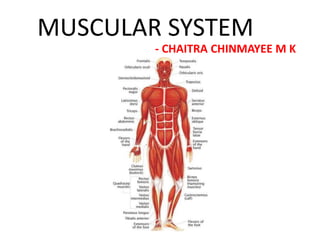
- 1. MUSCULAR SYSTEM - CHAITRA CHINMAYEE M K
- 2. MUSCLE •Muscle is a contractile tissue which brings about body movements by contraction and relaxation of its fibers. • PARTS OF A MUSCLE:- i. Two ends: Origin – the end of the muscle which remains fixed during its contraction. Insertion – the end which moves during its contraction. ii. Two parts: Fleshy part - contractile, and is called the 'belly'. Fibrous part - non-contractile and inelastic. It is called TENDON.
- 3. PROPERTIES OF MUSCLE FIBRES •CONTRACTABILITY: the muscle fibers contract lengthwise, to bring body movements when stimulated. •EXCITABILITY: the muscle fibers are sensitive and respond to stimulus. • EXTENSIBILITY: the muscle fibers return to their original length on removing the stimulus. • ELASTICITY: the muscle fibers are elastic and can be stretched.
- 4. Types of Muscles • There are three types of muscles: i. Skeletal/Striated Muscle – the muscles attached to skeleton ii. Smooth/Non- striated Muscle – the muscles attaches to viscera (abdominal organs) iii. Cardiac Muscle – the muscle of the heart
- 5. SKELETAL MUSCLE • The most abundant muscle in our body. • They are voluntary, that is under our control and striated. • Located superficially and mostly attached to the skeleton. • They are unbranched, cylindrical, multinucleated muscle fibers.
- 6. Functions i. Body Movement – contraction of these muscle fibers cause body movement. ii. Heat Production – metabolism within the muscle cells releases heat as end product, which increases during strenuous exercises. iii. Posture and Body support – These muscles maintain body posture by providing support around flexible joints. Types of skeletal muscle fibres Red Fibres White Fibres Their speed of contraction is less but more sustained. They are fatigue resistant hence present in postural muscles and long muscles of the back. Their speed of contraction is fast but less sustained. They are easily fatigued hence present in the muscles of the eyeball, etc.
- 7. SARCOMERE • The structural and functional unit of a striated muscle fibre, and it is due to the presence of this the muscle appears striated. • It is the basic unit of contraction, that is it is the contraction of this, which brings about the contraction of a muscle fibre. • it comprises of two types of filaments, called myofilaments:- i. Thick filaments: it is composed of a protein called myosin ii. Thin filaments: it is composed of protein called actin • The sliding of thin filament over the thick filament brings about the contraction of sarcomere, and hence the contraction of muscle fibre.
- 8. Smooth Muscle • They are present in the walls of the hollow organs, blood vessels, glands and skin. • They are involuntary, unstriated muscle fibres. • They have unbranched, spindle shaped muscle fibres. • Functions: i. Movement of organs to facilitate proper mixing of food ii. Regulation of blood flow. iii. Propulsion of urine
- 9. Cardiac Muscle • It is also called myocardium. • It is present only in the walls of the heart. • Its major function is pumping of blood from the heart • It is involuntary and striated muscle. • It has branched, cylindrical fibres with intercalated discs and they are uninucleated. • It is auto-rhythmic, that is it generates its own contractions and does not require an external stimulus for its activity.
- 10. Muscles of the Upper Limb Muscles of Arm:- Anterior: (Flexion of Arm) Posterior: (Extension of Arm) • Biceps Brachii Triceps Brachii • Coraco-brachialis • Brachialis Muscles of Forearm:- Anterior: (Flexion of Forearm) Posterior: (Extension of Forearm) • Pronator Teres Anconeus • Flexor carpi radialis Brachio-radialis • Palmaris longus superficial Extensor carpi radialis longus • Flexor digitorum superficialis Extensor carpi radialis brevis • Flexor carpi ulnaris Extensor digitorum • Flexor digitorum profundus Extensor digiti minimi • Flexor pollicis longus deep Extensor carpi ulnaris • Pronator quadrus Supinator Abductor pollicis longus Extensor pollicis brevis deep Extensor pollicis longus Extensor indices Superficial
- 13. Muscles of the hand • Abductor pollicis brevis • Flexor pollicis brevis •Opponens pollicis • Adductor pollicis • Palmaris brevis • Abductor digiti minimi • Flexor digiti minimi • Opponens digiti minimi • Lumbricals(4) • Palmar interossei(4) • Dorsal Interossei(4) F
- 14. Muscles of lower limb Muscles of thigh:- Anterior: (Extension of thigh) • Sartorius • Quadriceps Femoris- it has, Rectus Femoris Vastus lateralis Vastus intermedius Vastus medialis • Articularis genu • Tensor fascia lata Medial: (Adduction of thigh) • Adductor brevis • Adductor longus • Adductor magnus
- 15. Muscles of thigh(cont’d) Posterior:- (Flexion of thigh) HAMSRTING MUSCLES: • Semi-tendinosus • Semi-membranosus • Short head of biceps femoris • Ischial fibres of Adductor magnus NON-HAMSTRING MUSCLES: • Long head of biceps femoris
- 16. MUSCLES OF FRONT OF TRUNK •Pectoralis Major • Pectoralis minor Pectoral Region • Subclavius • Deltoid • External Intercostal • Internal intercostal • Transversus thoracis Subcostalis Thorax Intercostalis intimi Sternocostalis deltoid
- 17. MUSCLES OF BACK OF TRUNK • Trapezius • Lattisimus dorsi • Levator scapulae Back • Rhombois major • Rhomboid minor • Supraspinatous • Infraspinatus • Subscapularis Scapular • Teres minor Muscles • Teres major
- 18. Nerve-Muscle Junction (NMJ) • Neuromuscular junction (NMJ) refers to the intimate contact of the nerve endings with the muscle fibre to which they innervate. • It is a microstructure through which the process of contraction is initiated or halted in the muscles by the neurons. • Components of a NMJ are:- i) Pre - synaptic terminal (Axon terminal) ii) Synaptic Cleft iii) Post - synaptic terminal
- 19. COMPONENTS OF NMJ Presynaptic Terminal: The presynaptic terminal is an axonal terminal of a motor neuron. The axonal terminal contains a number of synaptic vesicles. These vesicles contain the neurotransmitters that are released upon receiving a nerve impulse. Synaptic Cleft: It is the space between the presynaptic terminal and the postsynaptic cell. It is roughly the size of 30 nm. The synaptic cleft allows the neurotransmitters to diffuse and reach the other side of the synapse or the neuromuscular junction. It also contains enzymes for the degradation of the excess or extra neurotransmitters. Post Synaptic Cell: The postsynaptic cell is the skeletal muscle fiber. The motor neurons make synapse on the membrane of the skeletal muscle fibers, whose membrane has receptors for binding to the released neurotransmitters.
- 20. Mechanism of Contraction • When a nerve impulse reaches the presynaptic axonal terminal, it causes depolarization. As a result, the voltage-gated calcium channels open. The calcium ions from the surrounding environment diffuse into the presynaptic axon. • These calcium ions activate the membrane proteins. These proteins mediate the fusion of synaptic vesicles to the cell membrane of the neuron, resulting in the release of neurotransmitters into the synaptic cleft. • They diffuses across the synaptic cleft and binds to the receptors. This results in the opening of the cation channels. These channels are open to both sodium and potassium ions.
- 21. • As the concentration of sodium ions is higher in the extracellular space, the sodium ions enter through these open cation channels. As a result, depolarization of the skeletal muscle increases.. The depolarization of post – synaptic membrane results in the opening of voltage-gated calcium channels locate on the membrane as well as the membrane of the smooth endoplasmic reticulum. • As the calcium ions enter the cell, it initiates the cycle of skeletal muscle contraction. The actin-myosin bridges of the sarcomere are formed, and the result in contraction of the skeletal muscles. Mechanism of Contraction CONT’D
- 22. THANK YOU