
Recommended
Recommended
More Related Content
Similar to MUSCLE.ppt
Similar to MUSCLE.ppt (20)
More from MujahedAldhabyani
More from MujahedAldhabyani (11)
Recently uploaded
Recently uploaded (20)
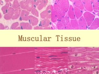
MUSCLE.ppt
- 2. MUSCLES • Muscle is one of the 4 basic tissue types. • Muscle cells are contractile cells. What are other contractile cells? • They are elongate elements Contraction long axis of cell • What do you understand by the following terms? • MUSCLE FIBER • SARCOPLASM • SARCOLEMMA • SARCOPLAMIC RETICULUM • MYOFIBRILS • MYOFILAMENTS
- 3. Muscle Functions 1. Production of Movement 2. Maintenance of posture 3.Thermogenesis 4. Stabilization of joints
- 4. Muscle Functions 1. Production of Movement – Movement of body parts and of the environment – Movement of blood through the heart and the circulatory vessels. – Movement of lymph through the lymphatic vessels – Movement of food (and, subsequently, food waste) through the GI tract – Movement of bile out of the gallbladder and into the digestive tract – Movement of urine through the urinary tract – Movement of semen through the male reproductive tract and female reproductive tract – Movement of a newborn through the birth canal
- 5. Muscle Functions 2. Maintenance of posture – Muscle contraction is constantly allowing us to remain upright. – The muscles of your neck are keeping your head up right now. – As you stand, your leg muscles keep you on two feet. 3. Thermogenesis – Generation of heat. Occurs via shivering – an involuntary contraction of skeletal muscle.
- 6. Muscle Functions 4. Stabilization of joints – Muscles keep the tendons that cross the joint nice and taut. This does a wonderful job of maintaining the integrity of the joint.
- 7. 3 TYPES OF MUSCLE
- 8. CLASSIFICATION OF MUSCLE STRUCTURAL and FUNCTIONAL Functional: Voluntary or Involuntary Structural: Striated or Smooth Combined: Smooth involuntary muscle = smooth muscle Striated involuntary muscle = cardiac muscle Striated voluntary muscle = skeletal muscle
- 9. Classification Functional type Skeletal m. Cardiac m. Smooth m Striated m. Unstriated m. Voluntary m. Involuntary m. Morphologic type
- 10. The muscle cells can be classified into 3 major groups based on their: 1. shape, 2. number and position of nuclei, 3. presence of striations, and 4. whether they are under voluntary or involuntary control. •Elongated cell •Multiple peripheral nuclei •Visible striations •voluntary •Branching cell •Single central nucleus •Visible striations •involuntary •Spindle shaped •Single central nucleus •Lack of visible striations •involuntary
- 11. Characteristics of Muscle Tissue 1. Excitability – The ability to receive and respond to a stimulus • In skeletal muscle, the stimulus is a neurotransmitter (chemical signal) release by a neuron (nerve cell). • In smooth muscle, the stimulus could be a neurotransmitter, a hormone, stretch, pH, Pco2, or Po2. (the symbol means “a change in”) • In cardiac muscle, the stimulus could be a neurotransmitter, a hormone, or stretch. – The response is the generation of an electrical impulse that travels along the plasma membrane of the muscle cell.
- 12. Characteristics of Muscle Tissue 2. Contractility – The ability to shorten forcibly when adequately stimulated. – This is the defining property of muscle tissue. 3. Extensibility – The ability to be stretched 4. Elasticity – The ability to recoil and resume original length after being stretched.
- 13. MUSCLE TISSUE 4 basic tissues. Contractile cells, Elongate elements, muscle fibers, Excitability and contractitility, Origin Terminology: Functions: movement, maintenance of posture, thermogenesis, stabilization of joint. Classification: 3 major groups based on their: shape, nuclei (position and number), striations, voluntary or not
- 14. Skeletal muscle
- 18. Muscle fiber PM is known as sarcolemma Muscle fiber cytoplasm is known as sarcoplasm Sarcoplasm has lots of mitochondria (why?), lots of glycogen granules (to provide glucose for energy needs) as well as myofibrils and sarcoplasmic reticuli. Sarcolemma has invaginations that penetrate through the cell called transverse tubules or T tubules.
- 19. Skeletal muscle LM: Shape: long cylindrical;diameters : 10 to 100 µm. Nuclei: ovoid & flattened; many peripherally placed nuclei. Sarcoplasm: * regular transverse / longitudinal striation * myofibrils myofibril Muscular cell nucleus
- 20. EM: 1. Myofibril 2.Transverse tubule 3. Sarcoplasmic reticulum 4. Other inclusions mitochondria myoglobin glycogen ribosome 1 2 3
- 21. Muscle fiber & myofibril ———muscular fiber (muscular cell) myofibril dark band (A band) Light band (I band)
- 22. Myofibril * long , parallel , cylindrical filamentous bundles *consisting of two types of myofilament (thin & thick).
- 23. Sarcomere: A segment between two adjacent Z line. It is a structural and basic contractile unit.
- 24. • Z line: zone of apposition of actin filaments belonging to two neighbouring sarcomeres • M-line: band of connections between myosin filaments
- 25. Banding I band:only thin filament H band (central region of A band): only thick filament without cross bridges Extremities of A band:both thin & thick filament Z Z
- 26. Molecular configuration of myofilaments * thick filament ----myosin * thin filament: actin, tropomyosin, troponin.
- 27. Myosin (thick filament) —head (adenosine triphosphatase, ATPase) —shaft
- 28. Myosin 肌球蛋白 (thick filament) —head (adenosine triphosphatase, ATPase) —shalf 横桥 Cross bridge
- 29. Myosin 肌球蛋白 (thick filament) —head (adenosine triphosphatase, ATPase) —shalf 横桥 Cross bridge
- 30. Thin filament
- 31. Troponin Actin 肌 Tropomyosin Components of a thin filament
- 32. —binding site to myosin Two strands of actin molecule arranged in a helix Actin Actin
- 37. Transverse tubule * invaginations of the surface sarcolemma * lying as rings around each myofibril * located at A-I junction * providing for rapid spread of surface membrane excitation throughout the entire muscle fiber A I Z myofibril
- 38. Sarcoplasmic reticulun * SER forming cylindrical sheaths around each myofibril (longitudinal tubule) * at A-I junction, tubules connect to channels of larger caliber called terminal cisternae Triad * A T tubule is sandwiched between two terminal cisternae.
- 39. Muscle Fiber Types 2 main types: 1. Slow fibers 2. Fast fibers
- 40. Slow Fibers • Contract slowly because its myosin ATPases work slowly. • Depends on oxygen delivery and aerobic metabolism. • Is fatigue resistant and has high endurance. • Is thin in diameter – large amt of cytoplasm impedes O2 and nutrient diffusion. • Cannot develop high tension – small diameter means few myofibrils. • Has rich capillary supply and lots of mitochondria. • Contains lots of the O2-storing protein, myoglobin which gives it a red color. • Uses lipids, carbs, and amino acids as substrates for it aerobic metabolism. • Best suited for endurance type activities. • A.k.a. red fibers, slow oxidative fibers, type I fibers.
- 41. Fast Fibers • So named because they can contract in 0.01 seconds or less after stimulation. • Fast fibers are large in diameter; they contain densely packed myofibrils, large glycogen reserves, and relatively few mitochondria. • Able to develop a great deal of tension b/c they contain a large number of sarcomeres. • Use ATP in massive amounts. Supported by anaerobic metabolism. Fatigue rapidly. • A.k.a., fast fatigue (FF) fibers, fast glycolytic (FG) fibers, white fibers. • Best suited for short term, power activities.
- 42. Myasthenia Gravis • My=muscle, asthen=weakness, gravi=heavy • Autoimmune disease where antibodies attack the ACh receptors on neuromuscular junctions. • Results in progressive weakening of the skeletal muscles. Why? • Treated w/ anticholinesterases such as neostigmine or physostigmine. These decrease the activity of acteylcholinesterase. – Why would this help someone with myasthenia gravis?
- 43. Muscular Dystrophy - a group of diseases involving muscle deterioration. -Duchenne muscular dystrophy related to deletions in a huge gene coding for the protein dystrophin. -The gene is linked to the X chromosome, so it appears predominantly in males.
- 44. ROLE OF MOTOR NEURON In order for skeletal muscle to contract, each cell must be stimulated by a process of a motor neuron
- 45. Excitation • In general each muscle is served by one nerve – a bundle of axons carrying signals from the spinal cord to the muscle. • W/i the muscle, each axon will go its own way and eventually branch into multiple small extensions called telodendria. Each telodendrium ends in a bulbous swelling known as the synaptic end bulb. The site of interaction btwn a neuron and any other cell is known as a synapse. The synapse btwn a neuron and a muscle is known as the neuromuscular junction.
- 48. ARRIVAL OF ACTION POTENTIAL AT AXON TERMINAL
- 49. FUSON OF SYNAPTIC VESICLES
- 51. ACETYLCHOLINE BINDS TO RECEPTOR SITES
- 52. ACETYLCHOLINE BINDS TO RECEPTOR SITES
- 56. CALCIUM RELEASE FROM TERMINAL CISTERNAE
- 57. CALCIUM RELEASE FROM TERMINAL CISTERNAE
- 58. SUMMARY
- 59. CONTRACTION OF THE MUSCLE CELL
- 60. CARDIAC MUSCLE
- 61. LM - short cylindrical shape - branched -central nucleus, one or two nucleus per fiber - cross striation - myofibril - intercalated disks Cardiac muscle
- 65. EM: *myofibril is not well defined; *sarcoplasmic reticulum less developed; *T tubules much wider; located in the Z lines; *Diad * intercalated disk T tubule z z terminal cisternae M
- 66. Intercalated disc *Specialized cell junctions located at Z lines *Stepwise pattern: transverse region: intermidiate/desmosomes j. longitudinal region: gap junction z G.J. I.J. D.J.
- 67. Intercalated disk: Stepwise pattern Transverse region : desmosome, intermediate J. Longitudinal region : Gap J
- 69. Specialized cardiac muscle fibers called purkinje fibers (green) are located throughout the heart ventricle. They conduct the electrical impulse from the pacemaker region of the heart to cause the ventricle to contract forcefully. Like all healthy tissue, heart muscle cells need an abundance of oxygen and nutrients. Tiny capillaries (orange), containing red blood cells, course through the muscle carrying nutrients, oxygen, and fluids. Other capillaries carry out carbon dioxide and waste materials to veins and the lymph system.
- 70. SMOOTH MUSCLE
- 71. Smooth muscle * spindle-shape with single central nucleus * sarcoplasm appears acidophilic * no transverse striation and intercalated disc.
- 73. EM: * invagination of sarcoplasm * sarcoplasmic reticulum poorly developed * there are myofilaments, but no myofibrils caveola Dense patch Dense body myofilaments
- 74. SMOOTH MUSCLE • LOCATION OF SMOOTH MUSCLE • Involuntary, non-striated muscle tissue • Occurs within almost every organ, forming sheets, bundles, or sheaths around other tissues. • Cardiovascular system: – Smooth muscle in blood vessels regulates blood flow through vital organs. Smooth muscle also helps regulate blood pressure. • Digestive systems: – Rings of smooth muscle, called sphincters, regulate movement along internal passageways. – Smooth muscle lining the passageways alternates contraction and relaxation to propel matter through the alimentary canal.
- 75. Smooth Muscle • Integumentary system: – Regulates blood flow to the superficial dermis – Allows for piloerection • Respiratory system – Alters the diameter of the airways and changes the resistance to airflow • Urinary system – Sphincters regulate the passage of urine – Smooth muscle contractions move urine into and out of the urinary bladder
- 76. Smooth Muscle • Reproductive system – Males • Allows for movement of sperm along the male reproductive tract. • Allows for secretion of the non-cellular components of semen • Allows for erection and ejaculation – Females • Assists in the movement of the egg (and of sperm) through the female reproductive tract • Plays a large role in childbirth
- 77. Smooth Muscle • Elongate, tapering spindle cells: – Are smaller: 5-10um in diameter and 30-200um in length – Are uninucleate: contain 1 centrally placed nucleus – Lack any visible striations – Lack T-tubules – Have a scanty sarcoplasmic reticulum • Smooth muscle tissue is innervated by the autonomic nervous system unlike skeletal muscle which is innervated by the somatic nervous system (over which you have control) • Only the endomysium is present. Nor perimysium or epimysium.
- 78. EM: * invagination of sarcoplasm * sarcoplasmic reticulum poorly developed * there are myofilaments, but no myofibrils caveola Dense patch Dense body myofilaments
- 80. Types of Smooth Muscle • Smooth muscle varies widely from organ to organ in terms of: – Fiber arrangement – Responsiveness to certain stimuli • How would the types of integral proteins that a smooth muscle cell contained contribute to this? • Broad types of smooth muscle: – Single unit (a.k.a. visceral) – Multi unit
- 81. Single Unit Smooth Muscle • More common • Cells contract as a unit because they are all connected by gap junctions - protein complexes that span the PM’s of 2 cells allowing the passage of ions between them, i.e., allowing the depolarization of one to cause the depolarization of another. • Some will contract rhythmically due to pacemaker cells that have a spontaneous rate of depolarization.
- 82. Single Unit Smooth Muscle • Not directly innervated. Diffuse release of neurotransmitters at varicosities (swellings along an axon). • Responsive to variety of stimuli including stretch and concentration changes of various chemicals • Found in the walls of the digestive tract, urinary bladder, and other organs
- 83. Multi-Unit Smooth Muscle • Innervated in motor units comparable to those of skeletal muscles • No gap junctions. Each fiber is independent of all the others. • Responsible to neural & hormonal controls • No pacemaker cells • Less common • Found in large airways to the lungs, large arteries, arrector pili, internal eye muscles (e.g., the muscles that cause dilation of the pupil) • Why is good to have the digestive smooth muscle single unit and the internal eye muscles multi-unit?
- 84. SMOOTH • Shape: Elongate, tapering spindle cells: • Size: Are smaller: 5-10um in diameter and 30-200um in length • Nucleus: 1 centrally placed nucleus • Striations: Lack any visible striations • T-tubules • Sarcoplasmic reticulum • Involuntary, • Locations: • Cardiovascular system: • Digestive systems: • Integumentary system: • Respiratory system • Urinary system • Males • Females hypertrophy &hyperplasia, pregnancy, oxytocin