
Recommended
More Related Content
Similar to muskular system anatomy.pptx
Similar to muskular system anatomy.pptx (20)
Recently uploaded
Recently uploaded (20)
muskular system anatomy.pptx
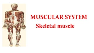
- 2. Skeletal Muscle • Skeletal muscles are muscles which are attached to the skeleton. • 40% of human body mass • Skeletal muscles are mainly responsible for locomotion, and voluntary contraction and relaxation.
- 3. Structure of Skeletal muscles • Cell structure – Muscles cells contain many nuclei – The plasma membrane→ sarcolemma – The cytoplasm→ sarcoplasm – Length – ranges from 0.1cm to more the 30cm in length – Diameter – ranges from 0.001cm to 0.01cm in diameter
- 4. Structure of skeletal muscle • Muscles are made of bundles of muscle fibers that are held together by connective tissue
- 5. Cont… Epimysium: This is the connective tissue wrap just under the deep fascia that surrounds the entire muscle Perimysium: This connective tissue surrounds each individual fascicle (bundle of muscle fibers). Endomysium: This is the connective tissue wrapped around each individual muscle cell (fiber).
- 7. Structure of Skeletal muscles • The myofibril consists of protein chains called myofilaments – Myofilaments have a symmetrical, alternating pattern of thick and thin elements.
- 8. Skeletal Muscle Myosin • Thick myofilament • consists of a large number of bundled myosin molecules aligned in overlapping arrays.
- 9. Skeletal Muscle Actin • The thin myofilament (F-actin, filamentous actin) – made up of two helically intertwined chains of G-actin (globular actin) units. • Other proteins that bind to the actin molecules: • Tropomyosin • The Troponin complex→ made up of three members
- 10. MUSCLE ARRANGEMENTS • Muscles are arranged around the skeleton to bring movements. • Two general types of arrangements oOpposing (antagonists) o Cooperative (synergists)
- 11. Synergistic Muscles • Synergistic muscles are those with the same function, or those that work together to perform a particular function. • biceps brachii flexes the forearm. • Brachioradialis and brachialis also flexes the forearm.
- 12. Naming Skeletal Muscles 1-Direction of Muscle Fibers Relative to the Midline • RECTUS: parallel to the midline • Eg: Rectus Abdominus • TRANSVERSE: perpendicular to midline • Eg: Transverse Abdominus • OBLIQUE: diagonal to midline • Eg: External Oblique
- 13. 2. Location • Structure near which muscle is found • FRONTALIS near FRONTAL bone • OCCIPITALIS near OCCIPITAL bone
- 14. 3. Size • Relative Size of Muscle • MAXIMUS: largest • Gluteus Maximus • MEDIUS: middle • Gluteus Medius • MINIMUS: smallest • Gluteus Minimus • LONGUS: longest • Fibularis Longus • BREVIS: short • Fibularis Brevis • TERTIUS: shortest • Fibularis Tertius
- 15. 4. Number of Origins • Number of tendons of origin • BICEPS: Two • Biceps Brachii • Biceps Femoris • TRICEPS: Three • Triceps Brachii • QUADRICEPS: Four • Quadriceps Femoris
- 16. 5. Shape Relative Shape of the Muscle • DELTOID: triangular shape • TRAPEZIUS: trapezoid shape • SERRATU: saw-toothed • RHOMBOIDEUS: rhomboid shape • TERES: round
- 18. • Pennate (penniform) muscle is a muscle with fascicles that attach obliquely to its tendon. allow better stabilization and force production but less flexibility.
- 20. Muscles of facial expression • Are located within the layers of superficial fascia. • Developed from the mesoderm of the 2nd pharyngeal arch, • Innervated by facial nerve. • Functionally, they perform an all important function of non-verbal communication in addition to closing and opening the orifices in the region of of face. Grouped into: (a) muscles of the scalp, (b) muscles of the external ear (c) muscles of the eyelid (d) the nasal muscles (e) the oral muscles.
- 29. Platysma is a large, thin sheet of muscle in the superficial fascia of the neck. It arises below the clavicle in the upper part of the thorax and ascends through the neck to the mandible. At this point, the more medial fibers insert on the mandible, whereas the lateral fibers join with muscles around the mouth. Platysma tenses the skin of the neck and can move the lower lip and corners of the mouth down. Platysma
- 30. FACIAL MUSCLES AND EMOTIONAL EXPRESSIONS The facial muscles responsible for important emotional expressions are presented in Table 3.3.
- 32. Muscles of the neck
- 39. Blood supply of face and neck
- 41. Nerve supply of muscles of the face
- 42. Nerve supply of muscles of the neck
- 43. Muscle of pectoral region
- 47. Posterior Axioappendicular Muscles • Attach the superior appendicular skeleton to the axial skeleton • Divided into three groups • Superficial (extrinsic shoulder) muscles : • trapezius and • latissimus dorsi • Deep (extrinsic shoulder) muscles : • levator scapulae and • rhomboids • Scapulohumeral (intrinsic shoulder) muscles: • deltoid, teres major, and • the four rotator cuff muscles (supraspinatus, infraspinatus, teres minor, and subscapularis)
- 52. Scapulohumeral(intrinsic shoulder) Muscles • Six muscles • Deltoid • Teres major • Supraspinatus • Infraspinatus • Subscapularis • Teres minor • Short muscles that pass from scapula to humerus • Act on glenohumeral joint
- 55. Rotator Cuff Muscles (SITS) • Teres Minor • External Rotation • Subscapularis • Internal Rotation • Supraspinatus • Abduction • Infraspinatus • External Rotation
- 57. MUSCLES OF THE UPPER LIMB • MUSCLES OF THE ARM • MUSCLES OF THE FOREARM • MUSCLES OF THE HAND
- 58. MUSCLES OF THE ARM Anterior (flexor ) group • Coracobrachialis • Biceps brachi • Long head • Short head • Brachialis Posterior(extensor)group • Triceps Brachai • long head • Lateral head • medial head • Anconeus
- 59. Coracobrachialis • an elongated muscle in the superomedial part of the arm • Origin: coracoid process of the scapula • Insertion: medial shaft of the humerus at about its middle part • Innervation : musculocutaneous nerve, C5,6,(C7) • Action: flexes the humerus and assists to adduct the humerus
- 60. Coracobrachialis
- 61. Biceps brachii • Although the biceps is located in the arm it has no attachment to the humerus • Origin: - long head- supraglenoid tubercle -short head- tip of the coracoid process of the scapula • Insertion: radial tuberosity • Innervation: musculocutaneous nerve, C5,6 • Action : Supinates forearm and when it is supine flexes forearm
- 62. • is the main flexor of the forearm • Origin: lower 1/2 of anterior humerus, both intermuscular septa • Insertion: ulnar tuberosity and ulnar coronoid process • Innervation :musculocutaneous nerve, C5,6 • Action : elbow flexion (prime mover) Brachialis
- 63. Triceps brachi • The main extensor of the forearm • Because the long head of the triceps crosses the shoulder joint, it also aids in extension and adduction of the arm. Origin: - long head - infraglenoid tubercle of the scapula - lateral head - upper half of the posterior surface of the shaft of the humerus - medial head - posterior shaft of humerus, distal to radial groove (deep to the long & lateral heads) Insertion: olecranon process • Innervation: radial nerve, C6,7 • Action: chief extensor of forearm
- 64. Anconeus • small triangular muscle and is on the lateral part of the posterior aspect of the elbow. • Origin: posterior surface of the lateral epicondyle of the humerus • Insertion: lateral aspect of olecranon • Innervation: radial nerve, C6,7 • Action :assists triceps in extension of elbow
- 65. Cubital fossa • Shallow triangular depression on anterior surface of the elbow • Boundaries • Superiorly, an imaginary line connecting the medial and lateral epicondyles • Medially, pronator teres • Laterally, brachioradialis • Floor • brachialis and supinator muscles • Roof • the continuity of brachial and antebrachial (deep) fascia, reinforced by the bicipital aponeurosis, subcutaneous tissue and skin
- 66. • Contents • Brachial artery and branches, radial and ulnar arteries • Accompanying veins of the arteries • Biceps brachii tendon • Median nerve • Radial nerve, dividing into its superficial and deep branches • Median cubital vein • Medial and lateral cutaneous nerves of the forearm
- 69. • The flexor muscles are arranged in three layers: 1. Superficial layer • Pronartor teres • Flexor carpi radialis • Palmaris longus • Flexor carpi ulnaris These muscles are attached to the medial epicondyle of the humerus , the common flexor attachment.
- 72. 2. Intermediate layer_ consists of one muscle • Flexor digitorum superficialis 3. Deep layer- three muscles • Flexor digitorum profundus • Flexor pollicis longus • Pronator quadrates
- 73. Pronator teres • Most lateral of the superficial muscles • Its lateral border forms the medial boundary of the cubital fossa. • Origin: - humeral head: medial epicondyle via the common flexor tendon (CFT) ,medial intermuscular septum - ulnar head - coronoid process of ulna • Insertion: lateral aspect of radius at the middle of the shaft (pronator tuberosity) • Innervation : median nerve, C6,7 • Action: pronates forearm & weakly flexes the elbow
- 74. Flexor carpi radialis • a long fusiform muscle located medial to the pronator teres • Its tendon is a good guide to the radial artery, which lies lateral to it • Origin :Medial epicondyle of humerus • Insertion: base of 2nd metacarpal • Innervation: median nerve • Action: flexes and abducts hand
- 75. Palmaris longus • has a short belly and long cord- like tendon that is a useful guide to the median nerve • Oirgin: Medial epicondyle of humerus • Insertion: distal half of flexor retinaculum and palmar aponeurosis • Innervation: median nerve • Action: flexes hand and tenses palmar aponeurosis
- 76. Flexor carpi ulnaris • This is the most medial of the superficial flexor muscles in the forearm. • It has 2 heads of proximal attachment, between which the ulnar nerve passes distally in the forearm • Origin: humeral head -Medial epicondyle of humerus • Ulnar head-medial aspect of olecranon • Insertion: pisiform, hook of hamate and base of 5th metacarpal • Innervation: ulnar nerve • Action: flexes and adducts hand
- 77. Flexor Digitorum Superficialis • The median nerve and ulnar artery enter the forearm by passing its humeroulnar and radial heads. • Near the wrist, gives 4 tendons which are enclosed (along the 4 tendons of FDP) in a common flexor synovial sheath. • Origin: humeroulnar head-medial epicondyle and coronoid process • Radial head-superior half of anterior border • Insertion: shafts of middle phalanges of medial 4 fingers • Innervation: median nerve • Action: flexes middle phalanges at PIP
- 78. Flexor Digitorum profundus • It is the only one that can flex the DIP joints of the digits • Origin: proximal ¾ of medial and anterior surface of ulna and IO membrane • Insertion: bases of distal phalanges of 2nd -5th fingers • Innervation: median nerve (lateral two heads) and ulnar nerve (medial 2 heads) • Action: flexes distal phalanges
- 79. Flexor pollicis longus • Lies lateral to the FDP ,where it covers the anterior surface of radius • The only muscle that flexes the IP joint of the thumb • Its flat tendon passes deep to the flexor retinaculum, enveloped in its own synovial sheath • Origin: anterior surface of radius and adjacent IO membrane • Insertion: base of distal phalanx of thumb • Innervation: median nerve • Action: flexes phalanges of 1st digit (thumb)
- 80. Pronator quadratus • The deepest muscle in the anterior group • Origin: distal quarter of the ulna • Insertion: distal quarter of the radius • Innervation: median nerve • Action: pronates forearm
- 81. Extensor Muscles of the Forearm • are in the posterior (extensor-supinator) compartment of the forearm. • All extensors are innervated by muscular and deep branches of the radial nerve. • The extensor tendons are held in place in the wrist region by the extensor retinaculum.
- 82. Superficial layer of extensors • Include: • Brachioradialis, • Extensor Carpi radialis longus, • Extensor Carpi radialis brevis, • Extensor Digitorum, • Extensor digiti minimi, • Extensor carpi ulnaris
- 83. Deep layer • Supinator • Extensor indicis • Abductor Pollicis longus • Extensor pollicis logus • Extensor pollicis brevis
- 85. MUSCLES OF THE HAND • The intrinsic muscles of the hand are located in five compartments: 1. Thenar muscles in the thenar compartment • Abductor pollicis brevis • Flexor pollicis brevis • Opponens pollicis 2. Adductor pollicis in the adductor compartment 3. Hypothenar muscles in the hypothenar compartment: • Abductor digiti minimi , • Flexor digiti minimi brevis, and • Opponens digiti minimi. 4. Lumbricals in the central compartment with long flexor tendons 5. The interossei lie in separate interosseos compartments between the metacarpals
- 89. Muscle Origin Insertion Innervation Main action Palmar Interossei (3) Palmar surface of 2nd, 4thand 5thmetacarpals. As unipennate muscles Dorsal aponeurosis of the 2nd, 4th and 5th fingers. Ulnar nerve Adduction of the 2nd, 4th and 5th fingers, Flexion of the corresponding metacarpo-phlangeal joint, and extension of the interphalangeal joints. Dorsal interossei (4) Adjacent sides of two metacarpals as bipennate muscles Dorsal aponeurosis of 2nd - 4th fingers Ulnar nerve Abduction of the 2nd, 3rd and 4th fingers, Flexion of the corresponding metacarpo-phalangeal joint, and extension of the interphalangeal joints
- 92. Muscles of the anterior abdominal wall
- 93. 93
- 94. 1. External Oblique Muscle • Largest & most superficial flat muscle • Fibers run inferiomedially Origin:- - external surface of 5th -12th ribs • Insertion:- - linea alba - outer lip of iliac crest - anterior superior iliac spine - pubic tubercle and pubic crest 94
- 95. Cont… Innervation:- - Anterior rami of lower six thoracic spinal nerves (T7 to T12) Blood supply:- - lower intercostal arteries, deep circumflex iliac artery and iliolumbar artery. Action:- - unilateral contraction tilt the trunk - bilateral contraction flexes the trunk - elevation of the pelvis - compression of the abdomen - expiration or defecation - fixation of the trunk during carrying 95
- 96. 2. Internal Oblique Muscle • Intermediate of the three flat abdominal wall • Muscle fibers run superomedially Origin: - thoracolumbar fascia - lateral 2/3rd of inguinal ligament - anterior 2/3rd of iliac crest - anterior superior iliac spine Insertion:- linea alba - lower border of 10th-12th ribs - pubic crest & pectineal line 96
- 97. Cont…. Innervation:- - Anterior rami of lower six thoracic spinal nerves (T7 to T12) - iliohypogastric nerve - ilioinguinal nerve Blood supply:- Subcostal arteries Action:- - unilateral contraction tilt & rotate on the side - bilateral contraction flexes the trunk - elevation of the pelvis - compression of the abdomen - expiration or defecation - fixation of the trunk 97
- 98. 3. Transversus Abdominis Muscle • The innermost of the three flat muscles • run more or less transversely • B/n internal oblique & transversus abdominis muscles, there is neurovascular bundle Origin:- - internal surface of 7th-12th costal cartilages - thoracolumbar fascia - inner lip of iliac crest - lateral 1/3rd of inguinal ligament 98
- 99. Cont…. Insertion:- - linea alba - pubic crest - pectineal line - fused part of aponeurosis (conjoint tendon) Innervation:- - Anterior rami of lower six thoracic spinal nerves (T7 to T12) - ilioinguinal nerve & iliohypogastric nerve Blood supply:- Subcostal arteries Action:- - compress & support abdominal viscera - increase intra-abdominal pressure 99
- 100. 4. Rectus Abdominis Muscle • A long, broad, strap-like muscle • Principal vertical muscle of anterior abdominal wall • Paired rectus muscles are separated by linea alba • Wider superiorly & narrow inferiorly • Has three or more tendinous intersections • Enclosed by the rectus sheath 100
- 101. Origin:- - upper surface of pubic (symphysis, and crest) Insertion:- - xiphoid process & 5th-7th costal cartilage Innervation:- - Anterior rami of lower seven thoracic spinal nerves (T6 to T12) - Blood supply:- inferior and superior epigastric arteries Action:- - tenses the abdominal wall - flexes trunk & increase intra- abdominal pressure - elevate pelvis & expiration or defecation 101
- 103. Ligaments Of The Gluteal Region The two important ligaments in the gluteal region are The sacrotuberous ligament It connects the back of the sacrum to the ischial tuberosity. The sacrospinous ligament. It connects the back of the sacrum to the ischial spine They stabilize the sacrum and prevent its rotation at the sacroiliac joint by the weight of the vertebral column.
- 104. Foramina of the gluteal region The two important foramina in the gluteal region are The greater sciatic foramen The lesser sciatic foramen. Greater Sciatic Foramen The greater sciatic foramen is formed by the greater sciatic notch of the hip bone and the sacrotuberous and sacrospinous ligaments. It provides an exit from the pelvis into the gluteal region. The following structures exit through the foramen: Piriformis muscle Sciatic nerve Posterior cutaneous nerve of the thigh Superior and inferior gluteal nerves Nerves to the obturator internus and quadratus femoris Pudendal nerve Superior and inferior gluteal arteries and veins Internal pudendal artery and vein
- 106. Lesser Sciatic Foramen The lesser sciatic foramen is formed by the lesser sciatic notch of the hip bone and the sacrotuberous and sacrospinous ligaments. It provides an entrance into the perineum from the gluteal region. Its presence enables nerves and blood vessels that have left the pelvis through the greater sciatic foramen above the pelvic floor to enter the perineum below the pelvic floor. The structures pass through the foramen are: Tendon of obturator internus muscle Nerve to obturator internus Pudendal nerve Internal pudendal artery and vein
- 107. Muscles of the gluteal region • Organized into 2 layers: superficial and deep Superficial layer:consists of • Gluteus maximus, • Gluteus medius • Gluteus minimus and • The tensor of the fascia lata • All have proximal attachments to the posterolateral (external) surface and margins of the ala of the ilium and are mainly extensors, abductors, and medial rotators of the thigh
- 108. Deep layer: • piriformis, • obturator internus, • superior and inferior gemilli, and • quadratus femoris • all have distal attachments on or adjacent to the intertrochanteric crest of the femur. • These muscles are lateral rotators of the thigh but they also stabilize the hip joint, working with the strong ligaments of the hip joint to steady the femoral head in the acetabulum
- 109. Gluteus maximus Origin Outer surface of ilium( posterior gluteal line), sacrum, coccyx, Sacrotuberous ligament Insertion Iliotibial tract and gluteal tuberosity of femur Nerve Supply Inferior gluteal nerve L5; S1, 2 Action Extends and Laterally rotates hip joint Through iliotibial tract, it extends knee joint
- 110. Gluteus medius Origin Outer surface of ilium( between posterior and anterior gluteal line) Insertion Lateral surface of greater trochanter of femur Nerve Supply Superior gluteal nerve L5;S1 Action Abducts thigh at hip joint Tilts pelvis when walking to permit opposite leg to clear ground
- 111. Gluteus minimus Origin External surface of ilium between inferior and anterior gluteal lines Insertion Linear facet on the antero-lateral aspect of the greater trochanter Nerve Supply Superior gluteal nerve (L4,L5,S1) Action Abducts femur at hip joint Holds pelvis secure over stance leg and prevents pelvic drop on the opposite swing side during walking Medially rotates thigh
- 112. • Injury to Superior Gluteal Nerve • When a person who has suffered a lesion of the superior gluteal nerve is asked to stand on one leg, the pelvis on the unsupported side descends indicating that the gluteus medius and minimus on the supported side are weak or non-functional • Trendelenburg test
- 113. Tensor fasciae latae Origin Lateral aspect of crest of ilium between anterior superior iliac spine and tubercle of the crest Insertion Iliotibial tract of fascia lata Nerve Supply Superior gluteal nerve (L4,L5,S1) Action Stabilizes the knee in extension
- 114. • Common Sites of IM injections Deltoid region Gluteal region Anterolateral thigh The gluteal region is a common site for intramuscular (IM) injection of drugs. It is important to be aware of the extent of the gluteal region and the safe region for giving injections. Injections into the buttock are safe in the superolateral quadrant of the buttock or superior to a line extending from the PSIS to the superior border of the greater trochanter.
- 115. Piriformis Origin Anterior surface of sacrum between anterior sacral foramina Insertion Medial side of superior border of greater trochanter of femur Nerve Supply Branches from (S1,S2) Action Laterally rotates the extended femur at hip joint Abducts flexed femur at hip joint
- 116. Obturator internus Origin Anterolateral wall of true pelvis; deep surface of obturator membrane and surrounding bone Insertion Medial side of greater trochanter of femur Nerve Supply Nerve to obturator internus (L5,S1) Action Laterally rotates the extended femur at hip joint Abducts flexed femur at hip joint
- 117. Gemellus superior Origin External surface of ischial spine Insertion Along length of superior surface of the obturator internus tendon and into the medial side of greater trochanter of femur with obturator internus tendon Nerve Supply Nerve to obturator internus (L5,S1) Action Laterally rotates the extended femur at hip joint Abduction of flexed femur at hip joint
- 118. Gemellus inferior Origin Upper aspect of ischial tuberosity Insertion Along length of inferior surface of the obturator internus tendon and into the medial side of greater trochanter of femur with obturator internus tendon Nerve Supply Nerve to quadratus femoris (L5,S1) Action Laterally rotates the extended femur at hip joint Abducts flexed femur at hip joint
- 119. Quadratus femoris Origin Lateral aspect of the ischium just anterior to the ischial tuberosity Insertion Quadrate tubercle on the intertrochanteric crest of the proximal femur Nerve Supply Nerve to quadratus femoris (L5,S1) Action Laterally rotates femur at hip joint
- 120. The Thigh
- 121. • The thigh is divided into three compartments by intermuscular septa between the posterior aspect of the femur and the fascia lata: • The anterior compartment of thigh contains muscles that mainly extend the leg at the knee joint. • The posterior compartment of thigh contains muscles that mainly extend the thigh at the hip joint and flex the leg at the knee joint. • The medial compartment of thigh consists of muscles that mainly adduct the thigh at the hip joint. Cross section of left thigh
- 122. Superficial Fascia of the Thigh • The membranous layer of the superficial fascia of the anterior abdominal wall extends into the thigh and is attached to the deep fascia (fascia lata) about a finger breadth below the inguinal ligament. • The fatty layer of the superficial fascia on the anterior abdominal wall extends into the thigh and continues down over the lower limb without interruption.
- 123. Deep Fascia of the Thigh (Fascia Lata) • The deep fascia encloses the thigh like a trouser leg and at its upper end is attached to the pelvis and the inguinal ligament. • On its lateral aspect, it is thickened & is called the iliotibial tract. • In the gluteal region, the deep fascia forms sheaths, which enclose the tensor fasciae latae and the gluteus maximus muscles. • In front of the thigh just below the inguinal ligament, the deep fascia has a gap & is called as the saphenous opening. • It transmits the great saphenous vein, some small branches of the femoral artery, and lymph vessels.
- 124. Femoral Triangle • The femoral triangle is a triangular depressed area situated in the upper part of the medial aspect of the thigh just below the inguinal ligament. Boundaries: • Superiorly: • The inguinal ligament • Laterally: • The sartorius muscle • Medially: • The adductor longus muscle • Floor • It is gutter shaped and formed from lateral to medial by the iliopsoas, the pectineus, and the adductor longus.
- 126. • Roof • Is formed by the skin and fasciae of the thigh. • Contents • The terminal part of the femoral nerve and its branches. • The femoral sheath, • The femoral artery and its branches. • The femoral vein and its tributaries. • The deep inguinal lymph nodes.
- 127. Femoral Sheath • The femoral sheath is a downward protrusion into the thigh of the fascial envelope lining the abdominal walls. • Its anterior wall is continuous above with the fascia transversalis, and its posterior wall with the fascia iliaca. • The sheath surrounds the femoral vessels and lymphatics about 2.5 cm below the inguinal ligament. • The femoral sheath is adherent to the walls of the blood vessels and inferiorly blends with the tunica adventitia of these vessels.
- 128. • The femoral sheath is divided into three compartments by septa. • The femoral artery occupies the lateral compartment of the sheath. • The femoral vein occupies the intermediate compartment. • The lymph vessels the most medial compartment. The femoral canal • It is the small medial compartment of the femoral sheath. • It is about 1.3 cm long. • Its upper opening of femoral canal is called the femoral ring. • The femoral ring is closed by a condensation of extraperitoneal tissue called as the femoral septum.
- 129. Femoral hernia
- 130. Adductor Canal • The adductor canal is an intermuscular cleft situated on the medial aspect of the middle third of the thigh beneath the sartorius muscle. • It commences(begins) above at the apex of the femoral triangle and ends below at the opening in the adductor magnus. • It is triangular in cross section & has three walls. • Anteromedial wall • A posterior wall • A lateral wall.
- 131. • The anteromedial wall • sartorius muscle and fascia. • The posterior wall • the adductor longus and magnus muscles. • The lateral wall • the vastus medialis. • Contents: • The terminal part of the femoral artery. • The femoral vein. • The deep lymph vessels. • The saphenous nerve. • The nerve to the vastus medialis • The terminal part of the obturator nerve.
- 132. Thigh • Thigh muscles are organized into 3 compartments by intermuscular septa • Anterior or extensor compartment: This is composed of the quadriceps femoris, iliopsoas, sartorius, and pectineus, as well as the femoral artery, vein, and nerve, and the lateral femoral cutaneous nerve. • Medial or adductor compartment: This contains the gracilis, adductor longus, brevis, magnus, and obturator externus muscles along with the obturator artery, vein, and nerve, and the profunda femoris artery. • Posterior or flexor compartment: This includes the biceps femoris, semitendinosus, and semimembranosus, a portion of the adductor magnus muscle, branches of the profunda femoris artery, the sciatic nerve, and the posterior femoral cutaneous nerve.
- 133. Muscles of the Anterior Compartment of the Thigh Sartorius • Origin • Anterior superior iliac spine • Insertion • Upper medial surface of shaft of tibia • Nerve Supply • Femoral nerve L2, 3 • Action • Flexes, abducts, laterally rotates thigh at hip joint • Flexes and medially rotates leg at knee joint
- 134. Iliacus • Origin • Iliac fossa of hip bone • Insertion • With psoas major into lesser trochanter of femur • Nerve Supply • Femoral nerve L2,3 • Action • Flexes thigh on trunk • If thigh is fixed, it flexes the trunk on the thigh as in sitting up from lying down
- 135. Psoas major • Origin • Transverse processes, bodies, and intervertebral discs of the 12th thoracic to 5th lumbar vertebrae • Insertion • With iliacus into lesser trochanter of femur • Nerve Supply • Lumbar plexus L1, 2,3 • Action • Flexes thigh on trunk • If thigh is fixed, it flexes the trunk on thigh as in sitting up from lying down
- 136. Pectineus • Origin • Superior ramus of pubis • Insertion • Oblique line extending from base of lesser trochanter to linea aspera on posterior aspect of proximal femur • Nerve Supply • Femoral nerve L2,3 • Action • Flexes and adducts thigh at hip joint
- 137. Quadriceps Femoris. Rectus femoris • Origin • Straight head: • Anterior inferior iliac spine • Reflected head: • Ilium above acetabulum • Insertion • Quadriceps tendon into patella, then via ligamentum patellae into tubercle of tibia. • Nerve Supply • Femoral nerve L2, 3, 4 • Action • Extension of leg at knee joint • Flexes thigh at hip joint
- 138. Vastus lateralis • Origin • Lateral part of intertrochanteric line, margin of greater trochanter, lateral margin of gluteal tuberosity, lateral lip of the linea aspera. • Insertion • Quadriceps tendon into patella, then via ligamentum patellae into tubercle of tibia • Nerve Supply • Femoral nerve L2, 3, 4 • Action • Extension of leg at knee joint
- 139. Vastus medialis • Origin • Medial part of intertrochanteric line, pectineal line, medial lip of the linea aspera, medial supracondylar line • Insertion • Quadriceps tendon into patella, then via ligamentum patellae into tubercle of tibia. • Nerve Supply • Femoral nerve L2, 3, 4 • Action • Extension of leg at knee joint; stabilizes patella
- 140. Vastus intermedius • Origin • Upper 2/3rd of the anterior and lateral surfaces of shaft of femur • Insertion • Quadriceps tendon into patella, then via ligamentum patellae into tubercle of tibia • Nerve Supply • Femoral nerve L2, 3, 4 • Action • Extension of leg at knee joint; articularis genus retracts synovial membrane
- 141. The Medial Compartment of the Thigh • Collectively called adductor group • Attach from the pubis and ischium to the linea aspera • Adductor muscles are • Adductor magnus • Adductor longus • Adductor brevis • Gracilis • Obturator externus All are supplied by obturator nerve except hamstring part of adductor magnus Three layers • Superficial - gracilis and adductor longus • Middle - adductor brevis • Deep - adductor magnus & obturator externus
- 142. Gracilis • Origin • Inferior ramus of pubis, ramus of ischium • Insertion • Upper part of shaft of tibia on medial surface • Nerve Supply • Obturator nerve L2,3 • Action • Adducts thigh at hip joint • Flexes leg at knee joint
- 143. Adductor longus • Origin • Body of pubis, medial to pubic tubercle • Insertion • Linea aspera on the middle 1/3rd of the femur. • Nerve Supply • Obturator nerve L2, 3, 4 • Action • Adducts thigh at hip joint and assists in lateral rotation
- 146. Adductor brevis • Origin • External surface of the pubis & Inferior ramus of pubis • Insertion • Posterior surface of shaft of femur or the upper 1/3rd of the linea aspera • Nerve Supply • Obturator nerve L2, 3, 4 • Action • Adducts thigh at hip joint and assists in lateral rotation
- 147. Adductor magnus • Origin • Adductor part • Inferior ramus of pubis, ramus of ischium, • Hamstring part • Ischial tuberosity. • Insertion • Posterior surface of shaft of femur, linea aspera, medial supracondylar ridge. • Adductor tubercle of femur • Nerve Supply • Adductor portion: obturator nerve • Hamstring portion: sciatic nerve L2, 3, 4 • Action • Adducts thigh at hip joint and assists in lateral rotation • Hamstring portion extends thigh at hip joint
- 148. Obturator externus • Origin • Outer surface of obturator membrane and pubic and ischial rami • Insertion • Medial surface of greater trochanter • Nerve Supply • Obturator nerve L3, 4 • Action • Laterally rotates thigh at hip joint
- 149. The posterior compartment of Thigh • Three of the four muscles in posterior thigh are hamstrings • Semitendinosus • Semimembranosus • Biceps femoris (long head)
- 150. Hamstring muscles •They share common features • Arise from ischial tuberosity • Insert into tibia or fibula • Innervated by the tibial division of sciatic nerve • the short head of the biceps femoris fail to meet any of these condition innervation comes from the lateral (fibular) division whereas branches to other hamstrings arise from the medial (tibial) division •Extend hip and flex knee joint •The semimembranosus and semitendinosus also rotate the leg medially, being attached to the medial head of the tibia
- 151. Biceps femoris • Origin • Long head: • Ischial tuberosity • Short head: • Linea aspera, lateral supracondylar ridge of shaft of femur • Insertion • Head of fibula • Nerve Supply • Long head: • Tibial portion of sciatic nerve- L5; S1,2 • Short head: • Common peroneal portion of sciatic nerve • Action • Flexes and laterally rotates leg at knee joint; long head also extends thigh at hip joint
- 152. Semitendinosus • Origin • Ischial tuberosity • Insertion • Upper part of medial surface of shaft of tibia • Nerve Supply • Tibial portion of sciatic nerve L5; S1,2 • Action • Flexes and medially rotates leg at knee joint; extends thigh at hip joint
- 153. Semimembranosus • Origin • Ischial tuberosity • Insertion • Medial condyle of tibia • Nerve Supply • Tibial portion of sciatic nerve L5; S1,2 • Action • Flexes and medially rotates leg at knee joint • Extends thigh at hip joint
- 154. The Popliteal Fossa
- 155. • The popliteal fossa is a diamond shaped intermuscular space situated at the back of the knee. • The fossa is most prominent when the knee joint is flexed. • Contents • The popliteal vessels • The small saphenous vein • The common peroneal and tibial nerves • The posterior cutaneous nerve of the thigh • The genicular branch of the obturator nerve • Loose connective tissue, and lymph nodes
- 156. Boundaries • Laterally: • The biceps femoris above and the lateral head of the gastrocnemius and plantaris below • Medially: • The semimembranosus and semitendinosus above and the medial head of the gastrocnemius below.) • The anterior wall or floor • Formed by the popliteal surface of the femur, the posterior ligament of the knee joint, and the popliteus muscle. • The roof • Formed by skin, superficial fascia, and the deep fascia of the thigh.
- 157. Popliteus Muscle • Origin: • The lateral surface of the lateral condyle of the femur by a rounded tendon and by a few fibers from the lateral meniscus. • Insertion: • To the posterior surface of the tibia, above the soleal line. • The muscle arises within the capsule of the knee joint, and its tendon separates the lateral meniscus from the lateral ligament of the joint. • It emerges through the lower part of the posterior surface of the capsule of the joint to pass to its insertion. • Nerve supply: • Tibial nerve. • Action: • Medial rotation of the tibia on the femur or, if the foot is on the ground, lateral rotation of the femur on the tibia.
- 158. The Leg
- 159. Fascial Compartments of the Leg • The deep fascia surrounds the leg and is continuous above with the deep fascia of the thigh. • Below the tibial condyles, it is attached to the periosteum on the anterior and medial borders of the tibia. • The anterior & posterior inter muscular septa together with the interosseous membrane, divide the leg into three compartments— • The anterior compartment • The lateral compartment • The posterior compartment • Each having its own muscles, blood supply, and nerve supply.
- 160. Retinacula of the Ankle • The retinacula are thickenings of the deep fascia that keep the long tendons around the ankle joint in position and act as pulleys. • The retinacula around the leg are: • Superior Extensor Retinaculum • Inferior Extensor Retinaculum • Flexor Retinaculum • Superior Peroneal Retinaculum • Inferior Peroneal Retinaculum
- 161. • Structures that pass anterior to the extensor retinacula from medial to lateral • Saphenous nerve • The great saphenous vein • Superficial peroneal nerve (medial and lateral branches) • Structures that pass beneath or through the extensor retinacula from medial to lateral • Tibialis anterior tendon • Extensor hallucis longus tendon • Anterior tibial artery with venae comitantes • Deep peroneal nerve • Extensor digitorum longus tendons • Peroneus tertius
- 164. • Flexor Retinaculum • The flexor retinaculum extends from the medial malleolus downward and backward to be attached to the medial surface of the calcaneum. • Structures that Pass behind the Medial Malleolus beneath the Flexor Retinaculum From Medial to Lateral • Tibialis posterior tendon • Flexor digitorum longus • Posterior tibial artery with venae comitantes • Tibial nerve • Flexor hallucis longus
- 165. • Superior Peroneal Retinaculum • connects the lateral malleolus to the lateral surface of the calcaneum. • It binds the tendons of the peroneus longus and brevis to the back of the lateral malleolus. • Inferior Peroneal Retinaculum • binds the tendons of the peroneus longus and brevis muscles to the lateral side of the calcaneum.
- 166. Muscles of anterior compartment of leg Tibialis anterior • Origin • Lateral surface of shaft of tibia and interosseous membrane • Insertion • Medial cuneiform and base of 1st metatarsal bone • Nerve Supply • Deep peroneal nerve L4, 5 • Action • Extends foot at ankle joint • Inverts foot at subtalar and transverse tarsal joints • Holds up medial longitudinal arch of foot
- 167. Extensor digitorum longus • Origin • Upper half of anterior surface of shaft of fibula • Insertion • Extensor expansion of lateral four toes • Nerve Supply • Deep peroneal nerve L5; S1 • Action • Extends toes • Extends foot at ankle joint
- 168. Peroneus tertius • Origin • Lower half of anterior surface of shaft of fibula • Insertion • Base of 5th metatarsal bone • Nerve Supply • Deep peroneal nerve L5; S1 • Action • Extends foot at ankle joint • Everts foot at subtalar and transverse tarsal joints
- 169. Extensor hallucis longus • Origin • Anterior surface of shaft of fibula • Insertion • Base of distal phalanx of great toe • Nerve Supply • Deep peroneal nerve L5; S1 • Action • Extends big toe • Extends foot at ankle joint • Inverts foot at subtalar and transverse tarsal joints
- 170. The Lateral Compartment Of The Leg
- 171. Peroneus longus • Origin • Lateral surface of shaft of fibula • Insertion • Base of 1st metatarsal and the medial cuneiform • Nerve Supply • Superficial peroneal nerve L5; S1, 2 • Action • Plantar flexes foot at ankle join • Everts foot at subtalar and transverse tarsal joints • Supports lateral longitudinal and transverse arches of foot
- 172. Peroneus brevis • Origin • Lateral surface of shaft of fibula • Insertion • Base of 5th metatarsal bone • Nerve Supply • Superficial peroneal nerve L5; S1, 2 • Action • Plantar flexes foot at ankle joint; everts foot at subtalar and transverse tarsal joint; supports lateral longitudinal arch of foot
- 173. The posterior of the Leg
- 174. Gastrocnemius • Origin • Lateral head from lateral condyle of femur • Medial head from above medial condyle • Insertion • Via tendocalcaneus into posterior surface of calcaneum • Nerve Supply • Tibial nerve S1, 2 • Action • Plantar flexes foot at ankle joint • Flexes knee joint
- 175. Plantaris • Origin • Lateral supracondylar ridge of femur • Insertion • Posterior surface of calcaneum • Nerve Supply • Tibial nerve S1, 2 • Action • Plantar flexes foot at ankle joint • Flexes knee joint
- 176. Soleus • Origin • Soleal line, medal border of tibia Shafts of fibula • Insertion • Via tendo calcaneus into posterior surface of calcaneum • Nerve Supply • Tibial nerve S1, 2 • Action • Together with gastrocnemius and plantaris is powerful plantar flexor of ankle joint • Provides main propulsive force in walking and running
- 177. Flexor digitorum longus • Origin • Posterior surface of shaft of tibia • Insertion • Bases of distal phalanges of lateral four toes • Nerve Supply • Tibial nerve S2, 3 • Action • Flexes distal phalanges of lateral four toes • Plantar flexes foot at ankle joint • Supports medial and lateral longitudinal arches of foot
- 178. Flexor hallucis longus • Origin • Posterior surface of shaft of fibula • Insertion • Base of distal phalanx of big toe • Nerve Supply • Tibial nerve S2, 3 • Action • Flexes distal phalanx of big toe • Plantar flexes foot at ankle joint • Supports medial longitudinal arch of foot
- 179. Tibialis posterior • Origin • Posterior surface of shafts of tibia and fibula and interosseous membrane • Insertion • Tuberosity of navicular bone and other neighboring bones • Nerve Supply • Tibial nerve L4, 5 • Action • Plantar flexes foot at ankle joint • Inverts foot at subtalar and transverse tarsal joints • Supports medial longitudinal arch of foot
- 180. The Foot
- 181. • Deep Fascia • The plantar aponeurosis is a triangular thickening of the deep fascia that protects the underlying nerves, blood vessels, and muscles. • Its apex is attached to the medial and lateral tubercles of the calcaneum. • The base of the aponeurosis divides into five slips that pass into the toes.
- 182. Muscles of the Sole of the Foot The muscles of the sole are conveniently described in four layers from the inferior layer superiorly. • First layer: • Abductor hallucis • Flexor digitorum brevis • Abductor digiti minimi • Second layer: • Quadratus plantae • Lumbricals • Flexor digitorum longus tendon • Flexor hallucis longus tendon • Third layer: • Flexor hallucis brevis • Adductor hallucis • Flexor digiti minimi brevis • Fourth layer: • Interossei • Peroneus longus tendon • Tibials posterior tendon
- 183. Abductor hallucis • Origin • Medial tuberosity of calcaneum and flexor retinaculum • Insertion • Base of proximal phalanx of big toe • Nerve Supply • Medial plantar nerve S2, 3 • Action • Flexes and abducts big toe; braces medial longitudinal arch
- 184. Flexor digitorum brevis • Origin • Medial tubercle of calcaneum • Insertion • Four tendons to four lateral toes—inserted into borders of middle phalanx; tendons perforated by those of flexor digitorum longus • Nerve Supply • Medial plantar nerve S2, 3 • Action • Flexes lateral four toes; braces medial and lateral longitudinal arches
- 185. Abductor digiti minimi • Origin • Medial and lateral tubercles of calcaneum • Insertion • Base of proximal phalanx of fifth toe • Nerve Supply • Lateral plantar nerve S2, 3 • Action • Flexes and abducts fifth toe; braces lateral longitudinal arch
- 186. Second Layer Quadratus plantae • Origin • Medial and lateral sides of calcaneum • Insertion • Tendon of flexor digitorum longus • Nerve Supply • Lateral plantar nerve S2, 3 • Action • Assists flexor digitorum longus in flexing lateral four toes
- 187. Lumbricals • Origin • Tendons of flexor digitorum longus • Insertion • Dorsal extensor expansion • Bases of proximal phalanges of lateral four toes • Nerve Supply • First lumbrical by medial plantar nerve • 2nd ,3rd & 4th by lateral plantar nerve S2, 3 • Action • Extends toes at interphalangeal joints
- 188. Third Layer Flexor hallucis brevis • Origin • Cuboid, lateral cuneiform, tibialis posterior insertion • Insertion • Medial tendon into medial side of base of proximal phalanx of big toe • Lateral tendon into lateral side of base of proximal phalanx of big toe • Nerve Supply • Medial plantar nerve S2, 3 • Action • Flexes metatarsophalangeal joint of big toe • Supports medial longitudinal arch
- 189. Adductor hallucis • Origin • Oblique head from bases of 2nd, 3rd, and 4th metatarsal bones; transverse head from plantar ligaments • Insertion • Lateral side of base of proximal phalanx of big toe • Nerve Supply • Deep branch lateral plantar nerve S2, 3 • Action • Flexes metatarsophalangeal joint of big toe • Holds together metatarsal bones
- 190. Flexor digiti minimi brevis • Origin • Base of 5th metatarsal bone • Insertion • Lateral side of base of proximal phalanx of little toe • Nerve Supply • Lateral plantar nerve S2, 3 • Action • Flexes metatarsophalangeal joint of little toe
- 191. Fourth Layer Dorsal Interossei(4) • Origin • Adjacent sides of metatarsal bones • Insertion • Bases of proximal phalanges & dorsal extensor expansion • First: • Medial side of second toe • Remainder: • Lateral sides of second, third, and fourth toes • Nerve Supply • Lateral plantar nerve S2, 3 • Action • Abduction of toes • Flexes metatarsophalangeal joints and extends interphalangeal joints
- 192. Plantar interossei(3) • Origin • Inferior surfaces of 3rd, 4th, and 5th metatarsal bones • Insertion • Medial side of bases of proximal phalanges of lateral three toes • Nerve Supply • Lateral plantar nerve S2, 3 • Action • Adduction of toes; flexes metatarsophalangeal joints and extends interphalangeal joints
- 193. Extensor digitorum brevis • Origin • Anterior part of upper surface of the calcaneum and from the inferior extensor retinaculum • Insertion • By four tendons into the proximal phalanx of big toe and long extensor tendons to second, third, and fourth toes • Nerve supply • Deep peroneal nerve S1, S2 • Action • Extends toes