2. THINK ABOUT IT
When medics examine an unconscious accident victim, one of
the first things they do is check whether the person is breathing.
This is one way to determine whether there is still a life to save.
Why is there such a close connection between breathing and
life?
.
Respiratory System
4. Structures of the Respiratory System
What is the function of the respiratory system?
The human respiratory system picks up oxygen from the air we inhale
and releases carbon dioxide into the air we exhale.
Breathing is the process of gas exchange between a body and the
environment.
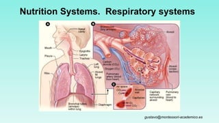
5. Respiratory System
• It is formed by
– Nostrils
– Pharynx
– Trachea
– Bronchi
– Bronchioles
– Pulmonary alveoli (which form the lungs)
• It performs two processes:
– Breathing (it includes breathing in and breathing out)
– Gases exchange (between the air in the alveoli and the blood)
8. NOSTRILS (the two orifices of the nose)
• They connect with the pharynx
through the choanas.
• Their functions are:
– Heating the air, thanks to
Folds in their wall
Lots of blood vessels
– Cleaning the air, by
Little hairs
•Mucus
– Humidify the air with the mucus
The sinuses are a connected system of hollow cavities in the skull. They’re lined with soft, pink tissue called mucosa.
Normally, the sinuses are empty except for a thin layer of mucus.
9. Pharynx, Larynx, and Trachea
Mucus produced in the trachea continues to trap inhaled particles.
Cilia lining the trachea sweep both mucus and trapped particles away
from the lungs toward the pharynx, where they can be swallowed or
spit out.
Humidification is also crucial for sustaining the integrity and survival
of the cilia blanket’s hairy layer, which covers the entire respiratory
tracts, including the nose.
10. LARYNX (the voice box)
• Its entrance is closed by the epiglottis during deglutition.
Epiglottis
The Adam's apple, or laryngeal prominence is a feature of the
human neck, and is the lump or protrusion that is formed by the angle of
the thyroid cartilage surrounding the larynx.
11. LARYNX
It contains the vocal folds. They vibrate to make sound.
Larynx seen from the pharynx
Epiglottis
Vocal folds
Vocal folds when
making low-
pitched sounds
Vocal folds when
making high-pitched
sounds
Larynx contains two highly elastic folds of
tissue known as the vocal cords.
When muscles pull the vocal cords
together, the air moving between them
causes the cords to vibrate and produce
sounds.
https://www.youtube.com/watch?v=P2pLJfWUjc8
12. Lungs
From the trachea, air moves into two large tubes in the chest cavity called
bronchi. Each bronchus leads to one lung.
Within each lung, the large bronchus divides into smaller bronchi, which lead to
even smaller passageways called bronchioles.
Bronchi and bronchioles are surrounded by smooth muscles, controlled by the
autonomic nervous system, that regulate the size of air passageways.
14. BRONCHI, BRONCHIOLES AND ALVEOLI
• Trachea, bronchi and bronchioles have cartilage rings.
• Bronchi branch out into bronchioles, which end in pulmonary alveoli,
forming the lungs.
15.
16. PLEURA
• They are two membranes with liquid
between them (in the pleural space or
cavity).
• They protect the lungs from friction and
shocks and make them move during
breathing.
17. Breathing
What mechanisms are involved in breathing?
Movements of the diaphragm and rib cage change air pressure in the chest
cavity during inhalation and exhalation.
18. BREATHING
Breathing in: The diaphragm and the intercostal and scalene (elevate ribs) muscles contract.
Breathing out: The diaphragm and the intercostal and scalene muscles relax.
In forced or active breathing out the abdominal muscles help expel the air.
19. Inhalation
The lungs are sealed in two sacs,
called pleural membranes, inside
the chest cavity.
At the bottom of the chest cavity is
a large dome-shaped muscle
known as the diaphragm.
https://www.youtube.com/watch?v=hp-gCvW8PRY
https://www.youtube.com/watch?v=wc2K1Olt4Q8
20. Exhalation
Exhalation is usually a passive process,
but to blow out a candle, speak, sing, or
yell, you need more force than passive
exhalation provides.
The extra force is provided by muscles
between the ribs and abdominal
muscles, which contract vigorously as
the diaphragm relaxes.
21. Exhalation
During exhalation, both the rib cage and
the diaphragm relax, decreasing the
volume of the chest cavity and making
air pressure in the chest cavity greater
than atmospheric pressure.
Air rushes back out of the lungs.
22. Exhalation
Breathing works only because the
chest cavity is sealed.
If a wound punctures the chest—
even if it does not affect the lungs
directly—air may leak into the chest
cavity and make breathing
impossible. This is one reason
chest wounds are always serious.
25. GASES EXCHANGE
• Oxygen passes from the alveoli to theblood vessels,
• Carbon dioxide passes from the blood to the alveloli.
26. 26
Definitions
Tidal volume (resting)
amount of air one can move in or out of lungs in single respiratory cycle (resting conditions)
Inspiratory reserve volume (IRV)
amount of air one can take in over and above tidal volume
Expiratory reserve volume (ERV)
amount of air one can voluntarily expel after completed normal respiratory cycle.
Residual volume
amount of air remaining in lungs after maximal exhalation (1200 males; 1100 females)
Minimal volume – amt air left if lungs collapsed
Inspiratory capacity
tidal volume + inspiratory reserve volume
Vital capacity
Maximum amt of air one can take into or out of lungs during forced exhalation and inhalation
Total lung capacity
Total volume of lungs = vital capacity and residual capacity (avg = 6000ml males; 4200ml females