SlideShare a Scribd company logo
1 of 67
In-toeing, Out-toeing, and Limping—Making
Sense of Common Pediatric Gait
Abnormalities
HOSSEIN ASLANI MD
Pediatric orthopedic fellowship
Rotational & Angular Deformities in Children
Objectives:
1.) Review common physiologic and pathologic causes of in-toeing/out-toeing gait in
children
2.) Review diagnosis and physical examination techniques used in assessing pediatric
rotational and angular deformities
3.) Review the current management of pediatric rotational and angular deformities in
children
4.) Review the differential diagnosis of limping gait/gait abnormality in children
Rotational & Angular Deformities in
Children: Introduction
• Rotational and Angular
Deformities are quite common
in pediatrics
• Very diverse spectrum of
diagnoses: physiologic to
pathologic
• In-toeing/Out-toeing, Genu
varum (bowlegs)/valgum
(knock-knees)
The In-toeing Toddler or Child
• History: Inquire about onset, severity, progression, disability,
and previous treatment
• Always assess developmental history: when did child start
walking independently, gross and fine motor skills
• Screening examination to r/o hip dysplasia (DDH), other
neurological problems (cerebral palsy)
Causes of In-toeing Gait in Children
• The most frequent causes of childhood in-
toeing:
Femoral anteversion
Medial Tibial Torsion
Metatarsus adductus
The In-Toeing Toddler/Child:
Assessment
Rotational Profile: Evaluate in four steps
1.) Observe child walking and running.
Estimate the foot progression angle (FPA): angular
difference between the axis of the foot and the line of
progression
Assessing the Foot Progression Angle
• Nonspecific estimation
• Normal: usually -5 tp +20
degrees
• In-toeing -5 to-10 degrees: mild
-10 to -15 degrees moderate
>-15 degrees severe
The In-Toeing Toddler/Child:
Assessment
2.) Assess Femoral Version: Measure external and internal rotation of
the hips with the child prone and the knees flexed to 90 degrees.
Assess both sides simultaneously. Internal rotation usually less than
65-70 degrees
If greater than 70 degrees: in-toeing likely from femoral
anteversion/femoral torsion
If rotation is asymmetrical, evaluate with AP of pelvis to r/o DDH
or hip problem
Assessing Femoral Anteversion:
Internal Rotation of the Hip
Internal Femoral Torsion/Anteversion
• In standing position, patellae will point inwards
when feet are forward
• Compensatory external rotation of tibia
Femoral Anteversion: Clinical Assessment
“Kissing” patellae
Femoral Anteversion: Definitions
• Femoral version defined as the
angular difference between axis of
femoral neck and the transcondylar
axis of knee
• Femoral anteversion ranges from
30-40 degrees at birth and
decreases progressively to about
10-15 degrees at skeletal maturity
• Measurement:X-rays (biplane):
technically difficult
CT--most accurate method
Internal Femoral Torsion/Anteversion
• Usually first seen in the 3-5 year age
group, usually most severe b/w 4-6
years
• Almost always symmetrical
• Mechanism unknown, genetic factors
and position of fetus in uterus causing
increased rotation
• More common in females: approx. 2: 1
ratio, often familial
• Gait/running described as
awkward/clumsy by parents
Femoral Anteversion: Management
• Gait is often worse when running or when fatigued
• Children prefer the “W” sitting position because it is more
comfortable…should not be discouraged or avoided
• Reassurance and Observation!!
• Special shoes, twister cables, etc avoided….no difference in outcome!!
Internal Femoral Torsion:
Management
• Internal femoral torsion/antetorsion
• Mild: internal rotation of hip 70-80 degrees
• Moderate: internal rotation 80-90 degrees
• Severe: internal rotation > 90 degrees
• External hip rotation is usually reduced: total arch of rotation is usually
90-100 degrees
• Resolves spontaneously without treatment in overwhelming
majority of patients: most literature-- 98-99%
• Results from decrease in femoral anterversion
over time (age 8-9 years) and from a lateral rotation
of the tibia
Femoral Anteversion: Operative
Treatment
• Indications for Osteotomy: Individualized
• Tachdijian indications: femoral anteversion >45 degrees
hip unable to laterally rotate beyond neutral, functional
disability and severe cosmetic deformity
• Must weigh the benefits from procedure versus the morbidity of
surgical procedure
Surgical Treatment of Femoral
Anteversion: Derotational Femoral
Osteotomy
• Because of high
spontaneous resolution
rate...derotational
osteotomy is not done
before 8-9 years
• Very rare surgery: delayed
until adolescence to
determine if spontaneous
correction will occur
Internal (Medial) Tibial Torsion
• Toddler or young child often presents
with c/o “bowing legs”
• Usually symmetric in-toeing, if
unilateral--usually worse on left
• Often noticed when child is first
starting to walk
• With patellae facing forwards
(in neutral position), feet turn in
Measurement of Thigh Foot Angle:
Medial Tibial Torsion
3.) Quantify Tibial Version
Thigh Foot Angle: patient is
prone, knees flexed 90 degrees:
TFA is the angular difference
between the axis of the foot and the
axis of the thigh
• Allow foot to fall into natural
position, avoid manual positioning
of foot
• Medial Tibial Torsion: Negative
Thigh Foot Angle
Internal Tibial Torsion: Diagnosis
• Resolves spontaneously in 95-98%
of patients by age 4-6 years
• Stretching, special shoes are
inefffective…does not speed up
resolution and makes no clinical
difference
• Can occasionally have mild
persistence with no handicap or
functional significance
• Usually simple observation is best
treatment and all that is needed
Medial Tibial Torsion: Management
• CT Scan is the best diagnostic study to precisely diagnose the
degree of torsion
• Always pursue conservative treatment: OBSERVATION!!
• If medial tibial torsion is causing gait problems and significant
disability (usually > 40 degrees internal rotation)... can consider
derotational osteotomy after age 8 years (very rare!!)
Metatarsus Adductus in Infants
• Assess the foot for forefoot adductus
• Lateral border of foot should be straight
• Convexity of lateral border and forefoot adduction are
features of metatarsus adductus
Grading Severity of Forefoot Adductus
• Project a line that bisects the heel. Normally it falls on the 2nd toe
• Mild: falls through the 3rd toe
• Moderate: falls between toes 3-4
• Severe: falls between toes 4-5
Metarsus Adductus in Infant
• Most common foot deformity in
children: 1-3/1000
• Prognosis is directly related to the
degree of stiffness
• Differentiate between metatarsus
varus and talipes eqinovarus
• Associated with DDH in 2% of
cases--careful hip examination
Metatarsus Adductus: Assessment
• Exact cause is unknown
• Commonly believed to be
caused by intrauterine
positioning or crowding
• No correlation between
gestational age at birth,
maternal age, or birth
order
Metatarsus Adductus: Management
• Forefoot can gently be stretched passively with each diaper change
• Occasionally will use serial casting and reverse/straight last shoes to
correct deformity
• Observation and Reassurance: will resolve spontaneously in 90-95%
of patients (tends to persist until age 12-18 months)
Physiologic Infantile Out-Toeing
• Out-toeing in early infancy is usually due to a lateral rotation
contracture of the hips
• When infant is positioned upright, the feet will usually turn out
• Resolves spontaneously with ambulation…no treatment is
needed
Out-toeing: External Tibial Torsion
• Most common cause of out-toeing in
children
• May worsen over time because of the
normal lateral rotation of the tibia that
occurs with growth
• May be associated with patellofemoral
knee pain
• If combined with femoral anteversion (
knee internally rotated and ankle
externally rotated):“miserable
malalignment syndrome”…inefficient
gait and patellofemoral joint pain
Mean Tibio-Femoral Angle In Children
Lower Extremity Rotational Profile at
Various Ages
• Normal alignment progresses from 10-15 degrees of varus
at birth to maximum valgus angulation of 10-15 degrees at
3-4 years of age
Genu Valgum (“Knock-Knees):
• Physiologic knock-knee
deformity very common in
children aged 3-5 years
• Screening evaluation: normal
height and body proportions,
symmetrical, localized or
generalized, limb lengths equal
• Measure rotational profile,
measure inter-malleolar distance
with the knees together
• If generalized deformity, order
metabolic screening labs
Physiologic Genu Valgum: Assessment
Pathologic Causes of Genu Valgum
• Post-traumatic (most
common)
• Dysplasias
• Primary tibial valga
• Tumor
• Infection
• Rickets
• Renal osteodystrophy
• Congenital deficiency of
fibula (fibular hemimelia)
Post-Traumatic Genu Valgum
• Usually results from overgrowth
following fracture of the proximal
tibial metaphysis in early childhood
• May also be due to malunion or soft
tissue interposition
• Valgus deformity develops during the
1st 12-18 months post-injury due to
tibial overgrowth
• Management: Most will correct
spontaneously over course of years
without operative treatment
• If deformity persists: osteotomy or
hemiepiphyseodesis
Rickets: Diagnosis and Management
• Suspect rickets in child with increasing genu varum/valgum, short
stature, poor nutritional status (vitamin D deficiency)
• Produces generalized genu varum/valgum with bowing of the
diaphysis and distinctive cupping and widening of the epiphysis
• Refer to endocrinologist for medical management: Ca, Phos
supplementation (Vitamin D resistant form possible)
• Correction (if necessary) usually delayed until the end of growth
as recurrence of deformity is quite common
Clinical Assessment: Rickets
Costochondral “beading” Severe genu valgum
Clinical Assessment: Genu Valgum-Rickets
Family with Rickets
increased varus in toddler, valgus in 5 and 12 year old females
Genu Valgum: General Management
• Age 2-6 years: 95-98% will resolve spontaneously
• If intermalleolar distance is > 8-10 cm at age 10
1.) Hemiephiphyseodesis of distal femur and/or proximal
tibia
2.) If skeletally mature: a.) tibial varus osteotomy
b.) femoral osteotomy: medial
closing wedge: if genu valgum
> 12-15 degrees and superolateral
tilt of joint > 10 degrees
Physiologic Genu Varum: Assessment
• Parents will often note bow leg
deformity, usually recognized
when child starts to walk (12-18
months)
• Commonly bilateral and
symmetric bowing
• Seldom causes functional
disability [X-rays unnecessary
until at least 18 months of age]
• Physiologic bowing usually
spontaneously resolves by the age
of two years
Differentiating physiological Genu
Varum vs. Blount’s disease
• Physiological: bilateral,
symmetrical, metaphyseal-
diaphyseal angle <15 degrees,
upper tibial metaphysis/
epiphysis normal
• Blount’s: unilateral/ bilateral,
asymmetrical, metaphyseal-
diaphyseal angle > 15 degrees,
fragmentation of upper tibial
metaphysis, tibial epiphysis
slopes medially, norrowing of
tibial physis medially, widening
laterally
Diagnosis of Blount’s cannot be made before age 2 years
Infantile Blount’s Disease: Radiographs
Pathological Conditions Causing
Varus Deformity of the Legs
• Metabolic bone disease: Vitamin
D deficiency, Vitamin D refractory
rickets
• Asymmetrical growth arrest or
retardation: Blount’s disease,
Trauma, Infection, Tumor
• Bone dysplasia: metaphyseal
dysplasia
• Congenital
• Neuromuscular
Infantile Blount’s Disease:
Epidemiology
• Risk factors: Obesity, African
American,Walking at early age, +
Family history
• Differential Diagnosis:
Physiologic genu varum
(metaphyseal-diaphyseal angle less than
15 degrees)
Rickets
Osteomyelitis,
Trauma, Tumor
Fibrous dysplasia
Metaphyseal chondrodysplasia
Blount’s Disease: Classification
• Infantile: (early onset)
onset between 1-3 years, bilateral, usually symmetric, pts often
large for age, etiology is abnormal compression on medial
proximal tibial physis, may feel bony prominence or “beak”
over the medial tibial condyle, often have lateral thrust to gait
Very difficult to differentiate from physiologic varus/
bowlegs in patients < 2 years
Adolescent: (onset over 11 years)
often presents with tenderness/pain over the medial prominence
of the proximal tibia, pts often obese
Prevalence: General population <0.3%, Obese African
American male:2.5%
Metaphyseal/Diaphyseal Varus Angle
• Often used to differntiate
physiologic from Blount’s
disease
• If greater than 15-17 degrees,
tibia vara or Blount’s disease
is likely
• Follow radiographs every six
months….physiologic varus
will gradually improve after
age 2 years while Blount’s will
progressively worsen
Measuring the Tibial-Femoral Angle
• Line is drawn down center
of tibia and femur….
Intersecting angle is the
tibio-femoral angle
Blount’s Disease: Radiographs
Adolescent Blount’s Disease
• Definition: Growth disorder involving
the medial portion of the proximal
tibial growth plate that produces a
localized varus deformity
• More often unilateral, usually seen in
obese individuals, slightly more males
than females, African American,
certain geographic regions
• Definite cause unknown:
biomechanical overload to proximal
tibia physis due to varus alignment
and excessive body weight
Adolescent Blount’s: Theorized Cause of
Progressive Varus Deformity
Childhood Varus
Rapid Weight Gain
Medial Growth Plate Injury
Progressive Varus Knee
Adolescent Blount’s Disease: Clinical
Assessment
AP Radiographs: Assessing the Tibia
Vara Deformity
Right
30
Left
26
Blount’s Disease: Non-Operative
Treatment
• Non-operative: Observation only until age 2 years
• Anti-Blount’s Brace: Usually used for pts aged 2-3 years with:
1.) metaphyseal/diaphyseal angle > 15-17 degrees
2.) tibial/femoral angle > 15 degrees
3.) brace is designed to provide rotational support, usually worn FT
4.) usually takes 1 year to determine effectiveness of brace
5.) brace is ineffective in adolescents
• If operative correction necessary in infantile Blount’s….results are
better when done before the age of 4 years
Blount’s Disease: Operative
Treatment
• For optimum correction and results in infantile tibia vara: Surgical
treatment in early stages is crucial
Avoids sequelae of joint incongruity, limb shortening, and persistent
angulation
• Proximal tibial osteotomy: distal to patellar tendon insertion (avoid
proximal physis (dome, closing or opening wedge)
• Adolescent tibia vara: predominately surgical treatment:
1.) Lateral epiphyseodesis: recommended as initial procedure if more
than one year of growth remaining
2.) High tibial osteotomy with internal fixation ( usually correct to
about 0-5 degrees of varus)
3.) Realignment by external fixation: Ilizarov, Dynamic Axial
External fixator, Taylor Spatial Frame
Surgical Correction: Proximal Tibial
Osteotomy
• Demonstration of opening
wedge tibial osteotomy
procedure for correction
of infantile blount’s
disease
• Usually recommend slight
overcorrection into mild
valgus (reverse excessive
compression forces
medially: avoid injury to
physis)
Adolescent Blount’s: Realignment by
External Fixation-Taylor Frame
Rotational and Angular deformities:
Summary
• Most rotational/alignment problems are physiological and
will resolve without intervention
• Good history and physical examination important
• Investigate more if asymmetrical, rapidly progressive
• Orthotics, special braces/shoes, twister cables are
frequently not helpful or necessary
• Most will never require surgery
The Limping Child….
• Relatively large differential diagnosis list
• Obtain good history: VERY important
• Observe child…do they look sick, do they have
fever, will he/she put weight on leg or let you move
extremity?---rule out septic arthritis/infection first!!
• Determine history of trauma, fall, or injury?
• Age of patient, duration of symptoms, onset of
symptoms, family history
The Limping Child
• Observe Child walking/running in hallway
• Generally 4 types of limping gait described
1.) Antalgic gait: shortened stance
2.) Abductor lurch: trendelenburg gait
3.) Equinus gait: toe toe progression
4.) Circumduction gait: leg length discrepency
Limping Gait in Child: Differential
• Fracture, Trauma, Overuse: MOST common
• Transient synovitis: Must differentiate from septic arthritis
• Discoid Lateral Meniscus
• Infection: septic arthritis, lyme disease, osteomyelitis
• LCPD, SCFE, DDH: Hip pathology
• Cerebral Palsy, Neurologic disorders
• Neoplasm/Tumor: benign, malignant
• JRA, other Rheum disorders
The cause of a limp can range from a life-threatening bone tumor to a pebble in a shoe!
The Limping Child:
Transient Synovitis vs Septic
Arthritis
Transient synovitis
•Child refuses to walk
•Movement of hip is painful
•May have fever
•Moderately elevated WBC
•Lasts a few days
•Disappears without
treatment
Bacteria
Enzymes
Destroy cartilage
Irreversable joint damage
White cells
Enzymes
The Limping Child: Septic Arthritis
The Worst Scenario….
• Destruction of articular
cartilage
• Destruction of femoral
head
• Destruction of femoral
neck
Septic Arthritis of the Hip
Treatment:
1. Kill the bacteria!
• IV Antibiotics
2. Eliminate the white cells
• Early Incision and drainage
3. Don’t delay!!!
The Limping Child: Septic
Arthritis
• How to tell the difference?
• Four predictors
– History of fever >101.5
– Refusal to weight-bear
– ESR > 40 mm/hr
– WBC > 12,000
• If in doubt…
– Review in 12 hours
– Do incision and drainage!
The Limping Child: Transient Synovitis
vs. Septic Arthritis
Questions?
Thank You!!!

More Related Content

What's hot

CONGENITAL TALIPES EQUINO VARUS
CONGENITAL TALIPES EQUINO VARUSCONGENITAL TALIPES EQUINO VARUS
CONGENITAL TALIPES EQUINO VARUSAbino David
 
supracondylar fracrture of humerus in children
supracondylar fracrture of humerus in childrensupracondylar fracrture of humerus in children
supracondylar fracrture of humerus in childrenHardik Pawar
 
Coxa Vara, Genu VArum & Valgum. Under Gradts.
Coxa Vara, Genu VArum & Valgum. Under Gradts.Coxa Vara, Genu VArum & Valgum. Under Gradts.
Coxa Vara, Genu VArum & Valgum. Under Gradts.Sarthy Velayutham
 
Developmental dysplasia of the hip
Developmental dysplasia of the hip Developmental dysplasia of the hip
Developmental dysplasia of the hip Diaa Srahin
 
Legg calve perthes disease
Legg calve perthes disease Legg calve perthes disease
Legg calve perthes disease Ratan Khuman
 
Congenital vertical talus
Congenital vertical talusCongenital vertical talus
Congenital vertical talusJoydeep Mandal
 
Ankle fractures
Ankle fracturesAnkle fractures
Ankle fracturesAnand Dev
 
Congenital Tallipes Equino Varus (CTEV)
Congenital Tallipes Equino Varus (CTEV)Congenital Tallipes Equino Varus (CTEV)
Congenital Tallipes Equino Varus (CTEV)Amalina Mohd Daud
 
Congenital anomalies of upper limb
Congenital anomalies of upper limbCongenital anomalies of upper limb
Congenital anomalies of upper limbVictor Effiom
 
The pathology and management of blount’s disease
The pathology and management of blount’s diseaseThe pathology and management of blount’s disease
The pathology and management of blount’s diseaseAsi-oqua Bassey
 
Osgood Schlatter Disease
Osgood Schlatter DiseaseOsgood Schlatter Disease
Osgood Schlatter DiseaseSayantika Dhar
 
Limb length discrepancy
Limb length discrepancyLimb length discrepancy
Limb length discrepancysudarshan731
 

What's hot (20)

CONGENITAL TALIPES EQUINO VARUS
CONGENITAL TALIPES EQUINO VARUSCONGENITAL TALIPES EQUINO VARUS
CONGENITAL TALIPES EQUINO VARUS
 
supracondylar fracrture of humerus in children
supracondylar fracrture of humerus in childrensupracondylar fracrture of humerus in children
supracondylar fracrture of humerus in children
 
Genu varus and valgus
Genu varus and valgusGenu varus and valgus
Genu varus and valgus
 
Polydactyly
PolydactylyPolydactyly
Polydactyly
 
BLOUNT’S DISEASE.pptx
BLOUNT’S DISEASE.pptxBLOUNT’S DISEASE.pptx
BLOUNT’S DISEASE.pptx
 
Coxa Vara, Genu VArum & Valgum. Under Gradts.
Coxa Vara, Genu VArum & Valgum. Under Gradts.Coxa Vara, Genu VArum & Valgum. Under Gradts.
Coxa Vara, Genu VArum & Valgum. Under Gradts.
 
Developmental dysplasia of the hip
Developmental dysplasia of the hip Developmental dysplasia of the hip
Developmental dysplasia of the hip
 
Ctev
CtevCtev
Ctev
 
Legg calve perthes disease
Legg calve perthes disease Legg calve perthes disease
Legg calve perthes disease
 
Congenital vertical talus
Congenital vertical talusCongenital vertical talus
Congenital vertical talus
 
Ankle fractures
Ankle fracturesAnkle fractures
Ankle fractures
 
Ctev
CtevCtev
Ctev
 
Flat foot
Flat footFlat foot
Flat foot
 
DDH
DDHDDH
DDH
 
Congenital Tallipes Equino Varus (CTEV)
Congenital Tallipes Equino Varus (CTEV)Congenital Tallipes Equino Varus (CTEV)
Congenital Tallipes Equino Varus (CTEV)
 
Congenital anomalies of upper limb
Congenital anomalies of upper limbCongenital anomalies of upper limb
Congenital anomalies of upper limb
 
Club foot
Club footClub foot
Club foot
 
The pathology and management of blount’s disease
The pathology and management of blount’s diseaseThe pathology and management of blount’s disease
The pathology and management of blount’s disease
 
Osgood Schlatter Disease
Osgood Schlatter DiseaseOsgood Schlatter Disease
Osgood Schlatter Disease
 
Limb length discrepancy
Limb length discrepancyLimb length discrepancy
Limb length discrepancy
 

Similar to gait abnormalities in children.ppt

Assessment and evaluation tools for pediatric and acquired congenital disorde...
Assessment and evaluation tools for pediatric and acquired congenital disorde...Assessment and evaluation tools for pediatric and acquired congenital disorde...
Assessment and evaluation tools for pediatric and acquired congenital disorde...Monisha devi
 
club-foot in children pediatric nursing.pptx
club-foot in children pediatric nursing.pptxclub-foot in children pediatric nursing.pptx
club-foot in children pediatric nursing.pptxaasthasubedi3
 
Normal variations in children
Normal variations in childrenNormal variations in children
Normal variations in childrenPuneeth Pai
 
Knee and Foot Deformities in pediatrics.pdf
Knee and Foot Deformities in pediatrics.pdfKnee and Foot Deformities in pediatrics.pdf
Knee and Foot Deformities in pediatrics.pdfPTMAAbdelrahman
 
An approach to limping child
An approach to limping childAn approach to limping child
An approach to limping childmanoj das
 
MSS FOR REGULAR BSC.pptx
MSS FOR REGULAR BSC.pptxMSS FOR REGULAR BSC.pptx
MSS FOR REGULAR BSC.pptxMohammedAbdela7
 
Metatarsus adductus
Metatarsus adductusMetatarsus adductus
Metatarsus adductusRziUllah
 
Angular deformities around the knee seminar
Angular deformities around the knee seminarAngular deformities around the knee seminar
Angular deformities around the knee seminarPrashanth Kumar
 
DDH BY DR AJAY SHAH
DDH BY DR AJAY SHAHDDH BY DR AJAY SHAH
DDH BY DR AJAY SHAHAjay Shah
 
8.CONGENITAL ANOMALIES PPT orthopedic nursing
8.CONGENITAL ANOMALIES PPT orthopedic nursing8.CONGENITAL ANOMALIES PPT orthopedic nursing
8.CONGENITAL ANOMALIES PPT orthopedic nursingRenjini R
 
Orthopedic + erb palsy.pptx
Orthopedic + erb palsy.pptxOrthopedic + erb palsy.pptx
Orthopedic + erb palsy.pptxAhmedMufleh1
 
Congenital tallipes equinovarus
Congenital tallipes equinovarusCongenital tallipes equinovarus
Congenital tallipes equinovarusPratikDhabalia
 
final limp approach brbk.pptx
final limp approach brbk.pptxfinal limp approach brbk.pptx
final limp approach brbk.pptxbishwokunwar3
 

Similar to gait abnormalities in children.ppt (20)

Assessment and evaluation tools for pediatric and acquired congenital disorde...
Assessment and evaluation tools for pediatric and acquired congenital disorde...Assessment and evaluation tools for pediatric and acquired congenital disorde...
Assessment and evaluation tools for pediatric and acquired congenital disorde...
 
Orthopedic problems in neonates
Orthopedic problems in neonatesOrthopedic problems in neonates
Orthopedic problems in neonates
 
club-foot in children pediatric nursing.pptx
club-foot in children pediatric nursing.pptxclub-foot in children pediatric nursing.pptx
club-foot in children pediatric nursing.pptx
 
Normal variations in children
Normal variations in childrenNormal variations in children
Normal variations in children
 
Knee and Foot Deformities in pediatrics.pdf
Knee and Foot Deformities in pediatrics.pdfKnee and Foot Deformities in pediatrics.pdf
Knee and Foot Deformities in pediatrics.pdf
 
An approach to limping child
An approach to limping childAn approach to limping child
An approach to limping child
 
Club foot
Club footClub foot
Club foot
 
MSS FOR REGULAR BSC.pptx
MSS FOR REGULAR BSC.pptxMSS FOR REGULAR BSC.pptx
MSS FOR REGULAR BSC.pptx
 
Club foot
Club footClub foot
Club foot
 
Metatarsus adductus
Metatarsus adductusMetatarsus adductus
Metatarsus adductus
 
Congenital hip dysplasia
Congenital hip dysplasiaCongenital hip dysplasia
Congenital hip dysplasia
 
Angular deformities around the knee seminar
Angular deformities around the knee seminarAngular deformities around the knee seminar
Angular deformities around the knee seminar
 
DDH BY DR AJAY SHAH
DDH BY DR AJAY SHAHDDH BY DR AJAY SHAH
DDH BY DR AJAY SHAH
 
8.CONGENITAL ANOMALIES PPT orthopedic nursing
8.CONGENITAL ANOMALIES PPT orthopedic nursing8.CONGENITAL ANOMALIES PPT orthopedic nursing
8.CONGENITAL ANOMALIES PPT orthopedic nursing
 
CTEV/ Clubfoot
CTEV/ ClubfootCTEV/ Clubfoot
CTEV/ Clubfoot
 
Orthopedic + erb palsy.pptx
Orthopedic + erb palsy.pptxOrthopedic + erb palsy.pptx
Orthopedic + erb palsy.pptx
 
Congenital tallipes equinovarus
Congenital tallipes equinovarusCongenital tallipes equinovarus
Congenital tallipes equinovarus
 
Club foot
Club footClub foot
Club foot
 
Ponceti techniqe
Ponceti techniqePonceti techniqe
Ponceti techniqe
 
final limp approach brbk.pptx
final limp approach brbk.pptxfinal limp approach brbk.pptx
final limp approach brbk.pptx
 

Recently uploaded

Interactive Powerpoint_How to Master effective communication
Interactive Powerpoint_How to Master effective communicationInteractive Powerpoint_How to Master effective communication
Interactive Powerpoint_How to Master effective communicationnomboosow
 
Paris 2024 Olympic Geographies - an activity
Paris 2024 Olympic Geographies - an activityParis 2024 Olympic Geographies - an activity
Paris 2024 Olympic Geographies - an activityGeoBlogs
 
Science 7 - LAND and SEA BREEZE and its Characteristics
Science 7 - LAND and SEA BREEZE and its CharacteristicsScience 7 - LAND and SEA BREEZE and its Characteristics
Science 7 - LAND and SEA BREEZE and its CharacteristicsKarinaGenton
 
KSHARA STURA .pptx---KSHARA KARMA THERAPY (CAUSTIC THERAPY)————IMP.OF KSHARA ...
KSHARA STURA .pptx---KSHARA KARMA THERAPY (CAUSTIC THERAPY)————IMP.OF KSHARA ...KSHARA STURA .pptx---KSHARA KARMA THERAPY (CAUSTIC THERAPY)————IMP.OF KSHARA ...
KSHARA STURA .pptx---KSHARA KARMA THERAPY (CAUSTIC THERAPY)————IMP.OF KSHARA ...M56BOOKSTORE PRODUCT/SERVICE
 
CARE OF CHILD IN INCUBATOR..........pptx
CARE OF CHILD IN INCUBATOR..........pptxCARE OF CHILD IN INCUBATOR..........pptx
CARE OF CHILD IN INCUBATOR..........pptxGaneshChakor2
 
microwave assisted reaction. General introduction
microwave assisted reaction. General introductionmicrowave assisted reaction. General introduction
microwave assisted reaction. General introductionMaksud Ahmed
 
Separation of Lanthanides/ Lanthanides and Actinides
Separation of Lanthanides/ Lanthanides and ActinidesSeparation of Lanthanides/ Lanthanides and Actinides
Separation of Lanthanides/ Lanthanides and ActinidesFatimaKhan178732
 
call girls in Kamla Market (DELHI) 🔝 >༒9953330565🔝 genuine Escort Service 🔝✔️✔️
call girls in Kamla Market (DELHI) 🔝 >༒9953330565🔝 genuine Escort Service 🔝✔️✔️call girls in Kamla Market (DELHI) 🔝 >༒9953330565🔝 genuine Escort Service 🔝✔️✔️
call girls in Kamla Market (DELHI) 🔝 >༒9953330565🔝 genuine Escort Service 🔝✔️✔️9953056974 Low Rate Call Girls In Saket, Delhi NCR
 
Hybridoma Technology ( Production , Purification , and Application )
Hybridoma Technology  ( Production , Purification , and Application  ) Hybridoma Technology  ( Production , Purification , and Application  )
Hybridoma Technology ( Production , Purification , and Application ) Sakshi Ghasle
 
Mastering the Unannounced Regulatory Inspection
Mastering the Unannounced Regulatory InspectionMastering the Unannounced Regulatory Inspection
Mastering the Unannounced Regulatory InspectionSafetyChain Software
 
URLs and Routing in the Odoo 17 Website App
URLs and Routing in the Odoo 17 Website AppURLs and Routing in the Odoo 17 Website App
URLs and Routing in the Odoo 17 Website AppCeline George
 
ECONOMIC CONTEXT - LONG FORM TV DRAMA - PPT
ECONOMIC CONTEXT - LONG FORM TV DRAMA - PPTECONOMIC CONTEXT - LONG FORM TV DRAMA - PPT
ECONOMIC CONTEXT - LONG FORM TV DRAMA - PPTiammrhaywood
 
MENTAL STATUS EXAMINATION format.docx
MENTAL     STATUS EXAMINATION format.docxMENTAL     STATUS EXAMINATION format.docx
MENTAL STATUS EXAMINATION format.docxPoojaSen20
 
18-04-UA_REPORT_MEDIALITERAСY_INDEX-DM_23-1-final-eng.pdf
18-04-UA_REPORT_MEDIALITERAСY_INDEX-DM_23-1-final-eng.pdf18-04-UA_REPORT_MEDIALITERAСY_INDEX-DM_23-1-final-eng.pdf
18-04-UA_REPORT_MEDIALITERAСY_INDEX-DM_23-1-final-eng.pdfssuser54595a
 
Sanyam Choudhary Chemistry practical.pdf
Sanyam Choudhary Chemistry practical.pdfSanyam Choudhary Chemistry practical.pdf
Sanyam Choudhary Chemistry practical.pdfsanyamsingh5019
 
Concept of Vouching. B.Com(Hons) /B.Compdf
Concept of Vouching. B.Com(Hons) /B.CompdfConcept of Vouching. B.Com(Hons) /B.Compdf
Concept of Vouching. B.Com(Hons) /B.CompdfUmakantAnnand
 
Alper Gobel In Media Res Media Component
Alper Gobel In Media Res Media ComponentAlper Gobel In Media Res Media Component
Alper Gobel In Media Res Media ComponentInMediaRes1
 

Recently uploaded (20)

Interactive Powerpoint_How to Master effective communication
Interactive Powerpoint_How to Master effective communicationInteractive Powerpoint_How to Master effective communication
Interactive Powerpoint_How to Master effective communication
 
Paris 2024 Olympic Geographies - an activity
Paris 2024 Olympic Geographies - an activityParis 2024 Olympic Geographies - an activity
Paris 2024 Olympic Geographies - an activity
 
Science 7 - LAND and SEA BREEZE and its Characteristics
Science 7 - LAND and SEA BREEZE and its CharacteristicsScience 7 - LAND and SEA BREEZE and its Characteristics
Science 7 - LAND and SEA BREEZE and its Characteristics
 
KSHARA STURA .pptx---KSHARA KARMA THERAPY (CAUSTIC THERAPY)————IMP.OF KSHARA ...
KSHARA STURA .pptx---KSHARA KARMA THERAPY (CAUSTIC THERAPY)————IMP.OF KSHARA ...KSHARA STURA .pptx---KSHARA KARMA THERAPY (CAUSTIC THERAPY)————IMP.OF KSHARA ...
KSHARA STURA .pptx---KSHARA KARMA THERAPY (CAUSTIC THERAPY)————IMP.OF KSHARA ...
 
CARE OF CHILD IN INCUBATOR..........pptx
CARE OF CHILD IN INCUBATOR..........pptxCARE OF CHILD IN INCUBATOR..........pptx
CARE OF CHILD IN INCUBATOR..........pptx
 
microwave assisted reaction. General introduction
microwave assisted reaction. General introductionmicrowave assisted reaction. General introduction
microwave assisted reaction. General introduction
 
Separation of Lanthanides/ Lanthanides and Actinides
Separation of Lanthanides/ Lanthanides and ActinidesSeparation of Lanthanides/ Lanthanides and Actinides
Separation of Lanthanides/ Lanthanides and Actinides
 
Staff of Color (SOC) Retention Efforts DDSD
Staff of Color (SOC) Retention Efforts DDSDStaff of Color (SOC) Retention Efforts DDSD
Staff of Color (SOC) Retention Efforts DDSD
 
call girls in Kamla Market (DELHI) 🔝 >༒9953330565🔝 genuine Escort Service 🔝✔️✔️
call girls in Kamla Market (DELHI) 🔝 >༒9953330565🔝 genuine Escort Service 🔝✔️✔️call girls in Kamla Market (DELHI) 🔝 >༒9953330565🔝 genuine Escort Service 🔝✔️✔️
call girls in Kamla Market (DELHI) 🔝 >༒9953330565🔝 genuine Escort Service 🔝✔️✔️
 
Hybridoma Technology ( Production , Purification , and Application )
Hybridoma Technology  ( Production , Purification , and Application  ) Hybridoma Technology  ( Production , Purification , and Application  )
Hybridoma Technology ( Production , Purification , and Application )
 
Mastering the Unannounced Regulatory Inspection
Mastering the Unannounced Regulatory InspectionMastering the Unannounced Regulatory Inspection
Mastering the Unannounced Regulatory Inspection
 
URLs and Routing in the Odoo 17 Website App
URLs and Routing in the Odoo 17 Website AppURLs and Routing in the Odoo 17 Website App
URLs and Routing in the Odoo 17 Website App
 
ECONOMIC CONTEXT - LONG FORM TV DRAMA - PPT
ECONOMIC CONTEXT - LONG FORM TV DRAMA - PPTECONOMIC CONTEXT - LONG FORM TV DRAMA - PPT
ECONOMIC CONTEXT - LONG FORM TV DRAMA - PPT
 
MENTAL STATUS EXAMINATION format.docx
MENTAL     STATUS EXAMINATION format.docxMENTAL     STATUS EXAMINATION format.docx
MENTAL STATUS EXAMINATION format.docx
 
Model Call Girl in Bikash Puri Delhi reach out to us at 🔝9953056974🔝
Model Call Girl in Bikash Puri  Delhi reach out to us at 🔝9953056974🔝Model Call Girl in Bikash Puri  Delhi reach out to us at 🔝9953056974🔝
Model Call Girl in Bikash Puri Delhi reach out to us at 🔝9953056974🔝
 
Model Call Girl in Tilak Nagar Delhi reach out to us at 🔝9953056974🔝
Model Call Girl in Tilak Nagar Delhi reach out to us at 🔝9953056974🔝Model Call Girl in Tilak Nagar Delhi reach out to us at 🔝9953056974🔝
Model Call Girl in Tilak Nagar Delhi reach out to us at 🔝9953056974🔝
 
18-04-UA_REPORT_MEDIALITERAСY_INDEX-DM_23-1-final-eng.pdf
18-04-UA_REPORT_MEDIALITERAСY_INDEX-DM_23-1-final-eng.pdf18-04-UA_REPORT_MEDIALITERAСY_INDEX-DM_23-1-final-eng.pdf
18-04-UA_REPORT_MEDIALITERAСY_INDEX-DM_23-1-final-eng.pdf
 
Sanyam Choudhary Chemistry practical.pdf
Sanyam Choudhary Chemistry practical.pdfSanyam Choudhary Chemistry practical.pdf
Sanyam Choudhary Chemistry practical.pdf
 
Concept of Vouching. B.Com(Hons) /B.Compdf
Concept of Vouching. B.Com(Hons) /B.CompdfConcept of Vouching. B.Com(Hons) /B.Compdf
Concept of Vouching. B.Com(Hons) /B.Compdf
 
Alper Gobel In Media Res Media Component
Alper Gobel In Media Res Media ComponentAlper Gobel In Media Res Media Component
Alper Gobel In Media Res Media Component
 

gait abnormalities in children.ppt

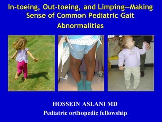
  • 1. In-toeing, Out-toeing, and Limping—Making Sense of Common Pediatric Gait Abnormalities HOSSEIN ASLANI MD Pediatric orthopedic fellowship
  • 2. Rotational & Angular Deformities in Children Objectives: 1.) Review common physiologic and pathologic causes of in-toeing/out-toeing gait in children 2.) Review diagnosis and physical examination techniques used in assessing pediatric rotational and angular deformities 3.) Review the current management of pediatric rotational and angular deformities in children 4.) Review the differential diagnosis of limping gait/gait abnormality in children
  • 3. Rotational & Angular Deformities in Children: Introduction • Rotational and Angular Deformities are quite common in pediatrics • Very diverse spectrum of diagnoses: physiologic to pathologic • In-toeing/Out-toeing, Genu varum (bowlegs)/valgum (knock-knees)
  • 4. The In-toeing Toddler or Child • History: Inquire about onset, severity, progression, disability, and previous treatment • Always assess developmental history: when did child start walking independently, gross and fine motor skills • Screening examination to r/o hip dysplasia (DDH), other neurological problems (cerebral palsy)
  • 5. Causes of In-toeing Gait in Children • The most frequent causes of childhood in- toeing: Femoral anteversion Medial Tibial Torsion Metatarsus adductus
  • 6. The In-Toeing Toddler/Child: Assessment Rotational Profile: Evaluate in four steps 1.) Observe child walking and running. Estimate the foot progression angle (FPA): angular difference between the axis of the foot and the line of progression
  • 7. Assessing the Foot Progression Angle • Nonspecific estimation • Normal: usually -5 tp +20 degrees • In-toeing -5 to-10 degrees: mild -10 to -15 degrees moderate >-15 degrees severe
  • 8. The In-Toeing Toddler/Child: Assessment 2.) Assess Femoral Version: Measure external and internal rotation of the hips with the child prone and the knees flexed to 90 degrees. Assess both sides simultaneously. Internal rotation usually less than 65-70 degrees If greater than 70 degrees: in-toeing likely from femoral anteversion/femoral torsion If rotation is asymmetrical, evaluate with AP of pelvis to r/o DDH or hip problem
  • 10. Internal Femoral Torsion/Anteversion • In standing position, patellae will point inwards when feet are forward • Compensatory external rotation of tibia
  • 11. Femoral Anteversion: Clinical Assessment “Kissing” patellae
  • 12. Femoral Anteversion: Definitions • Femoral version defined as the angular difference between axis of femoral neck and the transcondylar axis of knee • Femoral anteversion ranges from 30-40 degrees at birth and decreases progressively to about 10-15 degrees at skeletal maturity • Measurement:X-rays (biplane): technically difficult CT--most accurate method
  • 13. Internal Femoral Torsion/Anteversion • Usually first seen in the 3-5 year age group, usually most severe b/w 4-6 years • Almost always symmetrical • Mechanism unknown, genetic factors and position of fetus in uterus causing increased rotation • More common in females: approx. 2: 1 ratio, often familial • Gait/running described as awkward/clumsy by parents
  • 14. Femoral Anteversion: Management • Gait is often worse when running or when fatigued • Children prefer the “W” sitting position because it is more comfortable…should not be discouraged or avoided • Reassurance and Observation!! • Special shoes, twister cables, etc avoided….no difference in outcome!!
  • 15. Internal Femoral Torsion: Management • Internal femoral torsion/antetorsion • Mild: internal rotation of hip 70-80 degrees • Moderate: internal rotation 80-90 degrees • Severe: internal rotation > 90 degrees • External hip rotation is usually reduced: total arch of rotation is usually 90-100 degrees • Resolves spontaneously without treatment in overwhelming majority of patients: most literature-- 98-99% • Results from decrease in femoral anterversion over time (age 8-9 years) and from a lateral rotation of the tibia
  • 16. Femoral Anteversion: Operative Treatment • Indications for Osteotomy: Individualized • Tachdijian indications: femoral anteversion >45 degrees hip unable to laterally rotate beyond neutral, functional disability and severe cosmetic deformity • Must weigh the benefits from procedure versus the morbidity of surgical procedure
  • 17. Surgical Treatment of Femoral Anteversion: Derotational Femoral Osteotomy • Because of high spontaneous resolution rate...derotational osteotomy is not done before 8-9 years • Very rare surgery: delayed until adolescence to determine if spontaneous correction will occur
  • 18. Internal (Medial) Tibial Torsion • Toddler or young child often presents with c/o “bowing legs” • Usually symmetric in-toeing, if unilateral--usually worse on left • Often noticed when child is first starting to walk • With patellae facing forwards (in neutral position), feet turn in
  • 19. Measurement of Thigh Foot Angle: Medial Tibial Torsion 3.) Quantify Tibial Version Thigh Foot Angle: patient is prone, knees flexed 90 degrees: TFA is the angular difference between the axis of the foot and the axis of the thigh • Allow foot to fall into natural position, avoid manual positioning of foot • Medial Tibial Torsion: Negative Thigh Foot Angle
  • 20. Internal Tibial Torsion: Diagnosis • Resolves spontaneously in 95-98% of patients by age 4-6 years • Stretching, special shoes are inefffective…does not speed up resolution and makes no clinical difference • Can occasionally have mild persistence with no handicap or functional significance • Usually simple observation is best treatment and all that is needed
  • 21. Medial Tibial Torsion: Management • CT Scan is the best diagnostic study to precisely diagnose the degree of torsion • Always pursue conservative treatment: OBSERVATION!! • If medial tibial torsion is causing gait problems and significant disability (usually > 40 degrees internal rotation)... can consider derotational osteotomy after age 8 years (very rare!!)
  • 22. Metatarsus Adductus in Infants • Assess the foot for forefoot adductus • Lateral border of foot should be straight • Convexity of lateral border and forefoot adduction are features of metatarsus adductus
  • 23. Grading Severity of Forefoot Adductus • Project a line that bisects the heel. Normally it falls on the 2nd toe • Mild: falls through the 3rd toe • Moderate: falls between toes 3-4 • Severe: falls between toes 4-5
  • 24. Metarsus Adductus in Infant • Most common foot deformity in children: 1-3/1000 • Prognosis is directly related to the degree of stiffness • Differentiate between metatarsus varus and talipes eqinovarus • Associated with DDH in 2% of cases--careful hip examination
  • 25. Metatarsus Adductus: Assessment • Exact cause is unknown • Commonly believed to be caused by intrauterine positioning or crowding • No correlation between gestational age at birth, maternal age, or birth order
  • 26. Metatarsus Adductus: Management • Forefoot can gently be stretched passively with each diaper change • Occasionally will use serial casting and reverse/straight last shoes to correct deformity • Observation and Reassurance: will resolve spontaneously in 90-95% of patients (tends to persist until age 12-18 months)
  • 27. Physiologic Infantile Out-Toeing • Out-toeing in early infancy is usually due to a lateral rotation contracture of the hips • When infant is positioned upright, the feet will usually turn out • Resolves spontaneously with ambulation…no treatment is needed
  • 28. Out-toeing: External Tibial Torsion • Most common cause of out-toeing in children • May worsen over time because of the normal lateral rotation of the tibia that occurs with growth • May be associated with patellofemoral knee pain • If combined with femoral anteversion ( knee internally rotated and ankle externally rotated):“miserable malalignment syndrome”…inefficient gait and patellofemoral joint pain
  • 30. Lower Extremity Rotational Profile at Various Ages • Normal alignment progresses from 10-15 degrees of varus at birth to maximum valgus angulation of 10-15 degrees at 3-4 years of age
  • 31. Genu Valgum (“Knock-Knees): • Physiologic knock-knee deformity very common in children aged 3-5 years • Screening evaluation: normal height and body proportions, symmetrical, localized or generalized, limb lengths equal • Measure rotational profile, measure inter-malleolar distance with the knees together • If generalized deformity, order metabolic screening labs
  • 33. Pathologic Causes of Genu Valgum • Post-traumatic (most common) • Dysplasias • Primary tibial valga • Tumor • Infection • Rickets • Renal osteodystrophy • Congenital deficiency of fibula (fibular hemimelia)
  • 34. Post-Traumatic Genu Valgum • Usually results from overgrowth following fracture of the proximal tibial metaphysis in early childhood • May also be due to malunion or soft tissue interposition • Valgus deformity develops during the 1st 12-18 months post-injury due to tibial overgrowth • Management: Most will correct spontaneously over course of years without operative treatment • If deformity persists: osteotomy or hemiepiphyseodesis
  • 35. Rickets: Diagnosis and Management • Suspect rickets in child with increasing genu varum/valgum, short stature, poor nutritional status (vitamin D deficiency) • Produces generalized genu varum/valgum with bowing of the diaphysis and distinctive cupping and widening of the epiphysis • Refer to endocrinologist for medical management: Ca, Phos supplementation (Vitamin D resistant form possible) • Correction (if necessary) usually delayed until the end of growth as recurrence of deformity is quite common
  • 36. Clinical Assessment: Rickets Costochondral “beading” Severe genu valgum
  • 37. Clinical Assessment: Genu Valgum-Rickets Family with Rickets increased varus in toddler, valgus in 5 and 12 year old females
  • 38. Genu Valgum: General Management • Age 2-6 years: 95-98% will resolve spontaneously • If intermalleolar distance is > 8-10 cm at age 10 1.) Hemiephiphyseodesis of distal femur and/or proximal tibia 2.) If skeletally mature: a.) tibial varus osteotomy b.) femoral osteotomy: medial closing wedge: if genu valgum > 12-15 degrees and superolateral tilt of joint > 10 degrees
  • 39.
  • 40. Physiologic Genu Varum: Assessment • Parents will often note bow leg deformity, usually recognized when child starts to walk (12-18 months) • Commonly bilateral and symmetric bowing • Seldom causes functional disability [X-rays unnecessary until at least 18 months of age] • Physiologic bowing usually spontaneously resolves by the age of two years
  • 41. Differentiating physiological Genu Varum vs. Blount’s disease • Physiological: bilateral, symmetrical, metaphyseal- diaphyseal angle <15 degrees, upper tibial metaphysis/ epiphysis normal • Blount’s: unilateral/ bilateral, asymmetrical, metaphyseal- diaphyseal angle > 15 degrees, fragmentation of upper tibial metaphysis, tibial epiphysis slopes medially, norrowing of tibial physis medially, widening laterally Diagnosis of Blount’s cannot be made before age 2 years
  • 43. Pathological Conditions Causing Varus Deformity of the Legs • Metabolic bone disease: Vitamin D deficiency, Vitamin D refractory rickets • Asymmetrical growth arrest or retardation: Blount’s disease, Trauma, Infection, Tumor • Bone dysplasia: metaphyseal dysplasia • Congenital • Neuromuscular
  • 44. Infantile Blount’s Disease: Epidemiology • Risk factors: Obesity, African American,Walking at early age, + Family history • Differential Diagnosis: Physiologic genu varum (metaphyseal-diaphyseal angle less than 15 degrees) Rickets Osteomyelitis, Trauma, Tumor Fibrous dysplasia Metaphyseal chondrodysplasia
  • 45. Blount’s Disease: Classification • Infantile: (early onset) onset between 1-3 years, bilateral, usually symmetric, pts often large for age, etiology is abnormal compression on medial proximal tibial physis, may feel bony prominence or “beak” over the medial tibial condyle, often have lateral thrust to gait Very difficult to differentiate from physiologic varus/ bowlegs in patients < 2 years Adolescent: (onset over 11 years) often presents with tenderness/pain over the medial prominence of the proximal tibia, pts often obese Prevalence: General population <0.3%, Obese African American male:2.5%
  • 46. Metaphyseal/Diaphyseal Varus Angle • Often used to differntiate physiologic from Blount’s disease • If greater than 15-17 degrees, tibia vara or Blount’s disease is likely • Follow radiographs every six months….physiologic varus will gradually improve after age 2 years while Blount’s will progressively worsen
  • 47. Measuring the Tibial-Femoral Angle • Line is drawn down center of tibia and femur…. Intersecting angle is the tibio-femoral angle
  • 49. Adolescent Blount’s Disease • Definition: Growth disorder involving the medial portion of the proximal tibial growth plate that produces a localized varus deformity • More often unilateral, usually seen in obese individuals, slightly more males than females, African American, certain geographic regions • Definite cause unknown: biomechanical overload to proximal tibia physis due to varus alignment and excessive body weight
  • 50. Adolescent Blount’s: Theorized Cause of Progressive Varus Deformity Childhood Varus Rapid Weight Gain Medial Growth Plate Injury Progressive Varus Knee
  • 51. Adolescent Blount’s Disease: Clinical Assessment
  • 52. AP Radiographs: Assessing the Tibia Vara Deformity Right 30 Left 26
  • 53. Blount’s Disease: Non-Operative Treatment • Non-operative: Observation only until age 2 years • Anti-Blount’s Brace: Usually used for pts aged 2-3 years with: 1.) metaphyseal/diaphyseal angle > 15-17 degrees 2.) tibial/femoral angle > 15 degrees 3.) brace is designed to provide rotational support, usually worn FT 4.) usually takes 1 year to determine effectiveness of brace 5.) brace is ineffective in adolescents • If operative correction necessary in infantile Blount’s….results are better when done before the age of 4 years
  • 54. Blount’s Disease: Operative Treatment • For optimum correction and results in infantile tibia vara: Surgical treatment in early stages is crucial Avoids sequelae of joint incongruity, limb shortening, and persistent angulation • Proximal tibial osteotomy: distal to patellar tendon insertion (avoid proximal physis (dome, closing or opening wedge) • Adolescent tibia vara: predominately surgical treatment: 1.) Lateral epiphyseodesis: recommended as initial procedure if more than one year of growth remaining 2.) High tibial osteotomy with internal fixation ( usually correct to about 0-5 degrees of varus) 3.) Realignment by external fixation: Ilizarov, Dynamic Axial External fixator, Taylor Spatial Frame
  • 55. Surgical Correction: Proximal Tibial Osteotomy • Demonstration of opening wedge tibial osteotomy procedure for correction of infantile blount’s disease • Usually recommend slight overcorrection into mild valgus (reverse excessive compression forces medially: avoid injury to physis)
  • 56. Adolescent Blount’s: Realignment by External Fixation-Taylor Frame
  • 57. Rotational and Angular deformities: Summary • Most rotational/alignment problems are physiological and will resolve without intervention • Good history and physical examination important • Investigate more if asymmetrical, rapidly progressive • Orthotics, special braces/shoes, twister cables are frequently not helpful or necessary • Most will never require surgery
  • 58. The Limping Child…. • Relatively large differential diagnosis list • Obtain good history: VERY important • Observe child…do they look sick, do they have fever, will he/she put weight on leg or let you move extremity?---rule out septic arthritis/infection first!! • Determine history of trauma, fall, or injury? • Age of patient, duration of symptoms, onset of symptoms, family history
  • 59. The Limping Child • Observe Child walking/running in hallway • Generally 4 types of limping gait described 1.) Antalgic gait: shortened stance 2.) Abductor lurch: trendelenburg gait 3.) Equinus gait: toe toe progression 4.) Circumduction gait: leg length discrepency
  • 60. Limping Gait in Child: Differential • Fracture, Trauma, Overuse: MOST common • Transient synovitis: Must differentiate from septic arthritis • Discoid Lateral Meniscus • Infection: septic arthritis, lyme disease, osteomyelitis • LCPD, SCFE, DDH: Hip pathology • Cerebral Palsy, Neurologic disorders • Neoplasm/Tumor: benign, malignant • JRA, other Rheum disorders The cause of a limp can range from a life-threatening bone tumor to a pebble in a shoe!
  • 61. The Limping Child: Transient Synovitis vs Septic Arthritis Transient synovitis •Child refuses to walk •Movement of hip is painful •May have fever •Moderately elevated WBC •Lasts a few days •Disappears without treatment
  • 62. Bacteria Enzymes Destroy cartilage Irreversable joint damage White cells Enzymes The Limping Child: Septic Arthritis
  • 63. The Worst Scenario…. • Destruction of articular cartilage • Destruction of femoral head • Destruction of femoral neck Septic Arthritis of the Hip
  • 64. Treatment: 1. Kill the bacteria! • IV Antibiotics 2. Eliminate the white cells • Early Incision and drainage 3. Don’t delay!!! The Limping Child: Septic Arthritis
  • 65. • How to tell the difference? • Four predictors – History of fever >101.5 – Refusal to weight-bear – ESR > 40 mm/hr – WBC > 12,000 • If in doubt… – Review in 12 hours – Do incision and drainage! The Limping Child: Transient Synovitis vs. Septic Arthritis
  • 66.