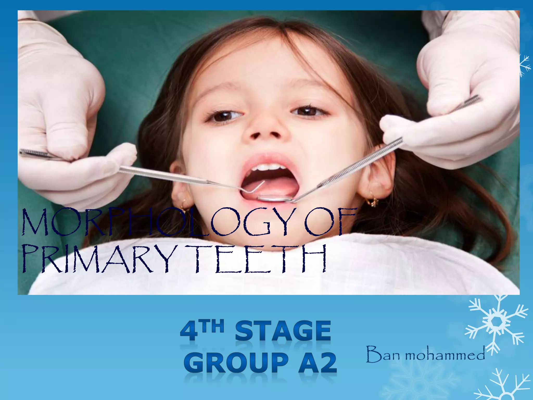
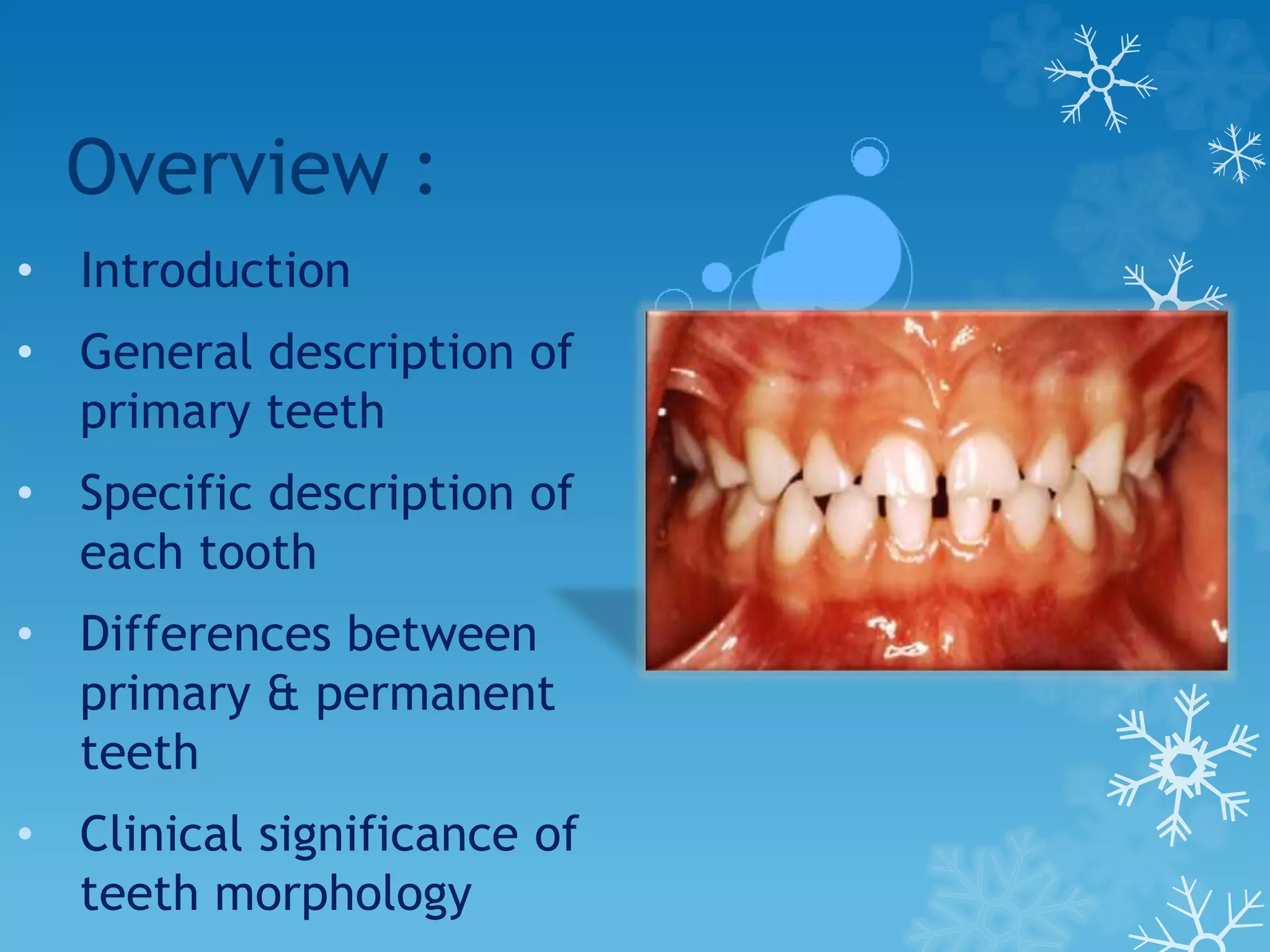
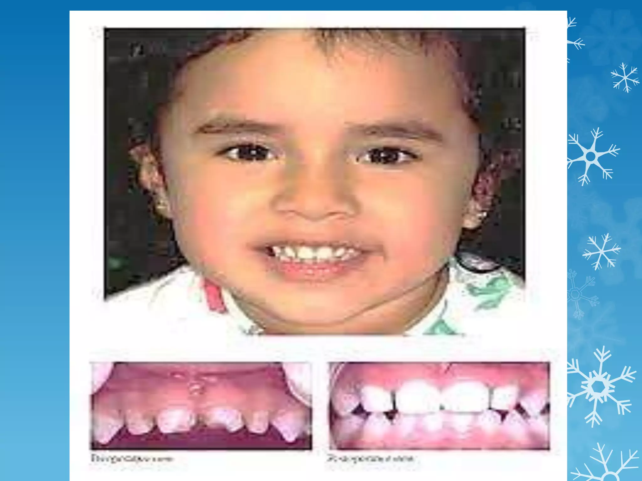
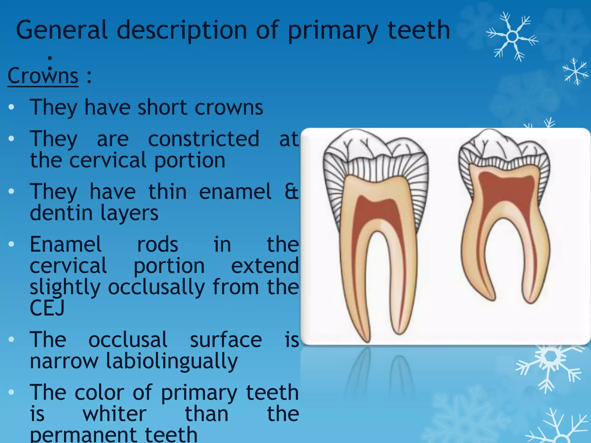
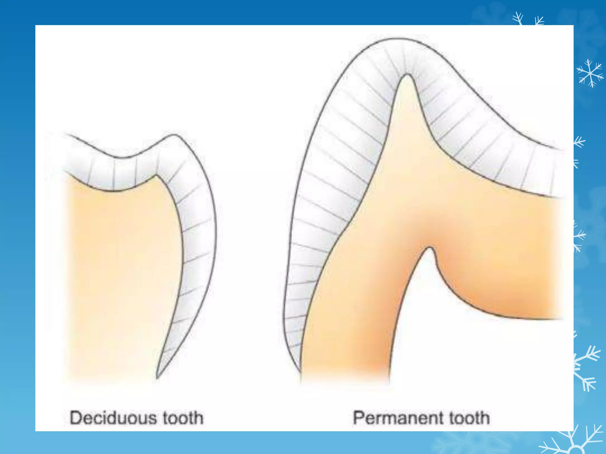
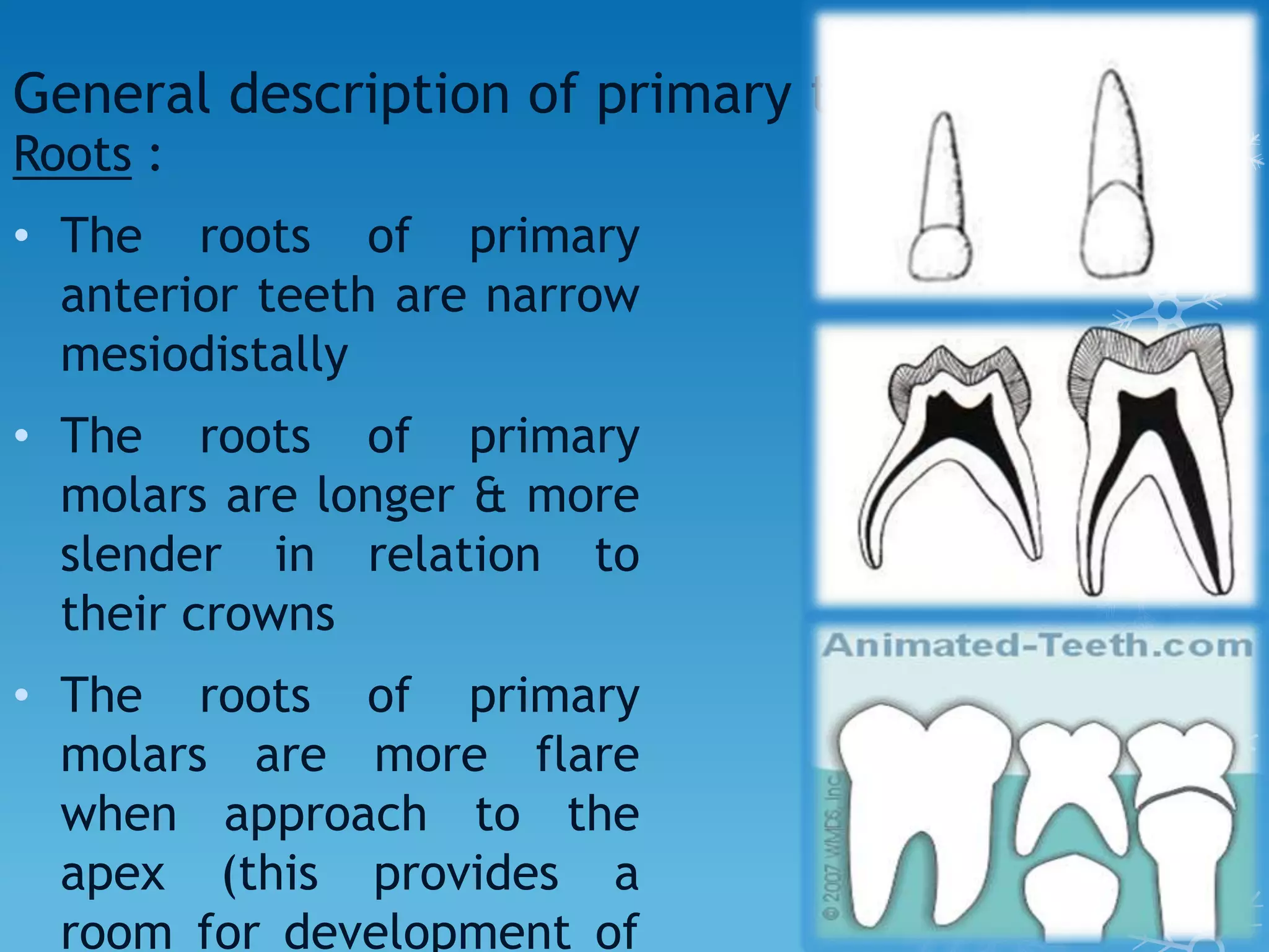
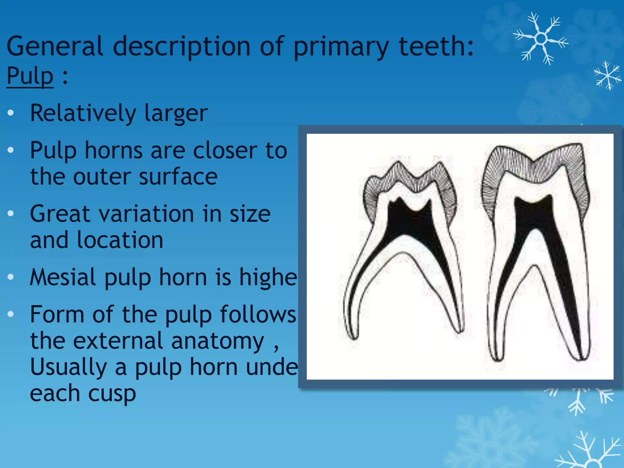
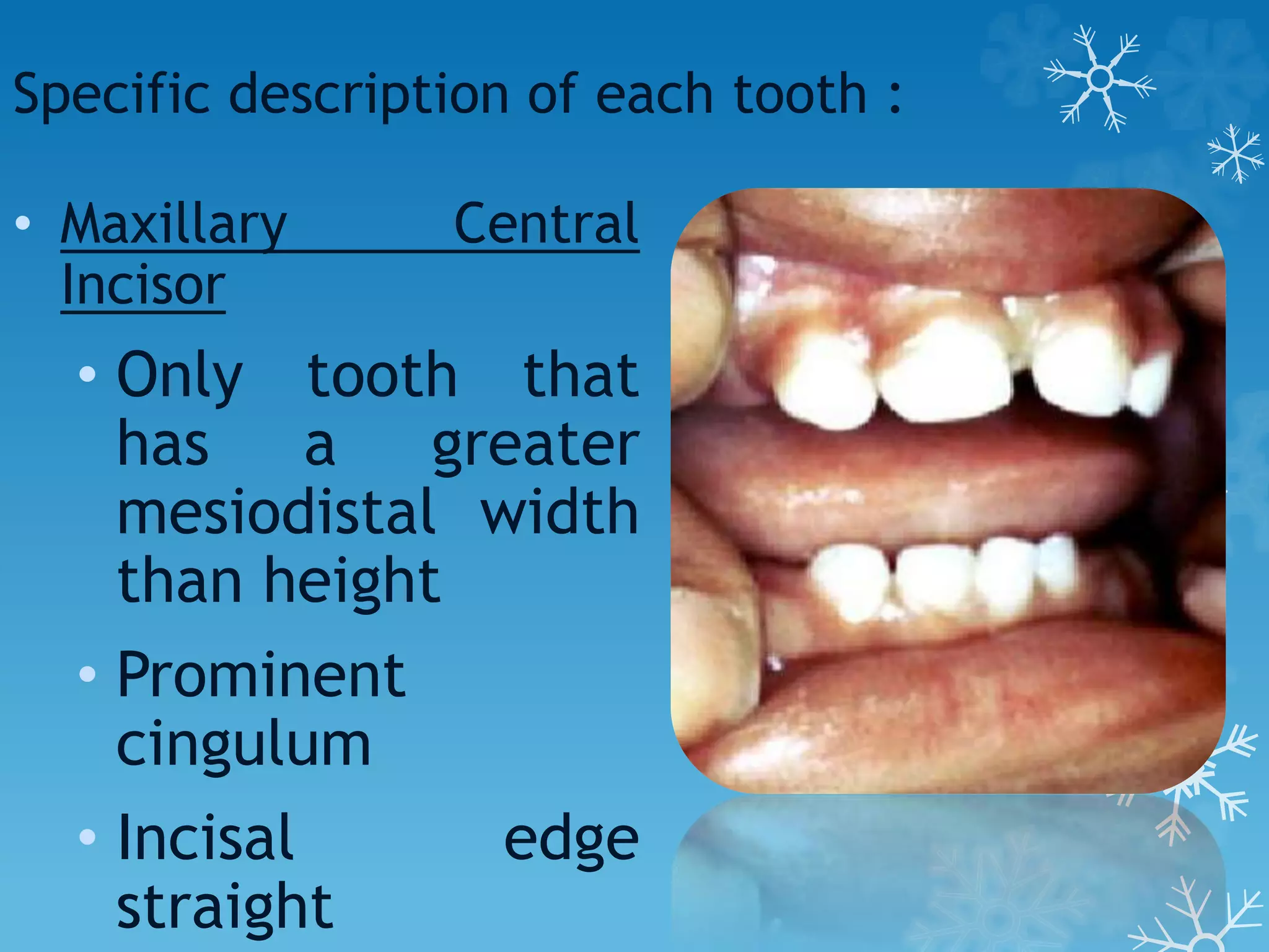
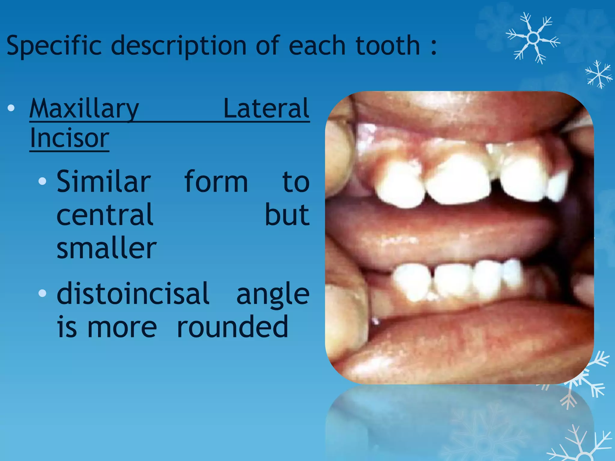
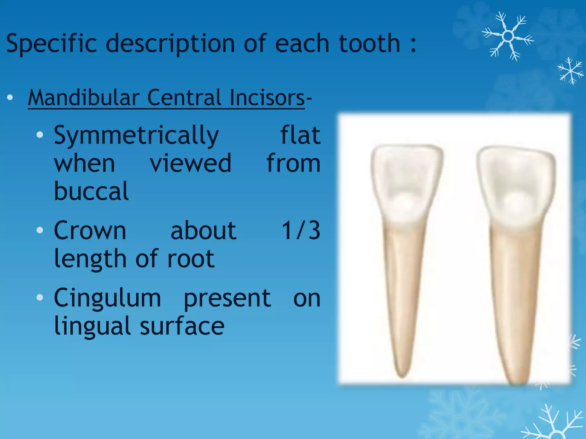
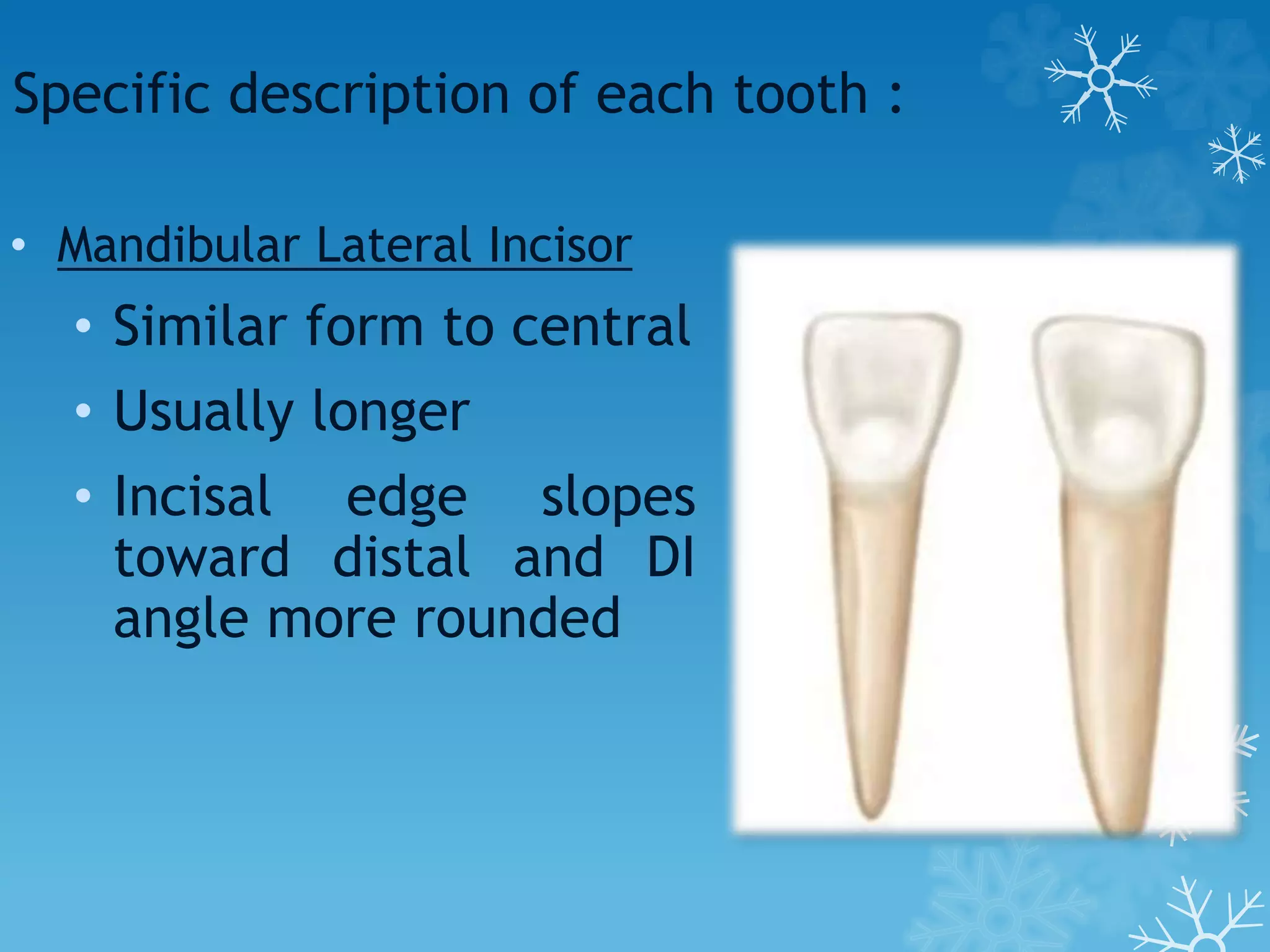
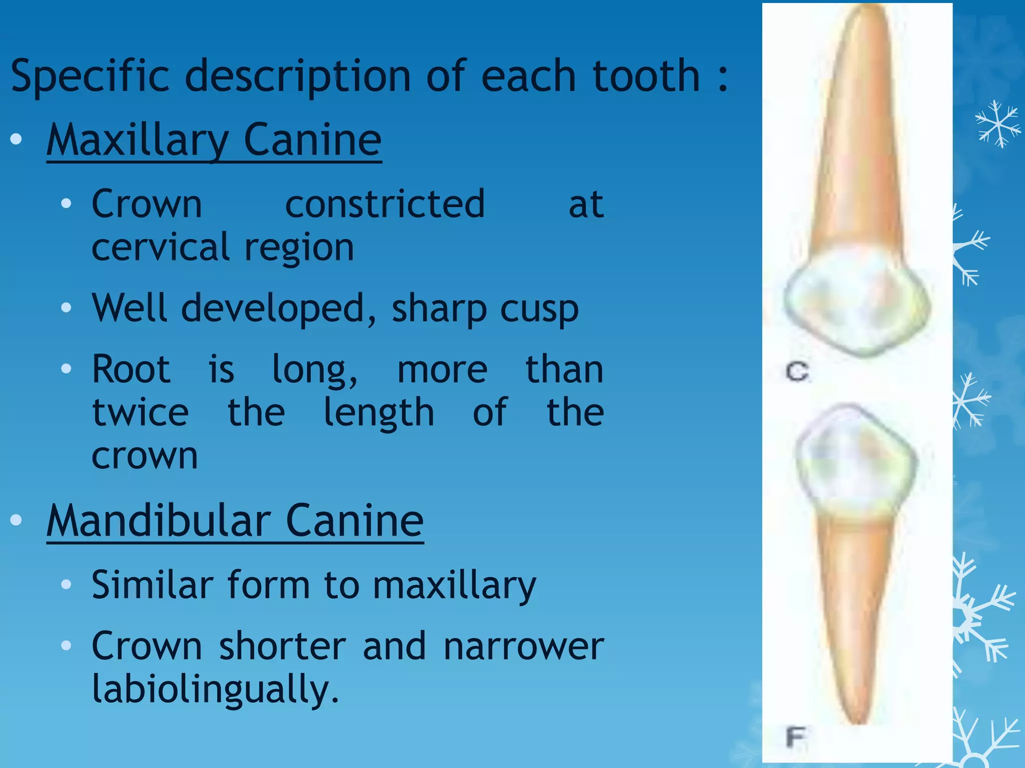
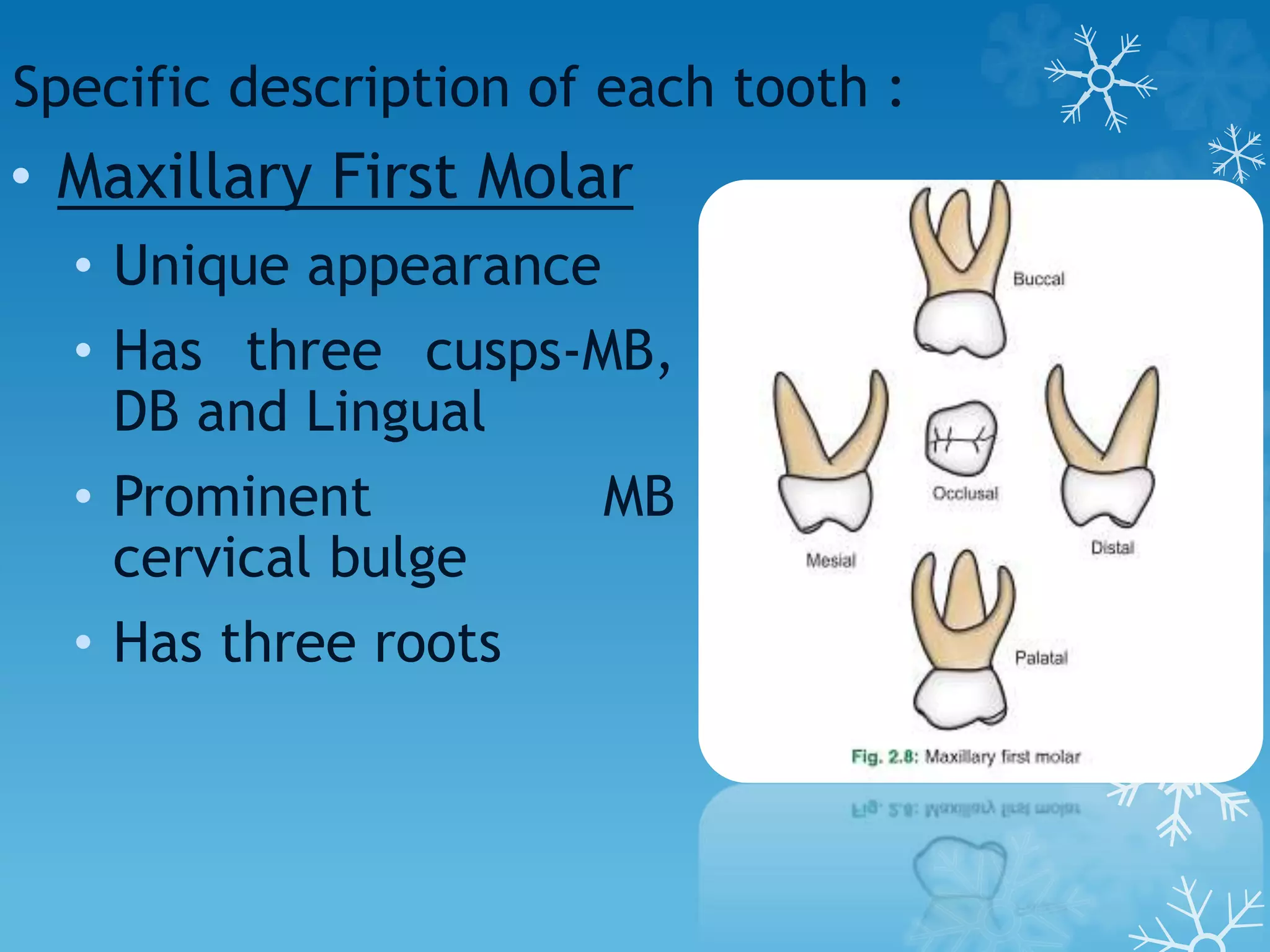
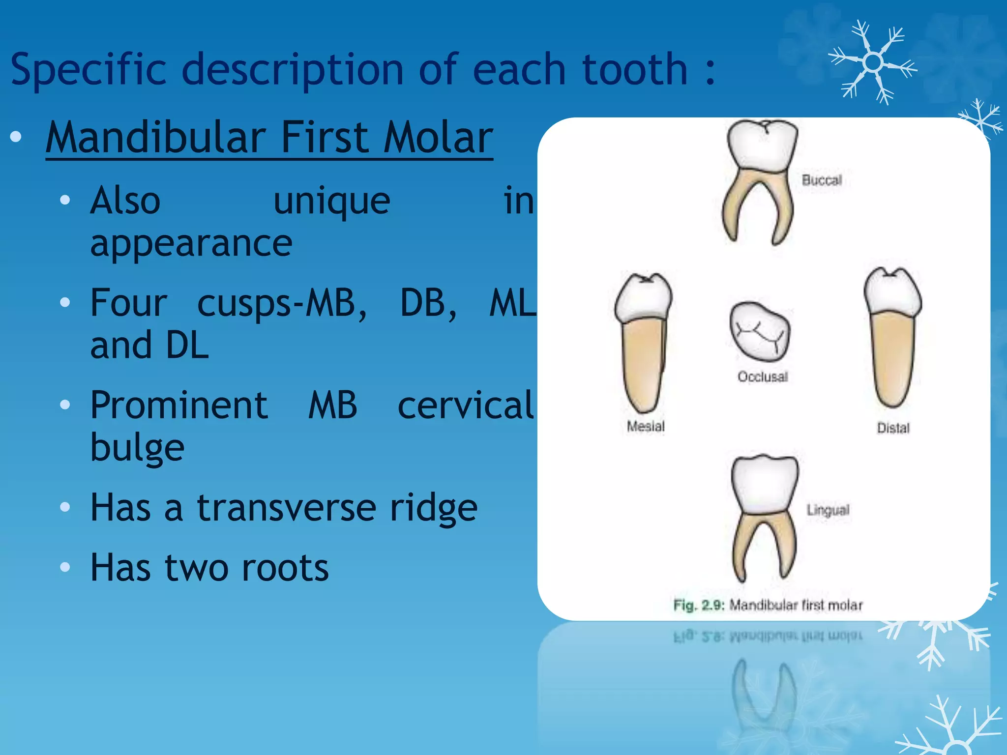
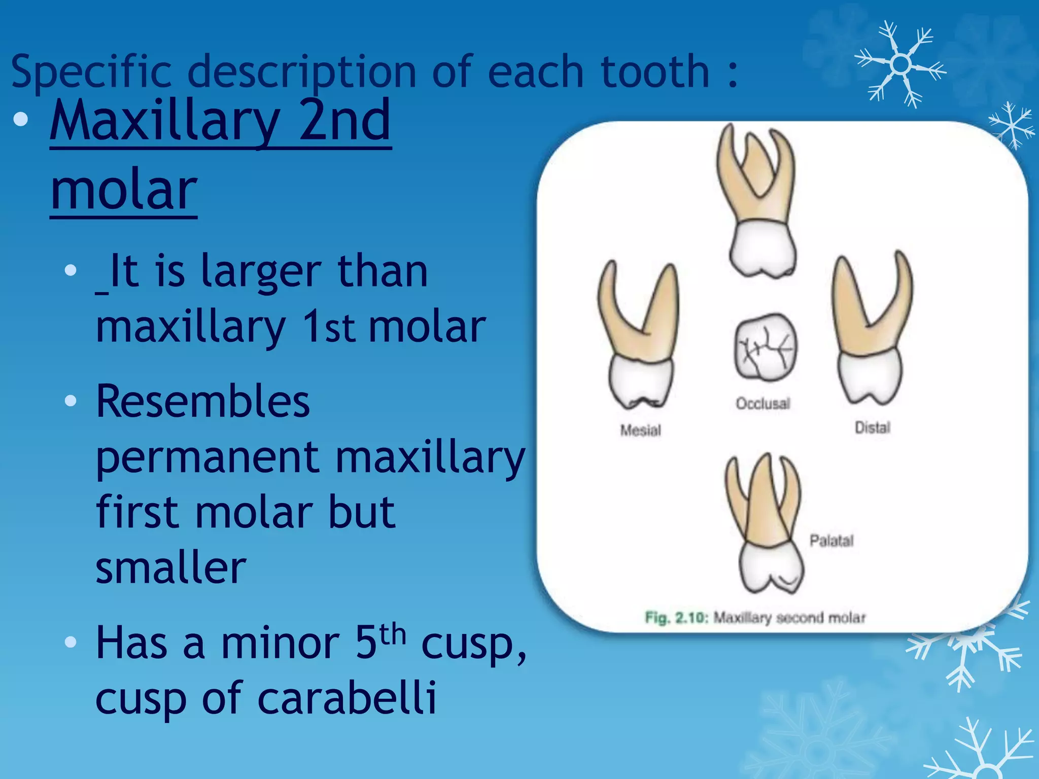
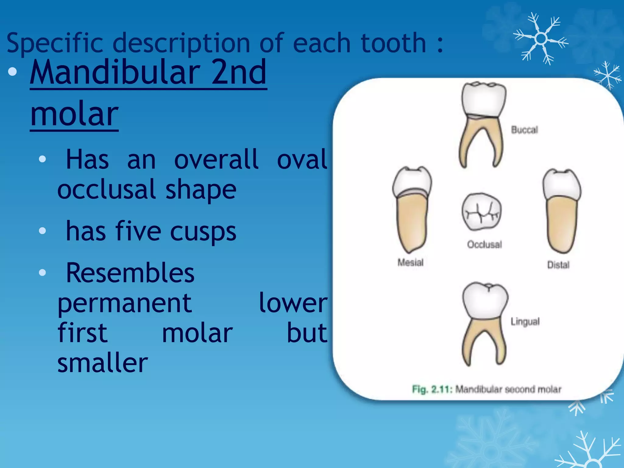
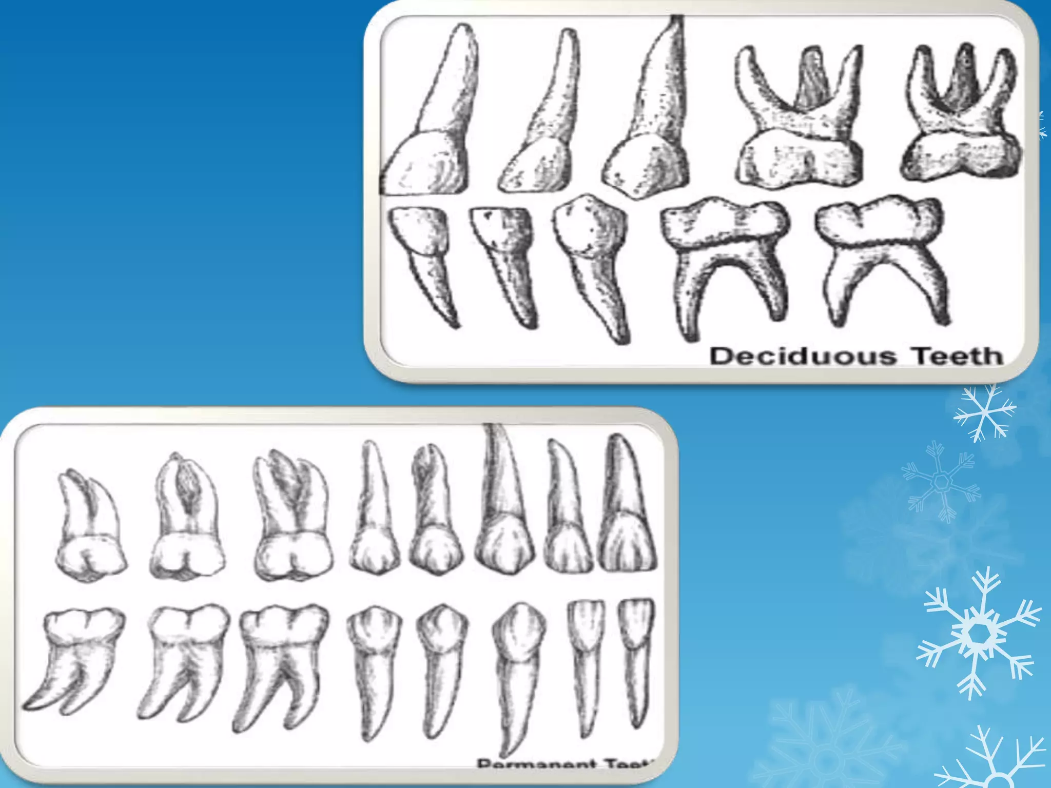
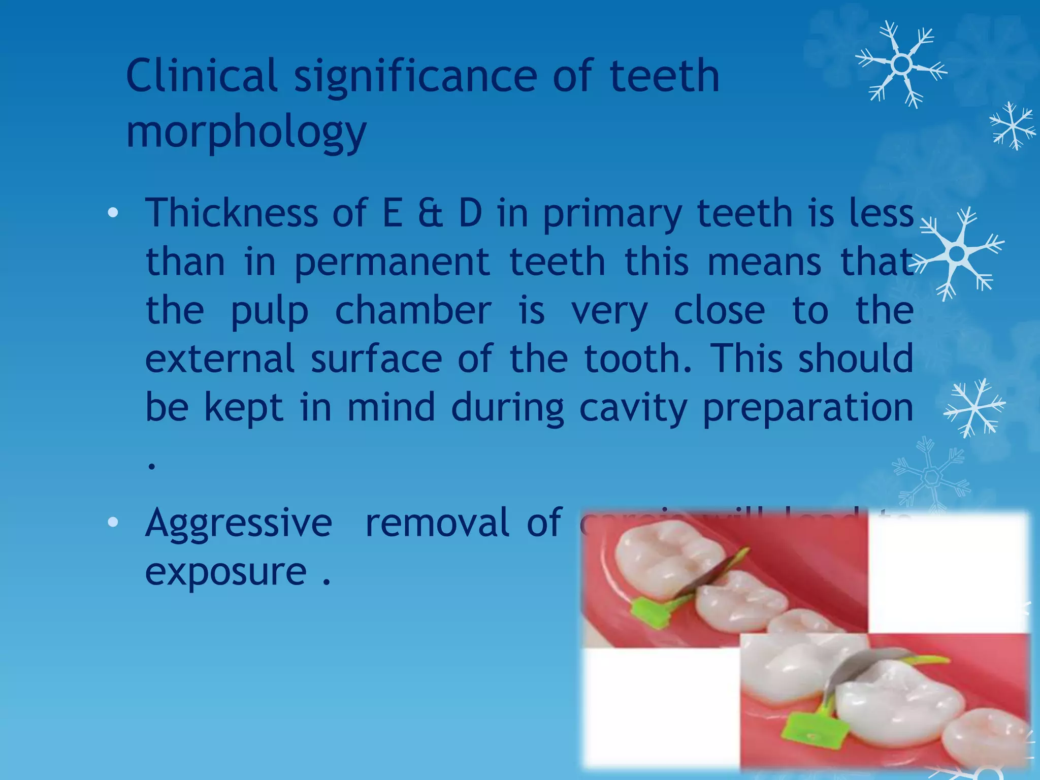
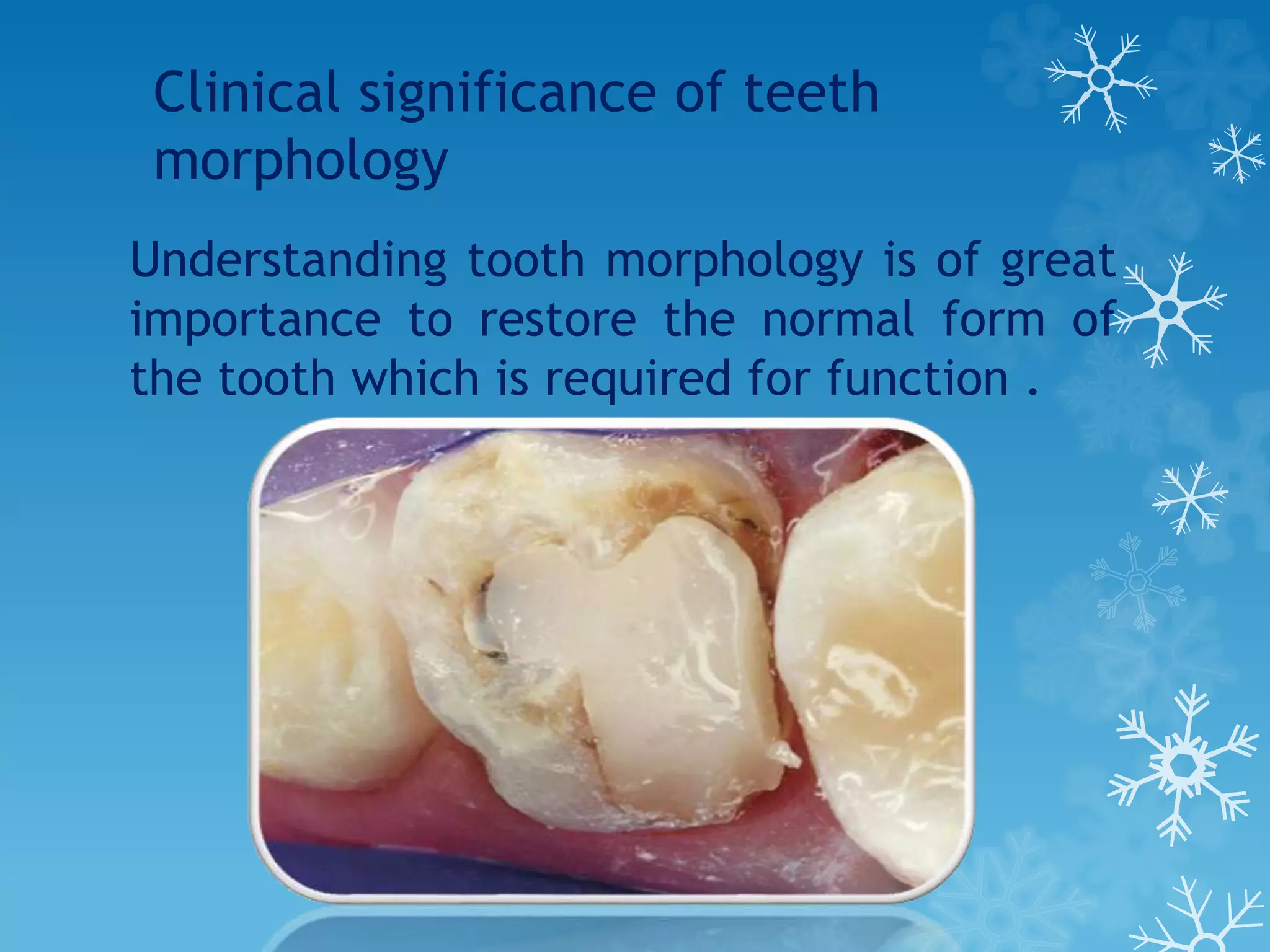
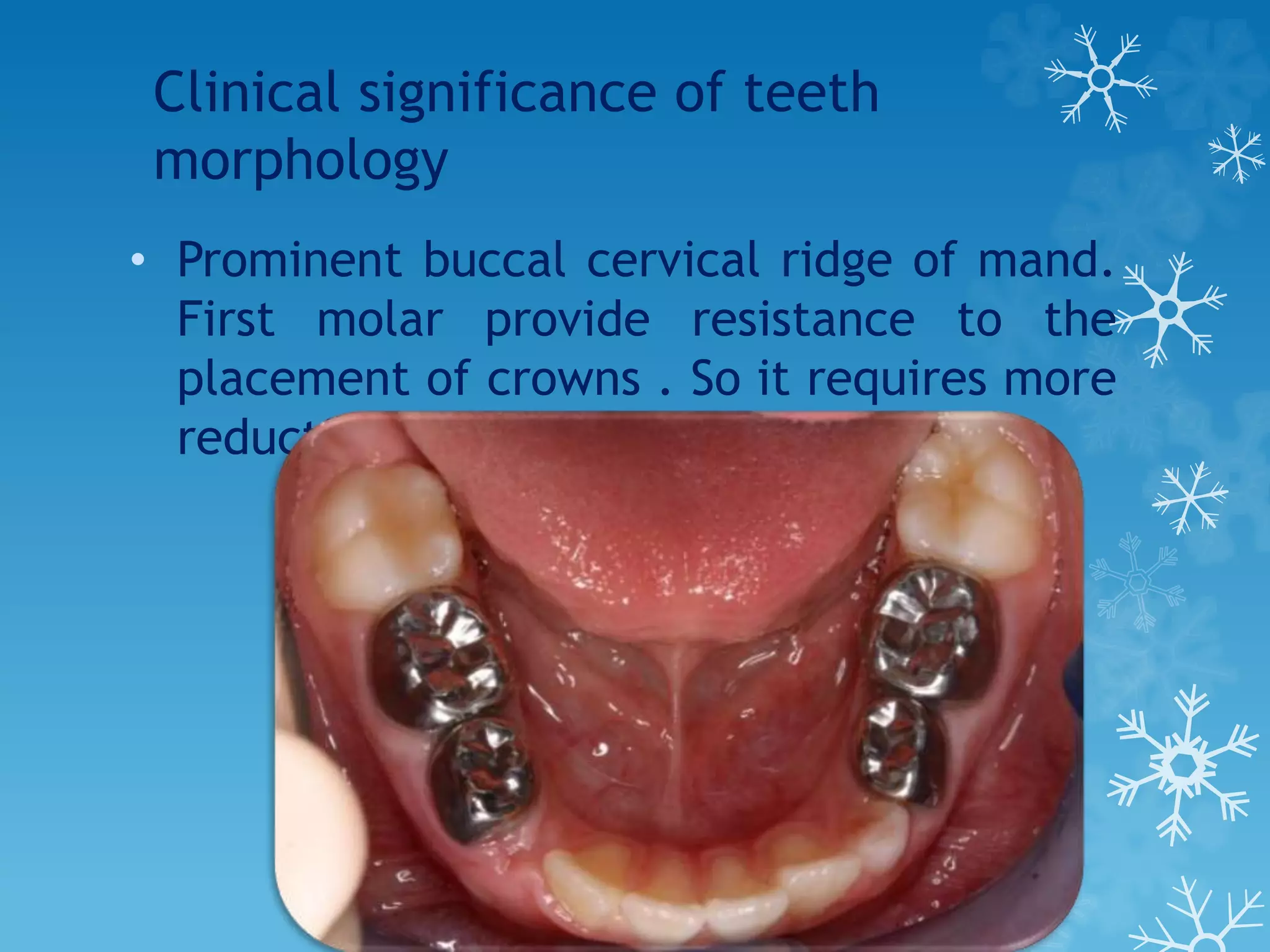
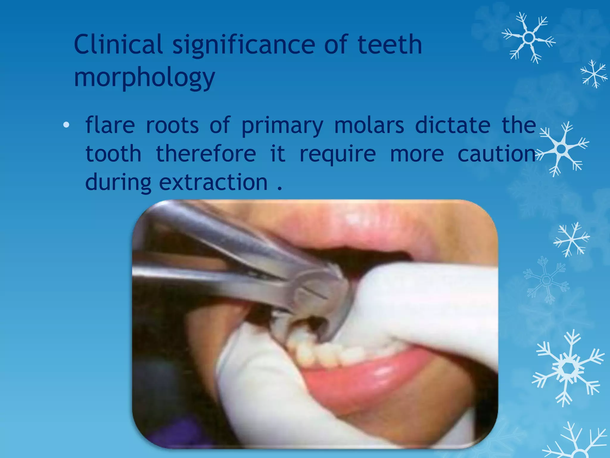
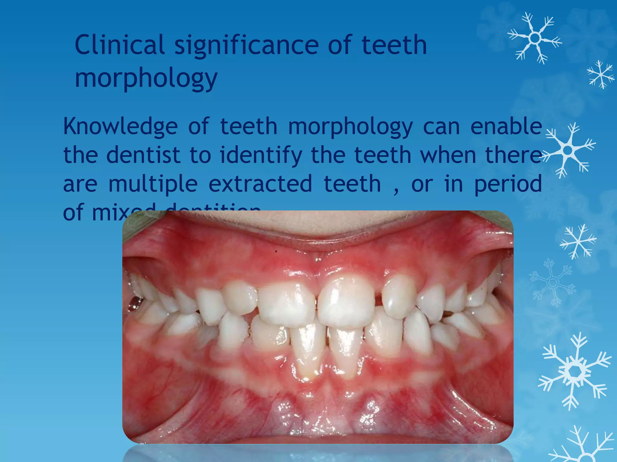
MORPHOLOGY OF
PRIMARY TEETH
Ban mohammed
Overview :
• Introduction
• General description of
primary teeth
• Specific description of
each tooth
• Differences between
primary & permanent
teeth
• Clinical significance of
teeth morphology
Introduction
The morphology of primary teeth is very
important clinically during performing
various procedures.
Cavity preparations must conform to the
thickness of enamel & dentin keeping in
mind the size & location of pulp horns .
Restoration of normal morphology of
primary teeth is needed for function , this
is can be achieved by good knowledge of
morphology
General description of primary teeth
:Crowns :
• They have short crowns
• They are constricted at
the cervical portion
• They have thin enamel &
dentin layers
• Enamel rods in the
cervical portion extend
slightly occlusally from the
CEJ
• The occlusal surface is
narrow labiolingually
• The color of primary teeth
is whiter than the
permanent teeth
General description of primary teeth:
Roots :
• The roots of primary
anterior teeth are narrow
mesiodistally
• The roots of primary
molars are longer & more
slender in relation to
their crowns
• The roots of primary
molars are more flare
when approach to the
apex (this provides a
room for development of
General description of primary teeth:
Pulp :
• Relatively larger
• Pulp horns are closer to
the outer surface
• Great variation in size
and location
• Mesial pulp horn is higher
• Form of the pulp follows
the external anatomy ,
Usually a pulp horn under
each cusp
Specific description of each tooth :
• Maxillary Central
Incisor
• Only tooth that
has a greater
mesiodistal width
than height
• Prominent
cingulum
• Incisal edge
straight
Specific description of each tooth :
• Maxillary Lateral
Incisor
• Similar form to
central but
smaller
• distoincisal angle
is more rounded
Specific description of each tooth :
• Mandibular Central Incisors-
• Symmetrically flat
when viewed from
buccal
• Crown about 1/3
length of root
• Cingulum present on
lingual surface
Specific description of each tooth :
• Mandibular Lateral Incisor
• Similar form to central
• Usually longer
• Incisal edge slopes
toward distal and DI
angle more rounded
Specific description of each tooth :
• Maxillary Canine
• Crown constricted at
cervical region
• Well developed, sharp cusp
• Root is long, more than
twice the length of the
crown
• Mandibular Canine
• Similar form to maxillary
• Crown shorter and narrower
labiolingually.
Specific description of each tooth :
• Maxillary First Molar
• Unique appearance
• Has three cusps-MB,
DB and Lingual
• Prominent MB
cervical bulge
• Has three roots
Specific description of each tooth :
• Mandibular First Molar
• Also unique in
appearance
• Four cusps-MB, DB, ML
and DL
• Prominent MB cervical
bulge
• Has a transverse ridge
• Has two roots
Specific description of each tooth :
• Maxillary 2nd
molar
• It is larger than
maxillary 1st molar
• Resembles
permanent maxillary
first molar but
smaller
• Has a minor 5th cusp,
cusp of carabelli
Specific description of each tooth :
• Mandibular 2nd
molar
• Has an overall oval
occlusal shape
• has five cusps
• Resembles
permanent lower
first molar but
smaller
Differences between primary & permanent teeth
Permanent dentitionPrimary dentitionfeature
3220number
I=2,C=1,P=2,M=3I=2,C=1,M=2 in each quad.formula
Larger in all dimensionSmaller in all dimensionsize
Darker , more pigmented &
have less translucent
enamel
Whiter , less pigmented &
have more translucent
enamel
color
Cusps are blunt & the
crowns are not bulbous
Cusps are more pointed &
the crowns are bulbous
shape
The enamel ends gradually
at the neck
The enamel ends abruptly
at the neck
Cervical
area
Longer, stronger, less
divergent away from the
crown
Shorter, weaker, more
divergent close to the
crown
roots
Differences between primary & permanent teeth
Permanent dentitionPrimary dentitionfeature
Relatively smaller, the
pulp horns are lower
Relatively larger, pulp
horns rise high in cusp
region
pulp
Placed obliquely on the
jaws
Placed perpendicularly on
the jaws
Placement
in jaws
Mamelons may presentNo mamelonsIncisal
edges
thickerthinnerdentin
more calcified, less
permeable, rods near the
cervical portion directed
apically
Less calcified, more
permeable, rods near the
cervical portion
perpendicular to the DEJ
enamel
Clinical significance of teeth
morphology
• Thickness of E & D in primary teeth is less
than in permanent teeth this means that
the pulp chamber is very close to the
external surface of the tooth. This should
be kept in mind during cavity preparation
.
• Aggressive removal of careis will lead to
exposure .
Clinical significance of teeth
morphology
Understanding tooth morphology is of great
importance to restore the normal form of
the tooth which is required for function .
Clinical significance of teeth
morphology
• Prominent buccal cervical ridge of mand.
First molar provide resistance to the
placement of crowns . So it requires more
reduction .
Clinical significance of teeth
morphology
• flare roots of primary molars dictate the
tooth therefore it require more caution
during extraction .
Clinical significance of teeth
morphology
Knowledge of teeth morphology can enable
the dentist to identify the teeth when there
are multiple extracted teeth , or in period
of mixed dentition .
REFERENCES :
• Wheeler’s of dental anatomy , physiology, and occlusion
• Pediatric Dentistry, By Muthu
• Textbook of Dental Anatomy and Tooth Morphology, By Kumar
• Anatomy of Primary Teeth , Steven Chussid D.D.S.
• UP TO DATE in
• DENTAL ANATOMY AND PHYSIOLOGY ,BY :Prof. Dr.
Nahed A. Khalil, Head of Oral Biology Departmen &
Contributing Staff Members ,Faculty of Oral and Dental Medicine, CairoUniversity
Morphology of primary teeth

Morphology of primary teeth

  • 1.
  • 3.
    Overview : • Introduction •General description of primary teeth • Specific description of each tooth • Differences between primary & permanent teeth • Clinical significance of teeth morphology
  • 4.
    Introduction The morphology ofprimary teeth is very important clinically during performing various procedures. Cavity preparations must conform to the thickness of enamel & dentin keeping in mind the size & location of pulp horns . Restoration of normal morphology of primary teeth is needed for function , this is can be achieved by good knowledge of morphology
  • 6.
    General description ofprimary teeth :Crowns : • They have short crowns • They are constricted at the cervical portion • They have thin enamel & dentin layers • Enamel rods in the cervical portion extend slightly occlusally from the CEJ • The occlusal surface is narrow labiolingually • The color of primary teeth is whiter than the permanent teeth
  • 8.
    General description ofprimary teeth: Roots : • The roots of primary anterior teeth are narrow mesiodistally • The roots of primary molars are longer & more slender in relation to their crowns • The roots of primary molars are more flare when approach to the apex (this provides a room for development of
  • 9.
    General description ofprimary teeth: Pulp : • Relatively larger • Pulp horns are closer to the outer surface • Great variation in size and location • Mesial pulp horn is higher • Form of the pulp follows the external anatomy , Usually a pulp horn under each cusp
  • 10.
    Specific description ofeach tooth : • Maxillary Central Incisor • Only tooth that has a greater mesiodistal width than height • Prominent cingulum • Incisal edge straight
  • 11.
    Specific description ofeach tooth : • Maxillary Lateral Incisor • Similar form to central but smaller • distoincisal angle is more rounded
  • 12.
    Specific description ofeach tooth : • Mandibular Central Incisors- • Symmetrically flat when viewed from buccal • Crown about 1/3 length of root • Cingulum present on lingual surface
  • 13.
    Specific description ofeach tooth : • Mandibular Lateral Incisor • Similar form to central • Usually longer • Incisal edge slopes toward distal and DI angle more rounded
  • 14.
    Specific description ofeach tooth : • Maxillary Canine • Crown constricted at cervical region • Well developed, sharp cusp • Root is long, more than twice the length of the crown • Mandibular Canine • Similar form to maxillary • Crown shorter and narrower labiolingually.
  • 15.
    Specific description ofeach tooth : • Maxillary First Molar • Unique appearance • Has three cusps-MB, DB and Lingual • Prominent MB cervical bulge • Has three roots
  • 16.
    Specific description ofeach tooth : • Mandibular First Molar • Also unique in appearance • Four cusps-MB, DB, ML and DL • Prominent MB cervical bulge • Has a transverse ridge • Has two roots
  • 17.
    Specific description ofeach tooth : • Maxillary 2nd molar • It is larger than maxillary 1st molar • Resembles permanent maxillary first molar but smaller • Has a minor 5th cusp, cusp of carabelli
  • 18.
    Specific description ofeach tooth : • Mandibular 2nd molar • Has an overall oval occlusal shape • has five cusps • Resembles permanent lower first molar but smaller
  • 20.
    Differences between primary& permanent teeth Permanent dentitionPrimary dentitionfeature 3220number I=2,C=1,P=2,M=3I=2,C=1,M=2 in each quad.formula Larger in all dimensionSmaller in all dimensionsize Darker , more pigmented & have less translucent enamel Whiter , less pigmented & have more translucent enamel color Cusps are blunt & the crowns are not bulbous Cusps are more pointed & the crowns are bulbous shape The enamel ends gradually at the neck The enamel ends abruptly at the neck Cervical area Longer, stronger, less divergent away from the crown Shorter, weaker, more divergent close to the crown roots
  • 21.
    Differences between primary& permanent teeth Permanent dentitionPrimary dentitionfeature Relatively smaller, the pulp horns are lower Relatively larger, pulp horns rise high in cusp region pulp Placed obliquely on the jaws Placed perpendicularly on the jaws Placement in jaws Mamelons may presentNo mamelonsIncisal edges thickerthinnerdentin more calcified, less permeable, rods near the cervical portion directed apically Less calcified, more permeable, rods near the cervical portion perpendicular to the DEJ enamel
  • 23.
    Clinical significance ofteeth morphology • Thickness of E & D in primary teeth is less than in permanent teeth this means that the pulp chamber is very close to the external surface of the tooth. This should be kept in mind during cavity preparation . • Aggressive removal of careis will lead to exposure .
  • 24.
    Clinical significance ofteeth morphology Understanding tooth morphology is of great importance to restore the normal form of the tooth which is required for function .
  • 25.
    Clinical significance ofteeth morphology • Prominent buccal cervical ridge of mand. First molar provide resistance to the placement of crowns . So it requires more reduction .
  • 26.
    Clinical significance ofteeth morphology • flare roots of primary molars dictate the tooth therefore it require more caution during extraction .
  • 27.
    Clinical significance ofteeth morphology Knowledge of teeth morphology can enable the dentist to identify the teeth when there are multiple extracted teeth , or in period of mixed dentition .
  • 28.
    REFERENCES : • Wheeler’sof dental anatomy , physiology, and occlusion • Pediatric Dentistry, By Muthu • Textbook of Dental Anatomy and Tooth Morphology, By Kumar • Anatomy of Primary Teeth , Steven Chussid D.D.S. • UP TO DATE in • DENTAL ANATOMY AND PHYSIOLOGY ,BY :Prof. Dr. Nahed A. Khalil, Head of Oral Biology Departmen & Contributing Staff Members ,Faculty of Oral and Dental Medicine, CairoUniversity