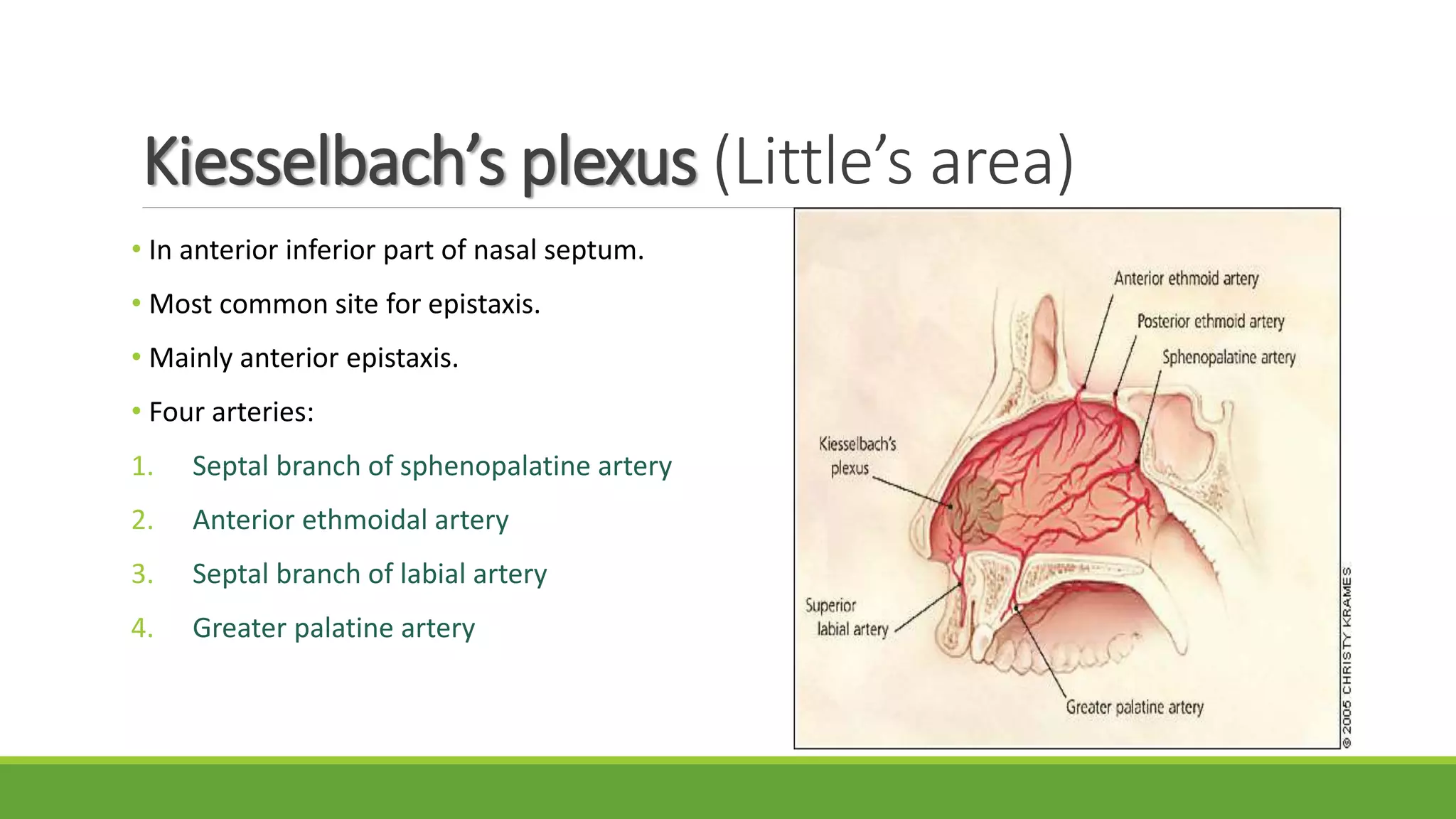
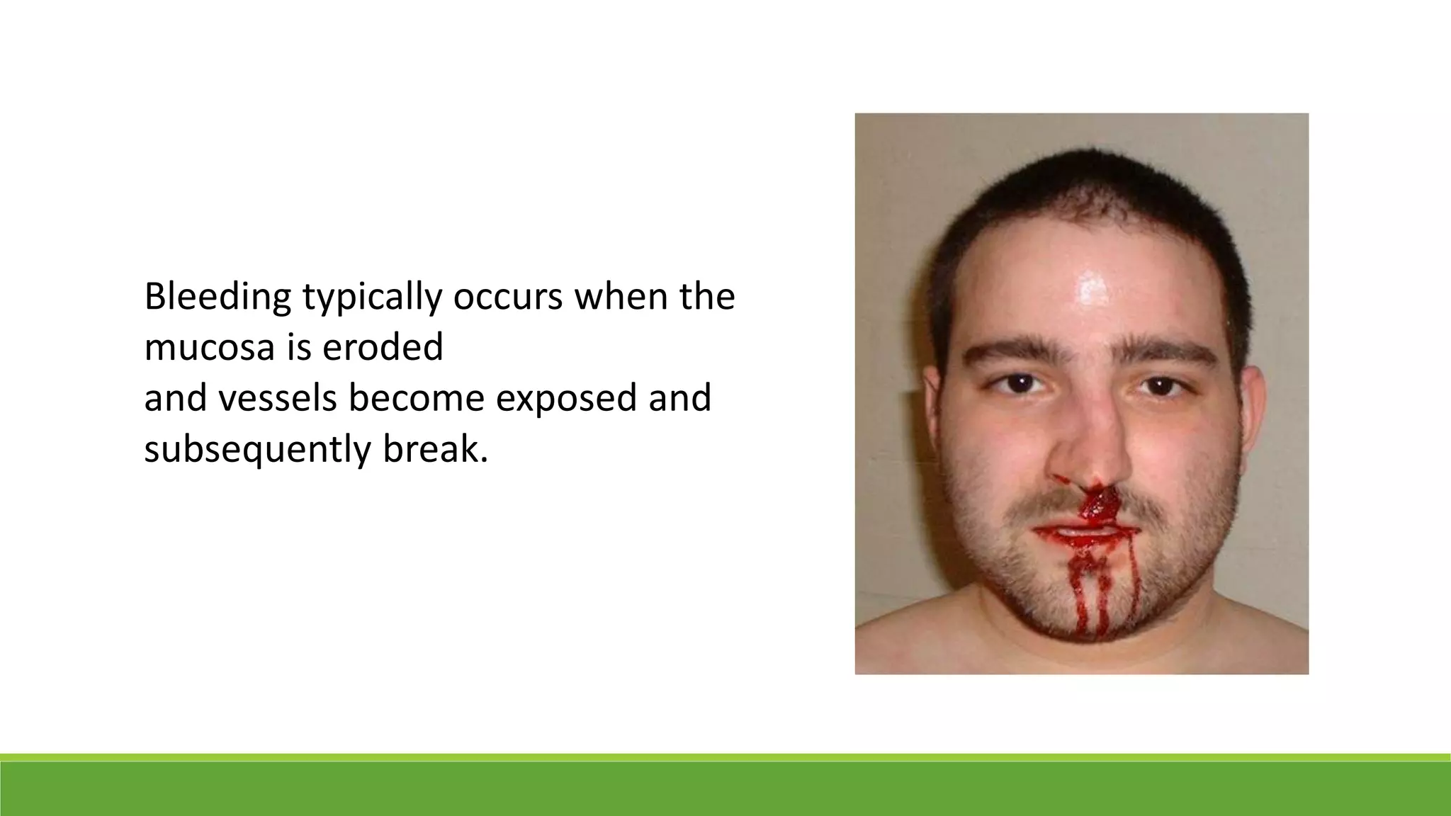
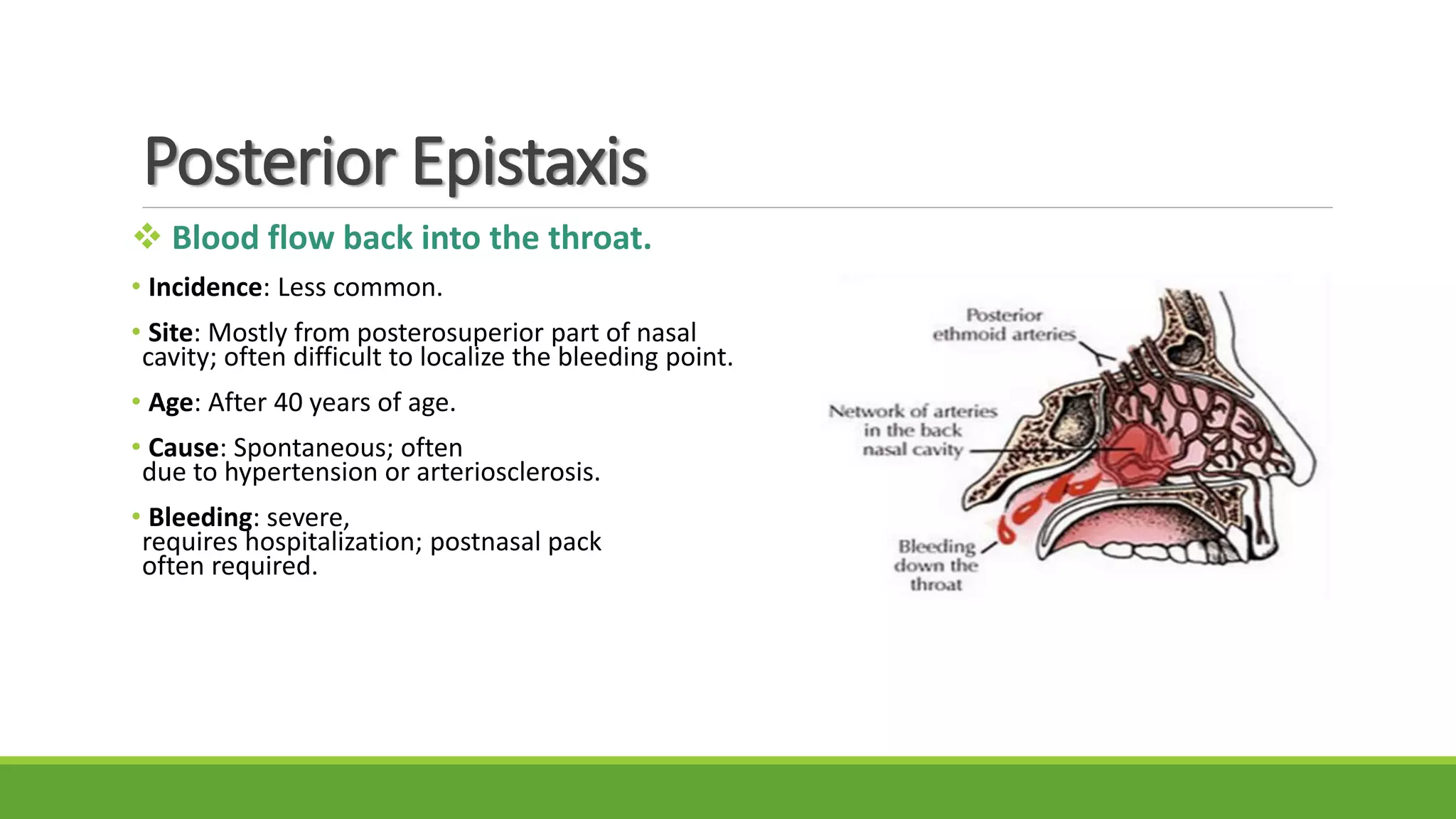
ASIM ABDU
AAY96@HOTMAIL.COM
KING KHALED UNIVERSITY
Introduction
Introduction
Definition
Epistaxis : Bleeding from inside the nose
Origin
From a Greek term “epistazo” which means “to bleed from nose”.
Introduction
• It is fairly common.
• It is seen in all age groups : children,
adults and older people.
• It often presents as an emergency.
• Epistaxis is a sign and not a disease.
Introduction
Why Nose?
• Situated in a vulnerable position as
it protrudes on the face.
• Has a very rich blood supply.
• Supplied by both internal and
external carotid system.
• Various anastomoses between
arteries and veins.
• Vasculature runs just under the mucosa.
• Exposed to the drying effect of inspiratory current.
Introduction
Epidemiology
• Lifelong incidence of epistaxis in general population is about 60%.
• Fewer than 60% seek medical attention.
• Peaks in young children (2 – 10 y) and older individuals (50 – 80 y)
• occur in males more than females.
The Blood Supply
of Nose
• Nose is richly supplied by both the external and internal carotid systems.
Superior part of the nose (Internal carotid artery):
• Ophthalmic artery
- Anterior ethmoidal artery
- Posterior ethmoidal artery
Inferior part of the nose (External carotid artery):
• Maxillary artery
- Greater palatine artery
- Sphenopalatine artery
• Facial artery
- Superior labial artery  vestibule of the nose
Kiesselbach’s plexus (Little’s area)
• In anterior inferior part of nasal septum.
• Most common site for epistaxis.
• Mainly anterior epistaxis.
• Four arteries:
1. Septal branch of sphenopalatine artery
2. Anterior ethmoidal artery
3. Septal branch of labial artery
4. Greater palatine artery
Woodruff’s Plexus
• Posterior end of middle turbinate.
• Most common site for posterior epistaxis.
• It is anastomoses of two arteries:
1. Sphenopalatine artery
2. posterior pharyngeal artery
Reterocolumellar Veins
• Runs vertically downwards just behind columella.
• Crosses the floor of nose and joints venus plexus on lateral nasal wall.
• Common site for venous bleed in young patient.
Pathophysiology
Bleeding typically occurs when the
mucosa is eroded
and vessels become exposed and
subsequently break.
Classification
Anterior Epistaxis
 Blood flow out from front nose with patient
in sitting position.
•Incidence: More common.
• Site: Mostly from Little’s area or anterior
part of lateral wall.
• Age: Mostly occurs in children or young adults.
• Cause: Mostly trauma.
• Bleeding: Usually mild, can be easily controlled
by local pressure or anterior pack.
Posterior Epistaxis
 Blood flow back into the throat.
• Incidence: Less common.
• Site: Mostly from posterosuperior part of nasal
cavity; often difficult to localize the bleeding point.
• Age: After 40 years of age.
• Cause: Spontaneous; often
due to hypertension or arteriosclerosis.
• Bleeding: severe,
requires hospitalization; postnasal pack
often required.
Site of Epistaxis
1. Little’s area: 90% of cases.
2. Above level of middle turbinate: anterior and posterior ethmoidal artery.
3. Below level of middle turbinate: from branch of sphenopalatine artery.
4. Posterior part of nasal cavity: blood to pharynx.
5. Diffuse: from septum and lateral nasal wall.
6. Nasopharynx.
CAUSES OF EPISTAXIS
1. Local (nose or nasopharynx).
2. General.
3. Idiopathic.
Local Causes
Trauma:
• Finger nail trauma
• Injury of nose
• Intranasal surgery
• Fracture of middle third of face and base of skull
• Hard blowing of nose
• Violent sneeze
Infection:
• Acute: viral rhinitis, acute sinusitis, nasal diphtheria
• Chronic: Crust forming Disease(rhinitis sicca, tuberculosis); Granulamatous lesion of nose(rhinosporidosis)
Local Causes
Foreign bodies:
• Nonliving: any neglected foreign body
• Living: Maggots, leeches
Neoplasms:
• Benign: Hemangioma, papilloma
• Malignant: Carcinoma, sarcoma
Atmospheric changes:
high altitudes, sudden decompression, Casisson disease.
Deviation of nasal septum.
General Causes
Cardiovascular System:
• Hypertension
• Arteriosclerosis
• Mitral stenosis
• Pregnancy (hypertension and hormonal)
Disorders of blood and blood vessels:
• Aplastic anemia
• Leukemia
• Thrombocytopenia
• Vascular purpura
General Causes
Liver: Hepatic cirrhosis.
Kidney: Chronic nephritis.
Mediastinal compression: tumor causing raised venous pressure.
Acute general infection: influenza, measules, whooping cough.
Vicarious menstruation.
Drug:
• Excessive use of salicylates and other analgesics.
• Anticoagulant therapy
Management
First Aid
Bleeding from Little’s area can be stopped by pinching the nose for 5 min.
Trotter’s method:
1. Patient is made to sit
2. Leaning a little forward
3. Breath quietly from the mouth
4. Cold compression is applied(vasoconstriction)
Cauterization
• Useful in anterior epistaxis when bleeding point has been located
• Topically anaesthetized
• Bleeding point cauterization with bead of
silver nitrate or
coagulated with electrocautery
Anterior Nasal Packing
• Done if localized bleeding is profuse or bleeding point is not localize
• Use of a ribbon gauze soaked with liquid paraffin(1 m gauze; 2.5 cm
gauze in adult and 12 mm in children)
• Can be done with vertical layer and horizontal layer
• Can be removed with 24 hour and can be kept upto 2-3 days
• Systemic antibiotic should be given to prevent sinus infection
and toxic shock syndrome
Posterior Nasal Packing
• For posterior nasal bleed
• Can be carried through different instrument:
1. Gauze
2. Foley’s Catheterization
3. Nasal balloon
Nasal packing by Gauze
Nasal packing by Foley’s catheterNasal Balloon
Elevation of Mucoperichondrial Flap and Submucous Resection(SMR)
Operation:
• For persistant or recurrent bleeds from septum
• Elevation of mucoperichondrial flap
• Reposining flap back help to cause fibrosis and constrict blood vessel
Endoscopic Cauterization
• Topical or general anesthesia, bleeding point is localized
• with rigid endoscopy and cauterized
• Procedure is effective with less morbidity and decreased stay
• With profuse bleeding it is very difficult to localize so this
• procedure can’t be carried out
Ligation of Vessels
Ligation can be done to
• External Carotid (ligation on origin of superior thyroid artery)
• Maxillary artery
• Ethmoidal artery
Other process
• Transnasal Endoscpoic Sphenopalatine Artery
Ligation(TESPAL)
• Embolization of artery
General Measures in Epistaxis
• Making patient sit up with back rest and record any blood loss taking place through spitting or vomiting
• Mild sedation should be given
• Checking pulse, BP and respiration
• Maintenance of hemodynamics (Blood transfusion may required)
• Antibiotics can be given to prevent sinusitis, if pack is to be kept beyond 24 hours
• Intermittent oxygen may be required in patients with bilateral packs because of increased pulmonary
resistance from nasopharynx reflex
• Investigation and treatment for any underlying local or general cause
References
• Disease of Ear, Nose and
Throat and head and neck
surgery. Dhingra PL,
Dhingra S. 6th ed.
Epistaxis.
• Disease of Ear, Nose and
Throat. Bhansal M. 1st ed.
Nose and Paranasal
Sinuses. =

Epistaxis

  • 1.
  • 2.
  • 3.
    Introduction Definition Epistaxis : Bleedingfrom inside the nose Origin From a Greek term “epistazo” which means “to bleed from nose”.
  • 4.
    Introduction • It isfairly common. • It is seen in all age groups : children, adults and older people. • It often presents as an emergency. • Epistaxis is a sign and not a disease.
  • 5.
    Introduction Why Nose? • Situatedin a vulnerable position as it protrudes on the face. • Has a very rich blood supply. • Supplied by both internal and external carotid system. • Various anastomoses between arteries and veins. • Vasculature runs just under the mucosa. • Exposed to the drying effect of inspiratory current.
  • 6.
    Introduction Epidemiology • Lifelong incidenceof epistaxis in general population is about 60%. • Fewer than 60% seek medical attention. • Peaks in young children (2 – 10 y) and older individuals (50 – 80 y) • occur in males more than females.
  • 7.
  • 8.
    • Nose isrichly supplied by both the external and internal carotid systems. Superior part of the nose (Internal carotid artery): • Ophthalmic artery - Anterior ethmoidal artery - Posterior ethmoidal artery Inferior part of the nose (External carotid artery): • Maxillary artery - Greater palatine artery - Sphenopalatine artery • Facial artery - Superior labial artery  vestibule of the nose
  • 9.
    Kiesselbach’s plexus (Little’sarea) • In anterior inferior part of nasal septum. • Most common site for epistaxis. • Mainly anterior epistaxis. • Four arteries: 1. Septal branch of sphenopalatine artery 2. Anterior ethmoidal artery 3. Septal branch of labial artery 4. Greater palatine artery
  • 10.
    Woodruff’s Plexus • Posteriorend of middle turbinate. • Most common site for posterior epistaxis. • It is anastomoses of two arteries: 1. Sphenopalatine artery 2. posterior pharyngeal artery
  • 11.
    Reterocolumellar Veins • Runsvertically downwards just behind columella. • Crosses the floor of nose and joints venus plexus on lateral nasal wall. • Common site for venous bleed in young patient.
  • 12.
  • 13.
    Bleeding typically occurswhen the mucosa is eroded and vessels become exposed and subsequently break.
  • 14.
  • 15.
    Anterior Epistaxis  Bloodflow out from front nose with patient in sitting position. •Incidence: More common. • Site: Mostly from Little’s area or anterior part of lateral wall. • Age: Mostly occurs in children or young adults. • Cause: Mostly trauma. • Bleeding: Usually mild, can be easily controlled by local pressure or anterior pack.
  • 16.
    Posterior Epistaxis  Bloodflow back into the throat. • Incidence: Less common. • Site: Mostly from posterosuperior part of nasal cavity; often difficult to localize the bleeding point. • Age: After 40 years of age. • Cause: Spontaneous; often due to hypertension or arteriosclerosis. • Bleeding: severe, requires hospitalization; postnasal pack often required.
  • 17.
  • 18.
    1. Little’s area:90% of cases. 2. Above level of middle turbinate: anterior and posterior ethmoidal artery. 3. Below level of middle turbinate: from branch of sphenopalatine artery. 4. Posterior part of nasal cavity: blood to pharynx. 5. Diffuse: from septum and lateral nasal wall. 6. Nasopharynx.
  • 19.
  • 20.
    1. Local (noseor nasopharynx). 2. General. 3. Idiopathic.
  • 21.
    Local Causes Trauma: • Fingernail trauma • Injury of nose • Intranasal surgery • Fracture of middle third of face and base of skull • Hard blowing of nose • Violent sneeze Infection: • Acute: viral rhinitis, acute sinusitis, nasal diphtheria • Chronic: Crust forming Disease(rhinitis sicca, tuberculosis); Granulamatous lesion of nose(rhinosporidosis)
  • 22.
    Local Causes Foreign bodies: •Nonliving: any neglected foreign body • Living: Maggots, leeches Neoplasms: • Benign: Hemangioma, papilloma • Malignant: Carcinoma, sarcoma Atmospheric changes: high altitudes, sudden decompression, Casisson disease. Deviation of nasal septum.
  • 23.
    General Causes Cardiovascular System: •Hypertension • Arteriosclerosis • Mitral stenosis • Pregnancy (hypertension and hormonal) Disorders of blood and blood vessels: • Aplastic anemia • Leukemia • Thrombocytopenia • Vascular purpura
  • 24.
    General Causes Liver: Hepaticcirrhosis. Kidney: Chronic nephritis. Mediastinal compression: tumor causing raised venous pressure. Acute general infection: influenza, measules, whooping cough. Vicarious menstruation. Drug: • Excessive use of salicylates and other analgesics. • Anticoagulant therapy
  • 25.
  • 26.
    First Aid Bleeding fromLittle’s area can be stopped by pinching the nose for 5 min. Trotter’s method: 1. Patient is made to sit 2. Leaning a little forward 3. Breath quietly from the mouth 4. Cold compression is applied(vasoconstriction)
  • 27.
    Cauterization • Useful inanterior epistaxis when bleeding point has been located • Topically anaesthetized • Bleeding point cauterization with bead of silver nitrate or coagulated with electrocautery
  • 28.
    Anterior Nasal Packing •Done if localized bleeding is profuse or bleeding point is not localize • Use of a ribbon gauze soaked with liquid paraffin(1 m gauze; 2.5 cm gauze in adult and 12 mm in children) • Can be done with vertical layer and horizontal layer • Can be removed with 24 hour and can be kept upto 2-3 days • Systemic antibiotic should be given to prevent sinus infection and toxic shock syndrome
  • 29.
    Posterior Nasal Packing •For posterior nasal bleed • Can be carried through different instrument: 1. Gauze 2. Foley’s Catheterization 3. Nasal balloon Nasal packing by Gauze Nasal packing by Foley’s catheterNasal Balloon
  • 30.
    Elevation of MucoperichondrialFlap and Submucous Resection(SMR) Operation: • For persistant or recurrent bleeds from septum • Elevation of mucoperichondrial flap • Reposining flap back help to cause fibrosis and constrict blood vessel Endoscopic Cauterization • Topical or general anesthesia, bleeding point is localized • with rigid endoscopy and cauterized • Procedure is effective with less morbidity and decreased stay • With profuse bleeding it is very difficult to localize so this • procedure can’t be carried out
  • 31.
    Ligation of Vessels Ligationcan be done to • External Carotid (ligation on origin of superior thyroid artery) • Maxillary artery • Ethmoidal artery
  • 32.
    Other process • TransnasalEndoscpoic Sphenopalatine Artery Ligation(TESPAL) • Embolization of artery
  • 33.
    General Measures inEpistaxis • Making patient sit up with back rest and record any blood loss taking place through spitting or vomiting • Mild sedation should be given • Checking pulse, BP and respiration • Maintenance of hemodynamics (Blood transfusion may required) • Antibiotics can be given to prevent sinusitis, if pack is to be kept beyond 24 hours • Intermittent oxygen may be required in patients with bilateral packs because of increased pulmonary resistance from nasopharynx reflex • Investigation and treatment for any underlying local or general cause
  • 34.
    References • Disease ofEar, Nose and Throat and head and neck surgery. Dhingra PL, Dhingra S. 6th ed. Epistaxis. • Disease of Ear, Nose and Throat. Bhansal M. 1st ed. Nose and Paranasal Sinuses. =