
Recommended
More Related Content
What's hot
What's hot (20)
Similar to cupressus.pptx
Similar to cupressus.pptx (20)
Recently uploaded
Recently uploaded (20)
cupressus.pptx
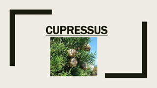
- 1. CUPRESSUS
- 2. SYSTEMATIC POSITION Division: Pinophyta Class: Pinopsida Order: Pinales Family: Cupressasceae Genus: Cupressus
- 3. ■ Occurrence ■ They are evergreen trees or large shrubs growing to 5- 4 m tall. ■ These are native to scattered localities in mainly warm temperate regions in the Northern Hemisphere. ■ All have decorative merit, particularly in a young state. ■ Common species planted in the plains and hills of India include: C. funebris , C.cashmeriana, C.sempervirens.
- 4. ■ Morphology ■ Tree is evergreen, large (5-40m ) tall, with pyramidal appearance. ■ The bark is thin, fibrous and greyish brown and peel off in long strips. ■ Branching is of 2 types: horizontal or erect main branches, which are spirally arranged on the main stem and drooping side branches. ■ Main branches are devoid of leaves, whereas the drooping branches bear small, greyish-green opposite and decussate leaves adpressed to the axis. ■ The leaves of the lateral pair are folded face to face and those of the facial pair are flattened and grooved in the middle . ■ 2 types of leaves are seen : The leaves of the apical zone are small, closely placed with acute apex and serrated margin. Mature leaves are longer, brownish and distantly placed due to elongation of the internode. ■ The leaves are characterized by a prominent midrib and scattered stomata on both the surfaces.
- 5. ■ The male cones (6-7mm long) , borne terminally on pendulous branch, are brownish when mature. ■ 6 to 8 pairs of microsporophylls, each bearing 2 to 6 sporangia abaxially, are arranged in a opposite and decussate manner on the cone axis. ■ The female cones (10-12 mm long) are axillary on the pendulous branches and each consist of four pairs of seed scale complexes arranged in a opposite and decussate manner. ■ The lower most pair is generally sterile. ■ The fertile scales are peltate bearing 3-7 ovules/seeds per scale. ■ The fertile seed scales complexes are thick, four to six sided and have a central pointed process called the boss. ■ The mature cone is brown and woody and persists on the tree long after the seed shed. ■ The seed are dark brown , orbicular, compressed and narrowly winged. The wings of successive seeds overlap.
- 6. ANATOMY STEM ■ Both cortex and pith are narrow and consist of parenchymatous cells in young stem. ■ After Secondary growth, distinct annual rings are formed. ■ Xylem Cylinder is thicker than the periderm, cortex and secondary phloem. ■ Secondary phloem is composed of sieve elements, phloem parenchyma, phloem fibres and resin ducts. ■ Sieve areas are confined to the radial walls. The resin canals are large, bounded by 2 or 3 layers of secertory cells. ■ Phloem fibres occur at regular intervals in single tangential layers. These alternative with rings of parenchyma and resin cells. ■ The phloem rays are uniseriate and 4 or 5 cell high . These are thin walled with simple pits . ■ The tracheids of Secondary xylem are long and narrow with circular to elliptical bordered pits on their radial walls.
- 7. ■ In the tracheids of summer wood , tangential pitting is also seen. ■ Parenchymatous cells are resinous. ■ Xylem rays are uniseriate, 1-5 cells high, comprising parenchymatous cells and showing simple pits. ■ The wood is devoid of resin canals. ■ Thd pith get obliterated and is seen as a dense region in the centre.
- 8. ■ LEAF ■ Epidermis is covered with a thin cuticle. ■ Single-layer of thick walled cells constituting the hypodermis which may be discontinous at places. ■ Mesophyll is differentiated into a layer of elongated palisade cells and spongy parenchyma. ■ The palisade cells are towards the abaxial side or lower surface and contain abdundant chloroplast. ■ The spongy tissue is composed of large irregular cells with numerous intercellular spaces. ■ This is single vascular bundle with a resin canal below it.
- 9. REPRODUCTION Microsporangium &Microsporogensis ■ Microsporangia arise on the abaxial side of the microsporophyll. ■ Below the epidermis few hypodermal archesporial cell differentiate which divide periclinally to give rise to primary perietal layer and primary sporogenous cells. ■ The former after division forms the middle layer towards outside and tapetum towards inside. ■ Primary sporogenous cells divide in all planes to form sporogenous tissue, the last cell generation of which eventually give rise to MMC. ■ MMC undergo reduction division followed by cytokinesis which is simultaneous.
- 10. ■ The microspore tetrads are isobilateral and tetrahedral. ■ A young microspore has a large nucleus, dense cytoplasm and numerous dtrach grains. ■ The pollen grains are shed at the uninucleate stage. ■ No prothallial cells ate formed. ■ Pollen wall consist of a fine granular, thin exine and a thick uniform intine. ■ Pollen are non-winged ■ Prior to meiosis , the strach grains which were present at the periphery get distrubuted throughout the cytoplasm of the microspore mother cell. ■ After meiosis 1 the strach grains get divided in 2 groups and after meiosis 2 four ssuch groups are seen. ■ There is thus a equal distribution of strach in the 4 microspore.
- 11. ■ MALE GAMETOPHYTE ■ The wind dispersed pollen grains are caught in the pollination drop and sucked in reaching nucellus. ■ The micropylar canal which is very wide during pollination is closed after pollination by the repeated division of the cells of the inner layer. ■ Also the edges of the seed scales complex give out teeth-like appendages which closely interlock with each other. ■ The pollen grains are aporate and germinate on the nucellar tip . ■ At the time of pollen germination , the exine is thrown off after it gets ruptured irregularly. ■ The microspore divides to form a small lenticular antheridial cell and a large tube cell.
- 12. ■ The tube nucleus moves into the pollen tube followed by the antheridial cell which divides to give rise to a large spermatogenous cell and a stalk cell. ■ The stalk cell soon loses its wall and comes to lie near the tip of the pollen tube along with the tube nucleus. ■ Just before the pollen tube reaches the archegonium the tube and the stalk nuclei degenerate and spermatagenous cell divides into 2 equal male cells. ■ Multiple male gamates have been reported. ■ The multiple male cells are produced by the super numerary division of the spermatogenous cells.
- 13. ■ MEGASPORANGIUM & MEGASPOROGENESIS ■ The ovules are unitegmic and crassinucellate. ■ Several deep-seated sporogenous cells have been reported. ■ The megaspore mother cell after meiosis gives rise to linear tetrad of megaspores. ■ Some times 2or more megaspore may start enlarging , but these become arrested soon.
- 14. ■ FEMALE GAMETOPHYTE ■ Nuclear divisions in the early stages are Simultaneous. ■ The gametophyte become cellular through alveoli formation. ■ A very conspicuous spongy tissue comprising 2or 3 layer surrounds the free nuclear gametophyte. It is derived from the non-functional sporogenous cells. ■ Few cells at the micropylar end of the female gametophyte become prominent and differentiate into archegonial initials. ■ They divide transversely to give rise to a small neck initials and a large central cell. ■ The neck is made up of 8 cells arranged in 2 tiers of four cells each. ■ The central cell divides to form an ephemeral ventral canal nucleus and a egg nucleus. ■ The latter enlarges and comes to lie in the centre of the devolping archegonium.
- 15. ■ A mature archegonium is oblong othe elongated a large egg nucleus , a ventral canal nucleus and eight neck cells. ■ The archegonia occur in archegonial complexes. A group of archegonia is surrounded by a common jacket. ■ With the development of the archegonial complex, the adjacent gametophytic tissue grows upward, resulting in the formation of an archegonial chamber. ■ The total number of archegonia is 10-13. ■ The archegonia differentiate in that region where the pollen tubes make contact with gametophyte. ■ The archegonial complexes are terminal at the micropylar end.
- 16. ■ FERTILIZATION ■ After traversing the nucellus, the pollen tube arrives in the archegonial chamber and releases the male cells. ■ Subsequent to the degeneration of neck cells, a passage is formed through which the male cells enter the archegonium. ■ Generally only one male cell finds its way into an archegonium. ■ The male nucleus is thus surrounded by its middle and inner cytoplasmic zones while moving through the egg or maternal cytoplasm. ■ As it reaches the egg nucleus, the male nucleus moves in advance of the cytoplasmic sheath. ■ The two fusing nuclei make contact with each other and take a turn of 180° so that the male nucleus comes to lie below the egg nucleus.
- 17. ■ The two inner zones of the male cytoplasm envelope the zygote and later become incorporated into the neocytoplasm. ■ After the fusion of the nuclear membrane , the zygote is formed surrounded by the neocytoplasm, the egg cytoplasm having degenerated.
- 18. ■ EMBRYOGENY ■ The zygote nucleus divides thrice to give rise to 8 free proembryonal nuclei at the archegonial base. ■ Wall formation take place resulting in primary proembryo comprising 2 tiers of 4 cells each.( Primary upper tier and Primary embryonal cells) ■ The primary upper tier again divides transversely to form an upper open tier and a middle suspensor tier. ■ The primary embryonal cells also divide to form embryonal cells resulting in a 16- celled secondary proembryo. ■ The cells of suspensor tier elongate to push the embryonal tier deep into the gametophytic tissue. ■ Each cell between the terminal embryonal cell and suspensor elongates forming embryonal suspensor.
- 19. ■ Both simple and cleavage polyembryonal are common. ■ The apical embryonal cells divides in all planes to form a meristamatic tissue. ■ The shoot apex and the root apex are differentiated by the activity of cells at proximal end and distal end , respectively. ■ The cells between the root and shoot apices elongate along the long axis of the embryo. ■ In peripheral region of the shoot apex , a group of cells become active and divides more rapidly than the rest resulting in 2- well developed cotyledons.