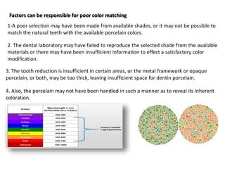
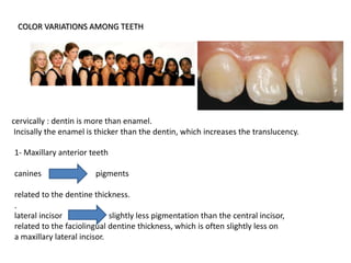
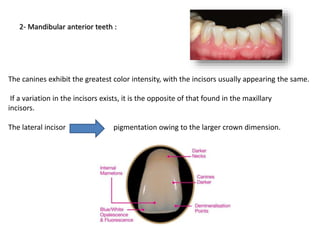
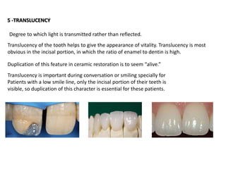
Asso.Prof.Dr. Ameer Hamdi Al-Ameedee
BDS, DDS, MsC, Ph.D, in Esthatic and Operative Dentistry.
ESTHETIC CONSIDERATIONS
Does beauty really come from the inside out or does your physical appearance play
the greater role?
Do we really think beauty is only skin deep or
are our brains hard wired to think otherwise?
What is beauty?
The secret of beauty and attractiveness has
been a quest of humans for as long as
we have been civilized
Many women – even some men - spend up
to one-third of their income on
improving their looks
4
Is that portion of the discipline that deals with achieving the ultimate in appearance.
Escape from artificiality.
Esthetic
Branch of philosophy dealing with beauty.
Art and science of dentistry applied to create or enhance the beauty of an individual within
functional and physiological limits.
Esthetic dentistry
Appearance Zone :
This is the anterior oral area where esthetics is of prime concern and which is visible on
smiling, from maxillary premolar to premolar (usually 1st molars also).
Depends on the person’s self-image, mouth size, teeth size, smile width, lip size and
tightness.
Factors Affecting Esthetics
1- SOFT TISSUE MANAGEMENT
A-FINISH LINE FORMATION.
B-IMP RESSION PROCEDUR.
C - THE TEMPORARY RESTORATION.
2- TOOTH REDUCTION
3- SHADE SELECTION
4 - COLOR VARIATION
5 -TRANSLUCENCY
6- SURFACE CHARACTERIZATION
7- DEGREE OF GLOSS
8-TOOTH FORM,SIZE AND ARCH POSITION
9- OPTICAL ILLUSON
1- SOFT TISSUE MANAGEMENT
Optimal soft tissue health should be established before any restorative procedures.
Many aspects of prosthetic treatment may cause esthetically detrimental changes in the
form of changes in gingival form ,color or position following cementation.
A- Finish line formation
With subgingival margins ….. every effort must be made to produce minimal soft tissue
injury .
Retraction Cord Technique
-The preparation is completely established - with a suprra-gingival finish line.
-Then a retraction cord is placed in the sulcus and temporarily displace the gingiva laterally
and apically
-Finally, the finish line can be lowered without soft tissue injury.
-Too large or too many cords --------- excessive trauma.
-Healthy gingival tissue, one thin cord -------- anterior teeth
-Single medium-sized ------------- posterior teeth.
-Excessive instrument pressure exerted in placing the excessively large cords----gingival
damage.
-Blanching (evidence of reduced blood supply) is often observed immediately after
placement of cord rapidly disappears.
An impression must provide detailed information about :
The prepared teeth, Surrounding intact teeth, Associated soft tissues
B-IMP RESSION PROCEDUR.
-Remove all cord from the sulcus as the impression material is syringed around the prepared
teeth.
-Removal of the impression from the mouth, then check the gingival sulcus with an explorer
and remove any remnants of retained impression material.
-Severe tissue reactions when the impression material is left in the sulcus.
Inadequate soft-tissue management
Causes of finish line not visible in the impression:
Bleeding from inflamed gingiva displacing the impression material
Tendency of the gingival cuff to recoil and displace
partially set impression material
because of inadequate bulk Sulcus impression tearing
Retraction cord
Two-cord technique
Rotary curettage
Electrosurgery Laser troughing
C - THE TEMPORARY RESTORATION
1-Properly contoured
2-Well adapted to the preparation margin
3-Should possess a very smooth surface
4-Establish cervical embrasures to provide access for oral hygiene aids
5-Left not more than two to three weeks
6-Overcontouring leads to food trap and hence complicating the periodontal status.
7-Interdental papilla is often neglected due to improper design of interdental space.
8-Crown contours should be such that it should not provide any niche for plaque
retention and should promote self-cleaning.
9-Open embrasures to allow easy access to the interproximal area
for plaque control.10-An over contoured embrasure will reduce the space intended for the gingival papilla
and causing pressure and irritation on the papilla, also inhibits effective oral hygiene
laceration of the gingiva with rotary instruments + poor temporary restoration.
Final restoration …………… fails to vertically reach the finish line of the prepared tooth
Overextended or under extended restoration
plaque accumulation at the margin of the restoration.
A uniform duplication of the form and contours of the natural dentition.
2- TOOTH REDUCTION
Insufficient tooth reduction poor esthetics
Development of adequate color requires a certain thickness of porcelain
The facial reduction should be 1 to 1.5 mm.
The areas to be considered during preparation :
1- labioincisal aspects.
2- Cervical portion of the facial surface.
A) The facial surface should be reduced in two planes; one nearly parallel with the path of
insertion, and one parallel with the incisal two-thirds of the facial surface of the tooth
B) One plane reduction parallel with the path of insertion may result in insufficient space
for porcelain in the incisal 1/3 of the tooth
C) One plane reduction which creates adequate space for the restoration both in the
shoulder and the incisal areas, will endanger the pulp entity and produce overtapered
restoration.
inadequate facial reduction, the laboratory fabrication can be handled in one of two ways
1- develop proper contour in the restoration, results in a lack of color vitality due to
insufficient porcelain thickness: External and internal color modification to enhance the
esthetics of the restoration.
2- over-contoured restoration to develop proper color leading to plaque accumulation
which affects the gingival health.
3- SHADE SELECTION
Shade interpretation
Difficulty in color matching …precise matching of a desired shade can be difficult.
The original color of a tooth is the color one sees as a result of the reflection,
refraction, deflection, and absorption of light by the enamel, the dentin, and possibly
the pulp.
The color seen in a tooth is the result of combined optical effects of the layers of
tooth structure, the translucency and thickness of the enamel and color of the
underlying dentin.
The esthetic dentistry, using a combination of science and art, involves the use of colors
to create a natural tooth like restoration, color and shade are very important because
teeth are multi-chromatic with color variations from cervical to incisal, every tooth in the
mouth from the central incisors to molars, both upper and lower, varies in color.
Color that is perceived is the result of a light source, the object that absorbs, transmits,
reflects or scatters the light from the source, and the interpretation of the result by the
human visual system
Light form of visible energy that is part of the radiant energy spectrum. Radiant energy
possesses specific wavelengths, which may be used to identify the type of energy
In 1666 Isaac Newton discovered that white light can be broken down into a rainbow of
color
In nineteenth century that German physiologist Ewald Hering first described the now
familiar color circle.
In 1905, Albert Henry Munsell, an American artist and art teacher, further modified the
color circle, devising a system of color organization that centered around three unique
aspects of color: hue, chroma, and value. Using these three aspects, was able to construct
a three-dimensional color wheel
Color Mixing
Light mixture- primary colors: red, green, blue.
Additive mixture system- mixing of two of the
light mixture primary colors
red + blue = magenta
red + green = yellow
green + blue = cyan
Pigment mixture system: yellow, cyan, magenta
Color of the Human Teeth
Clark was the first to accurately describe the color of the human teeth in 1931
Dr. E. B. Clark, a dentist, produced the first data in 1931.
He indicated the Hue ranged from 6 YR (yellow-red) to 9.3 Y (yellow).
The Value ranged from 4 to 8, and the Chroma ranged from 0 to 7.
Lemire and Burk found:
The Hue range from 8.9 Y to 3.3 Y, a Value range of 5.8 to 8, and a Chroma range from 0.8
to 3.4
Goodkind and Schwabacher:
Identified the Hue range as 4.5 YR to 2.6 Y, the Value range as 5.7 to 8.5, and the Chroma
range from 1.1 to 5.
Guidelines for Clinical Shade Selection
1.light
2.amount lighting
3.location of lighting
4.restricting light
5.surrounding colors
6.tone of selection
7.patient position
8.tooth condition
9.comparison prcds.
10.selectiodistance
11.verification
12.diagram
13.photograph
Vita 3D stapes 1 (Value)
1-Determine the lightness level (value)
2-Hold shade guide to patient’s mouth
3-Start with darkest group moving right to left
4-Select Value group 1, 2, 3, 4, or 5
Munsell color system extends from zero to ten, black is zero and white is ten
Vita 3D stapes 2 (Chroma)
Select the chroma
1-From your selected Value group, remove the
middle tab (M) and spread the samples out like
a fan
2-Select one of the three shade samples to
determine chroma
Vita 3D stapes 3 (Hue)
Determine the hue
Check whether the natural tooth is more yellowish
or more reddish than the shade ample selected
Shade matching using the VITA Easyshade Compact:
1-“Tooth areas” mode selected. Note the active dot appearing in the
cervical third of the tooth on the screen.
2-Wand tip on the buccal surface at the cervical third of the tooth.
3-“OK” signal in the cervical third and active dot in the middle third on the
screen.
4-Wand tip on the buccal surface at the middle third of the tooth.
Color research continued to evolve based on the
Munsell color model.
In 1976, The Commission Internationale de l’Eclairage
(CIE), an international color research group founded
in 1931, published the CIELAB color system.
In this 3-dimensional color system, L* refers to brightness (0 to 100), a* represents red
(+a*) vs. green (-a*) and b* indicates yellow (+b*) vs. blue (-b*). When a* and b* are
zero, the L value represents the continuum of black to white.
The CIELAB model offers some advantages over other color models. The L*a*b* color
space was designed to correlate with perceptions of color.
CIELAB COLOR SYSTEM
Color Differences CIELAB
Is often used to measure changes in color, including changes in tooth color from use of
whitening products. Color difference equations are used to quantify the color change. ΔL*,
the change in brightness,
5- OK signal in the cervical and middle thirds and active dot in the incisal third on the
screen.
6-Wand tip on the buccal surface at the incisal third of the tooth.
7- OK signal in the cervical, middle, and incisal thirds on the screen.
8-VITAPAN Classical and VITAPAN 3D-Master shades that are closest to the natural tooth
structure color shown on screen.
Note: The nonslip infection-control shield has been omitted from this series of photos
for demonstration clarity.
Factors can be responsible for poor color matching
1-A poor selection may have been made from available shades, or it may not be possible to
match the natural teeth with the available porcelain colors.
2. The dental laboratory may have failed to reproduce the selected shade from the available
materials or there may have been insufficient information to effect a satisfactory color
modification.
3. The tooth reduction is insufficient in certain areas, or the metal framework or opaque
porcelain, or both, may be too thick, leaving insufficient space for dentin porcelain.
4. Also, the porcelain may not have been handled in such a manner as to reveal its inherent
coloration.
COLOR VARIATIONS AMONG TEETH
1- Maxillary anterior teeth
canines pigments
related to the dentine thickness.
.
lateral incisor slightly less pigmentation than the central incisor,
related to the faciolingual dentine thickness, which is often slightly less on
a maxillary lateral incisor.
cervically : dentin is more than enamel.
Incisally the enamel is thicker than the dentin, which increases the translucency.
The canines exhibit the greatest color intensity, with the incisors usually appearing the same.
If a variation in the incisors exists, it is the opposite of that found in the maxillary
incisors.
The lateral incisor pigmentation owing to the larger crown dimension.
2- Mandibular anterior teeth :
5 -TRANSLUCENCY
Translucency of the tooth helps to give the appearance of vitality. Translucency is most
obvious in the incisal portion, in which the ratio of enamel to dentin is high.
Duplication of this feature in ceramic restoration is to seem “alive.”
Translucency is important during conversation or smiling specially for
Patients with a low smile line, only the incisal portion of their teeth is
visible, so duplication of this character is essential for these patients.
Degree to which light is transmitted rather than reflected.
Translucency also affects the esthetic quality of the restoration.
The degree of translucency is related to how deeply light penetrates into the tooth or
restoration before it is reflected outward.
Normally light penetrates through the enamel into dentin before being reflected outward.
This affords the lifelike esthetic vitality characteristic of normal, unrestored teeth.
Shallow penetration of light often results in a loss of esthetic vitality Illusions of
translucency also can be created to enhance the realism of a restoration.
Color modifiers (also referred to as tints) can be used to achieve apparent translucency
and tone down bright stains or characterize a restoration.
Translucency increases from cervical to incisal. Incisal edges, cusp tips and proximal
surfaces are areas of high translucency.
An anterior tooth sometimes has an area of slight
incisal opacity. This area is frequently
composed of enamel, the opacity is due to an
optical effect created by refraction of light as
it strikes the incisal edge (halo effect).
Reproduction of this effect by shaping the
incisal edge of the ceramic restoration so that
it possesses the exact lingual slope and
thickness of adjacent teeth.
little surface stain applied lingually or
incorporated internally, can enhance the
desired result. Surface stain located lingually
may wear off during function.
6- SURFACE CHARACTERIZATION
Young teeth characteristically exhibit significant
surface characterization, whereas teeth in older
individuals tend to possess a smoother surface
texture caused by a brasional wear. The surfaces
of natural teeth typically break up light and reflect
it in many directions .The restored areas of teeth should reflect light in
a similar manner to un restored adjacent surfaces.
surface texture controls the reflection of light
When light strikes a restoration surface, it should create a
reflection pattern similar to that of adjacent teeth, thus
enhancing the color match.
Developing the desired light reflection on a restoration’s surface by meticulous
duplication of the height of contours and depressions on the facial surface. The
number of depressions, their location, form, and depth can be recorded by close-up
photographs taken from different angles and by the working cast.
7- DEGREE OF GLOSS
1 - Surface gloss on ceramic restoration affects
the reflection of light, functions in conjunction
with surface characterization to enhance the
appearance of the restoration.
2 - Excessive gloss lightens the color
3 -Too long heating or heating at elevated
temperatures during glaze firing
can exhibit exaggerated gloss, in addition to
excessive flowing of the surface
and loss of surface characterization.
4 -Combining different degrees of gloss at
different areas creates the desirable natural
play of reflection of light.
5-Introducing highly glazed wear facets in
older patients improves esthetics.
8-TOOTH FORM,SIZE AND ARCH POSITION
Restoration of a single maxillary central incisor is one of the most difficult esthetic
situations. By contrast, if the restoration is slightly out of alignment with its contralateral
counterpart, , it is usually better for restorations to be shaped like their contralateral
counterpart when they are located adjacent to unrestored teeth.
But if all of the readily visible teeth are being restored and
there is no color-matching problem, it may be esthetically
advantageous to create slight alterations in form and position
to escape from artificiality.
Esthetics depend on proportion. An object is considered beautiful if it is properly
proportioned, Concepts of proportion are probably based on what is found in nature.
A ratio of approximately 1.619 to 1 between succeeding terms is considered pleasant,
and is known as the golden proportion.
Golden proportion of the tooth restoration
When a line is bisected in the golden proportion, the ratio
of the smaller section to the longer one is the same as the
ratio of the larger section to the whole line.
We dentists find the proportions of the central incisor very beautiful, but we have not been
able to find a Golden Proportion relationship between the obvious width and height. The
problem was recently solved when Dr Stephen Marquardt, an eminent Oral surgeon in
California, discovered that, “The HEIGHT of the central incisor is in the Golden Proportion to
the WIDTH of the TWO central incisors.” as below:
Golden proportion of the tooth restoration
The golden proportion is a
athematically constant ratio between
the larger and smaller length.
The ratio is approximately 1.618:1
In terms of proportion, the smaller
tooth is about 62% the size of the larger one.
The Golden Proportion results from the division of a straight line in
such a way that the shorter part is to the longer part as the longer
part is to the whole. Each ratio equals 0.618.
9- OPTICAL ILLUSON
Is the art of changing perception making an object appear different than it actually is.
Illusion is the art of changing the perception to cause an object to appear different than it
actually is.
This concept is particularly useful in solving problems
associated with presence of space limitations (too much
or too little space) or other problems that may make it
impossible to duplicate the original form.
The principles of illumination and
reflection can be manipulated by the
dentist and the technician to change
the apparent size and shape of a tooth
through illusion.
The law of the face:
The face of the silhouette of the tooth is the
area on the facial surface of anterior and
posterior teeth that is bounded by the
transitional line angles as viewed
from the facial (buccal) aspect.
The law of the face:
In anterior teeth, the transitional line angles mark the transition from the facial surface to
the mesial and distal surfaces, the incisal edge and cervically.
The tooth surface slopes lingually towards the mesial
and distal approximating surfaces and towards the
incisal edge and the cervical root surface from these
line angles, producing light reflections in different
directions corresponding to the sloping direction of
each surface, thus creating shadows in these areas.
The face of the tooth and
transitional line angles
Only the face of the tooth or the silhouette will
reflect the light forwards and anteriorly.
By reducing or increasing the portion of the tooth reflecting light forwards (the face)
we create the illusion of smaller or wider, shorter or longer teeth respectively
Creating equal apparent faces in two dissimilar adjacent teeth, makes dissimilarly
sized teeth look similar; as their faces reflect light in the same way .
Disharmony treated by optical illusion
The concept of the law of the face becomes apparent and more important when dealing
with canines and posterior teeth.
From the frontal view only a portion from the canine and posterior teeth are visible. In
this view, the canine face is bounded by the mesial transitional line angle, the cervical
transitional line angle and the midlabial ridge.
The distal half of the tooth is usually not visible from the frontal view. Moving the
midlabial ridge and the incisal tip mesially (a, b), will create the illusion of a narrower
tooth.
In addition, moving the distal transitional line angle more mesially ( c) will give the illusion
of equal mesial and distal faces and the tooth will look smaller both from frontal and side
views.
Principle of line
white hypoplastic lines, accentuated developmental
grooves, vertical texturing illusion of height
Stain lines, texturing,
straight incisal edges illusion of width
These lines create illusions by breaking up the smooth
reflecting surface causing ruptures in the continuity of the
linear reading of the surface making the tooth appear
longer or shorter, wider or narrower.
Characterized or textured surfaces produces shadows
and shadow position can determine how the mind will
interpret the form.
Arrangement of teeth
The position or arrangement of teeth can create illusion of decrease width.
When teeth are placed in linguo-version, not only its real width is masked by the
more prominent approximating teeth, but the effect of increased a hadowing also
its size.
Slight lingual rotation of anterior crowns may solve the problem of wide space by
narrowing the areas that reflects light forward; thus decreasing the apparent
width
Staining
Not only used to duplicate the natural variation of the tooth color, but also to
create and enhance illusion through manipulation of shape.
Darker stains optical illusion of smaller size
Increasing the value ( increase whiteness) closer area
Decreasing the value(increase grayness) less prominent area
Thank you

Esthetic considerations. lect

  • 2.
    Asso.Prof.Dr. Ameer HamdiAl-Ameedee BDS, DDS, MsC, Ph.D, in Esthatic and Operative Dentistry. ESTHETIC CONSIDERATIONS
  • 3.
    Does beauty reallycome from the inside out or does your physical appearance play the greater role? Do we really think beauty is only skin deep or are our brains hard wired to think otherwise? What is beauty?
  • 4.
    The secret ofbeauty and attractiveness has been a quest of humans for as long as we have been civilized Many women – even some men - spend up to one-third of their income on improving their looks 4
  • 5.
    Is that portionof the discipline that deals with achieving the ultimate in appearance. Escape from artificiality. Esthetic Branch of philosophy dealing with beauty. Art and science of dentistry applied to create or enhance the beauty of an individual within functional and physiological limits. Esthetic dentistry Appearance Zone : This is the anterior oral area where esthetics is of prime concern and which is visible on smiling, from maxillary premolar to premolar (usually 1st molars also). Depends on the person’s self-image, mouth size, teeth size, smile width, lip size and tightness.
  • 6.
    Factors Affecting Esthetics 1-SOFT TISSUE MANAGEMENT A-FINISH LINE FORMATION. B-IMP RESSION PROCEDUR. C - THE TEMPORARY RESTORATION. 2- TOOTH REDUCTION 3- SHADE SELECTION 4 - COLOR VARIATION 5 -TRANSLUCENCY 6- SURFACE CHARACTERIZATION 7- DEGREE OF GLOSS 8-TOOTH FORM,SIZE AND ARCH POSITION 9- OPTICAL ILLUSON
  • 7.
    1- SOFT TISSUEMANAGEMENT Optimal soft tissue health should be established before any restorative procedures. Many aspects of prosthetic treatment may cause esthetically detrimental changes in the form of changes in gingival form ,color or position following cementation.
  • 8.
    A- Finish lineformation With subgingival margins ….. every effort must be made to produce minimal soft tissue injury . Retraction Cord Technique -The preparation is completely established - with a suprra-gingival finish line. -Then a retraction cord is placed in the sulcus and temporarily displace the gingiva laterally and apically -Finally, the finish line can be lowered without soft tissue injury. -Too large or too many cords --------- excessive trauma. -Healthy gingival tissue, one thin cord -------- anterior teeth -Single medium-sized ------------- posterior teeth. -Excessive instrument pressure exerted in placing the excessively large cords----gingival damage. -Blanching (evidence of reduced blood supply) is often observed immediately after placement of cord rapidly disappears.
  • 9.
    An impression mustprovide detailed information about : The prepared teeth, Surrounding intact teeth, Associated soft tissues B-IMP RESSION PROCEDUR. -Remove all cord from the sulcus as the impression material is syringed around the prepared teeth. -Removal of the impression from the mouth, then check the gingival sulcus with an explorer and remove any remnants of retained impression material. -Severe tissue reactions when the impression material is left in the sulcus.
  • 10.
    Inadequate soft-tissue management Causesof finish line not visible in the impression: Bleeding from inflamed gingiva displacing the impression material Tendency of the gingival cuff to recoil and displace partially set impression material because of inadequate bulk Sulcus impression tearing Retraction cord Two-cord technique Rotary curettage Electrosurgery Laser troughing
  • 11.
    C - THETEMPORARY RESTORATION 1-Properly contoured 2-Well adapted to the preparation margin 3-Should possess a very smooth surface 4-Establish cervical embrasures to provide access for oral hygiene aids 5-Left not more than two to three weeks 6-Overcontouring leads to food trap and hence complicating the periodontal status. 7-Interdental papilla is often neglected due to improper design of interdental space. 8-Crown contours should be such that it should not provide any niche for plaque retention and should promote self-cleaning. 9-Open embrasures to allow easy access to the interproximal area for plaque control.10-An over contoured embrasure will reduce the space intended for the gingival papilla and causing pressure and irritation on the papilla, also inhibits effective oral hygiene
  • 12.
    laceration of thegingiva with rotary instruments + poor temporary restoration. Final restoration …………… fails to vertically reach the finish line of the prepared tooth Overextended or under extended restoration plaque accumulation at the margin of the restoration.
  • 13.
    A uniform duplicationof the form and contours of the natural dentition. 2- TOOTH REDUCTION Insufficient tooth reduction poor esthetics Development of adequate color requires a certain thickness of porcelain The facial reduction should be 1 to 1.5 mm.
  • 14.
    The areas tobe considered during preparation : 1- labioincisal aspects. 2- Cervical portion of the facial surface. A) The facial surface should be reduced in two planes; one nearly parallel with the path of insertion, and one parallel with the incisal two-thirds of the facial surface of the tooth B) One plane reduction parallel with the path of insertion may result in insufficient space for porcelain in the incisal 1/3 of the tooth C) One plane reduction which creates adequate space for the restoration both in the shoulder and the incisal areas, will endanger the pulp entity and produce overtapered restoration.
  • 15.
    inadequate facial reduction,the laboratory fabrication can be handled in one of two ways 1- develop proper contour in the restoration, results in a lack of color vitality due to insufficient porcelain thickness: External and internal color modification to enhance the esthetics of the restoration. 2- over-contoured restoration to develop proper color leading to plaque accumulation which affects the gingival health.
  • 17.
    3- SHADE SELECTION Shadeinterpretation Difficulty in color matching …precise matching of a desired shade can be difficult. The original color of a tooth is the color one sees as a result of the reflection, refraction, deflection, and absorption of light by the enamel, the dentin, and possibly the pulp. The color seen in a tooth is the result of combined optical effects of the layers of tooth structure, the translucency and thickness of the enamel and color of the underlying dentin.
  • 18.
    The esthetic dentistry,using a combination of science and art, involves the use of colors to create a natural tooth like restoration, color and shade are very important because teeth are multi-chromatic with color variations from cervical to incisal, every tooth in the mouth from the central incisors to molars, both upper and lower, varies in color.
  • 19.
    Color that isperceived is the result of a light source, the object that absorbs, transmits, reflects or scatters the light from the source, and the interpretation of the result by the human visual system Light form of visible energy that is part of the radiant energy spectrum. Radiant energy possesses specific wavelengths, which may be used to identify the type of energy
  • 20.
    In 1666 IsaacNewton discovered that white light can be broken down into a rainbow of color In nineteenth century that German physiologist Ewald Hering first described the now familiar color circle. In 1905, Albert Henry Munsell, an American artist and art teacher, further modified the color circle, devising a system of color organization that centered around three unique aspects of color: hue, chroma, and value. Using these three aspects, was able to construct a three-dimensional color wheel
  • 21.
    Color Mixing Light mixture-primary colors: red, green, blue. Additive mixture system- mixing of two of the light mixture primary colors red + blue = magenta red + green = yellow green + blue = cyan Pigment mixture system: yellow, cyan, magenta Color of the Human Teeth Clark was the first to accurately describe the color of the human teeth in 1931
  • 22.
    Dr. E. B.Clark, a dentist, produced the first data in 1931. He indicated the Hue ranged from 6 YR (yellow-red) to 9.3 Y (yellow). The Value ranged from 4 to 8, and the Chroma ranged from 0 to 7. Lemire and Burk found: The Hue range from 8.9 Y to 3.3 Y, a Value range of 5.8 to 8, and a Chroma range from 0.8 to 3.4 Goodkind and Schwabacher: Identified the Hue range as 4.5 YR to 2.6 Y, the Value range as 5.7 to 8.5, and the Chroma range from 1.1 to 5.
  • 24.
    Guidelines for ClinicalShade Selection 1.light 2.amount lighting 3.location of lighting 4.restricting light 5.surrounding colors 6.tone of selection 7.patient position 8.tooth condition 9.comparison prcds. 10.selectiodistance 11.verification 12.diagram 13.photograph
  • 27.
    Vita 3D stapes1 (Value) 1-Determine the lightness level (value) 2-Hold shade guide to patient’s mouth 3-Start with darkest group moving right to left 4-Select Value group 1, 2, 3, 4, or 5 Munsell color system extends from zero to ten, black is zero and white is ten
  • 28.
    Vita 3D stapes2 (Chroma) Select the chroma 1-From your selected Value group, remove the middle tab (M) and spread the samples out like a fan 2-Select one of the three shade samples to determine chroma
  • 29.
    Vita 3D stapes3 (Hue) Determine the hue Check whether the natural tooth is more yellowish or more reddish than the shade ample selected
  • 30.
    Shade matching usingthe VITA Easyshade Compact: 1-“Tooth areas” mode selected. Note the active dot appearing in the cervical third of the tooth on the screen. 2-Wand tip on the buccal surface at the cervical third of the tooth. 3-“OK” signal in the cervical third and active dot in the middle third on the screen. 4-Wand tip on the buccal surface at the middle third of the tooth.
  • 31.
    Color research continuedto evolve based on the Munsell color model. In 1976, The Commission Internationale de l’Eclairage (CIE), an international color research group founded in 1931, published the CIELAB color system. In this 3-dimensional color system, L* refers to brightness (0 to 100), a* represents red (+a*) vs. green (-a*) and b* indicates yellow (+b*) vs. blue (-b*). When a* and b* are zero, the L value represents the continuum of black to white. The CIELAB model offers some advantages over other color models. The L*a*b* color space was designed to correlate with perceptions of color. CIELAB COLOR SYSTEM
  • 32.
    Color Differences CIELAB Isoften used to measure changes in color, including changes in tooth color from use of whitening products. Color difference equations are used to quantify the color change. ΔL*, the change in brightness,
  • 33.
    5- OK signalin the cervical and middle thirds and active dot in the incisal third on the screen. 6-Wand tip on the buccal surface at the incisal third of the tooth. 7- OK signal in the cervical, middle, and incisal thirds on the screen. 8-VITAPAN Classical and VITAPAN 3D-Master shades that are closest to the natural tooth structure color shown on screen. Note: The nonslip infection-control shield has been omitted from this series of photos for demonstration clarity.
  • 34.
    Factors can beresponsible for poor color matching 1-A poor selection may have been made from available shades, or it may not be possible to match the natural teeth with the available porcelain colors. 2. The dental laboratory may have failed to reproduce the selected shade from the available materials or there may have been insufficient information to effect a satisfactory color modification. 3. The tooth reduction is insufficient in certain areas, or the metal framework or opaque porcelain, or both, may be too thick, leaving insufficient space for dentin porcelain. 4. Also, the porcelain may not have been handled in such a manner as to reveal its inherent coloration.
  • 35.
    COLOR VARIATIONS AMONGTEETH 1- Maxillary anterior teeth canines pigments related to the dentine thickness. . lateral incisor slightly less pigmentation than the central incisor, related to the faciolingual dentine thickness, which is often slightly less on a maxillary lateral incisor. cervically : dentin is more than enamel. Incisally the enamel is thicker than the dentin, which increases the translucency.
  • 36.
    The canines exhibitthe greatest color intensity, with the incisors usually appearing the same. If a variation in the incisors exists, it is the opposite of that found in the maxillary incisors. The lateral incisor pigmentation owing to the larger crown dimension. 2- Mandibular anterior teeth :
  • 37.
    5 -TRANSLUCENCY Translucency ofthe tooth helps to give the appearance of vitality. Translucency is most obvious in the incisal portion, in which the ratio of enamel to dentin is high. Duplication of this feature in ceramic restoration is to seem “alive.” Translucency is important during conversation or smiling specially for Patients with a low smile line, only the incisal portion of their teeth is visible, so duplication of this character is essential for these patients. Degree to which light is transmitted rather than reflected.
  • 38.
    Translucency also affectsthe esthetic quality of the restoration. The degree of translucency is related to how deeply light penetrates into the tooth or restoration before it is reflected outward. Normally light penetrates through the enamel into dentin before being reflected outward. This affords the lifelike esthetic vitality characteristic of normal, unrestored teeth. Shallow penetration of light often results in a loss of esthetic vitality Illusions of translucency also can be created to enhance the realism of a restoration. Color modifiers (also referred to as tints) can be used to achieve apparent translucency and tone down bright stains or characterize a restoration.
  • 39.
    Translucency increases fromcervical to incisal. Incisal edges, cusp tips and proximal surfaces are areas of high translucency. An anterior tooth sometimes has an area of slight incisal opacity. This area is frequently composed of enamel, the opacity is due to an optical effect created by refraction of light as it strikes the incisal edge (halo effect). Reproduction of this effect by shaping the incisal edge of the ceramic restoration so that it possesses the exact lingual slope and thickness of adjacent teeth. little surface stain applied lingually or incorporated internally, can enhance the desired result. Surface stain located lingually may wear off during function.
  • 40.
    6- SURFACE CHARACTERIZATION Youngteeth characteristically exhibit significant surface characterization, whereas teeth in older individuals tend to possess a smoother surface texture caused by a brasional wear. The surfaces of natural teeth typically break up light and reflect it in many directions .The restored areas of teeth should reflect light in a similar manner to un restored adjacent surfaces. surface texture controls the reflection of light When light strikes a restoration surface, it should create a reflection pattern similar to that of adjacent teeth, thus enhancing the color match. Developing the desired light reflection on a restoration’s surface by meticulous duplication of the height of contours and depressions on the facial surface. The number of depressions, their location, form, and depth can be recorded by close-up photographs taken from different angles and by the working cast.
  • 41.
    7- DEGREE OFGLOSS 1 - Surface gloss on ceramic restoration affects the reflection of light, functions in conjunction with surface characterization to enhance the appearance of the restoration. 2 - Excessive gloss lightens the color 3 -Too long heating or heating at elevated temperatures during glaze firing can exhibit exaggerated gloss, in addition to excessive flowing of the surface and loss of surface characterization. 4 -Combining different degrees of gloss at different areas creates the desirable natural play of reflection of light. 5-Introducing highly glazed wear facets in older patients improves esthetics.
  • 42.
    8-TOOTH FORM,SIZE ANDARCH POSITION Restoration of a single maxillary central incisor is one of the most difficult esthetic situations. By contrast, if the restoration is slightly out of alignment with its contralateral counterpart, , it is usually better for restorations to be shaped like their contralateral counterpart when they are located adjacent to unrestored teeth. But if all of the readily visible teeth are being restored and there is no color-matching problem, it may be esthetically advantageous to create slight alterations in form and position to escape from artificiality.
  • 43.
    Esthetics depend onproportion. An object is considered beautiful if it is properly proportioned, Concepts of proportion are probably based on what is found in nature. A ratio of approximately 1.619 to 1 between succeeding terms is considered pleasant, and is known as the golden proportion. Golden proportion of the tooth restoration When a line is bisected in the golden proportion, the ratio of the smaller section to the longer one is the same as the ratio of the larger section to the whole line.
  • 44.
    We dentists findthe proportions of the central incisor very beautiful, but we have not been able to find a Golden Proportion relationship between the obvious width and height. The problem was recently solved when Dr Stephen Marquardt, an eminent Oral surgeon in California, discovered that, “The HEIGHT of the central incisor is in the Golden Proportion to the WIDTH of the TWO central incisors.” as below: Golden proportion of the tooth restoration The golden proportion is a athematically constant ratio between the larger and smaller length. The ratio is approximately 1.618:1 In terms of proportion, the smaller tooth is about 62% the size of the larger one. The Golden Proportion results from the division of a straight line in such a way that the shorter part is to the longer part as the longer part is to the whole. Each ratio equals 0.618.
  • 45.
    9- OPTICAL ILLUSON Isthe art of changing perception making an object appear different than it actually is. Illusion is the art of changing the perception to cause an object to appear different than it actually is. This concept is particularly useful in solving problems associated with presence of space limitations (too much or too little space) or other problems that may make it impossible to duplicate the original form. The principles of illumination and reflection can be manipulated by the dentist and the technician to change the apparent size and shape of a tooth through illusion. The law of the face: The face of the silhouette of the tooth is the area on the facial surface of anterior and posterior teeth that is bounded by the transitional line angles as viewed from the facial (buccal) aspect.
  • 46.
    The law ofthe face: In anterior teeth, the transitional line angles mark the transition from the facial surface to the mesial and distal surfaces, the incisal edge and cervically. The tooth surface slopes lingually towards the mesial and distal approximating surfaces and towards the incisal edge and the cervical root surface from these line angles, producing light reflections in different directions corresponding to the sloping direction of each surface, thus creating shadows in these areas. The face of the tooth and transitional line angles Only the face of the tooth or the silhouette will reflect the light forwards and anteriorly.
  • 47.
    By reducing orincreasing the portion of the tooth reflecting light forwards (the face) we create the illusion of smaller or wider, shorter or longer teeth respectively Creating equal apparent faces in two dissimilar adjacent teeth, makes dissimilarly sized teeth look similar; as their faces reflect light in the same way . Disharmony treated by optical illusion The concept of the law of the face becomes apparent and more important when dealing with canines and posterior teeth.
  • 48.
    From the frontalview only a portion from the canine and posterior teeth are visible. In this view, the canine face is bounded by the mesial transitional line angle, the cervical transitional line angle and the midlabial ridge. The distal half of the tooth is usually not visible from the frontal view. Moving the midlabial ridge and the incisal tip mesially (a, b), will create the illusion of a narrower tooth. In addition, moving the distal transitional line angle more mesially ( c) will give the illusion of equal mesial and distal faces and the tooth will look smaller both from frontal and side views.
  • 49.
    Principle of line whitehypoplastic lines, accentuated developmental grooves, vertical texturing illusion of height Stain lines, texturing, straight incisal edges illusion of width These lines create illusions by breaking up the smooth reflecting surface causing ruptures in the continuity of the linear reading of the surface making the tooth appear longer or shorter, wider or narrower. Characterized or textured surfaces produces shadows and shadow position can determine how the mind will interpret the form.
  • 50.
    Arrangement of teeth Theposition or arrangement of teeth can create illusion of decrease width. When teeth are placed in linguo-version, not only its real width is masked by the more prominent approximating teeth, but the effect of increased a hadowing also its size. Slight lingual rotation of anterior crowns may solve the problem of wide space by narrowing the areas that reflects light forward; thus decreasing the apparent width
  • 51.
    Staining Not only usedto duplicate the natural variation of the tooth color, but also to create and enhance illusion through manipulation of shape. Darker stains optical illusion of smaller size Increasing the value ( increase whiteness) closer area Decreasing the value(increase grayness) less prominent area
  • 52.

Editor's Notes

  • #5 according to a study by the Federal Reserve Bank of St. Louis.