2. Introduction
Acid-base balance refers to the mechanisms
the body uses to keep its fluids close to neutral
pH (that is, neither basic nor acidic) so that the
body can function normally.
Acid-base balance is determined by Hydrogen
ion (pH).
The maintenance of a constant pH is important
because, the activities of almost all enzyme
systems in the body are influenced by
hydrogen ion concentration.
Therefore, changes in hydrogen ion
concentration alters virtually all cell and body
functions, the conformation of biological
2 9/30/2023
3. Terms
3
Acid- is defined as a substance that releases
protons or H+ ions e.g. Hydrochloric acid (HCl),
Carbonic acid (H2CO3).
HCl--------> H+ +Cl-
H2CO3 ---> H+ + CO3-
Base- is defined as a substance that accepts protons
or hydrogen ions e.g. Bicarbonate ion (HCO3-) and
Hydrogen phosphate (HPO4)
HCO3- + H+ -------> H2CO3
HPO4-- + H+ --------> H2PO4-
The relative strengths of acids and bases, their ability
to dissociate in water, are described by their
dissociation constant (also ionization constant K value).
9/30/2023
4. Terms
pK/ pKa
Negative log of the ionization constant of an acid
Strong acids would have a pKa <3
Strong base would have a pKa >9
pH
Negative log of the hydrogen ion concentration
Represents the hydrogen concentration
9/30/2023
4
5. Buffers
Buffer is a system that resists any alteration in
its pH when a small amount of acid or alkali is
added to it.
It consists of a mixture of a weak acid (HA) and
its conjugate base (A) or vice versa.
9/30/2023
5
6. Buffers
9/30/2023
6
A buffer system is most effective when (a) these
two components are present in equimolar
concentrations, and (b) pH (of the medium)
equals pK’ (of the acid-base pair)
A buffer remains effective when pH is within the
range of pK’ ±1.
7. Buffering system
9/30/2023
7
The pH of blood is 7.35-7.45 (7.4).
There are three primary systems that regulate the
hydrogen ion (pH) concentration in blood:
1. Buffer mechanism.
2.Respiratory mechanism (Lungs).
3.Renal mechanism.
The first two mechanism prevent the hydrogen ion
concentration from altering significantly until the
kidneys, which reacts more slowly, can remove the
excess acid or base from the body.
9. Action of a Buffer System
9/30/2023
9
When a small amount of acid is added, it is taken
up by the base component of the buffer (A), and
any pH change is averted.
Similarly, the acid component of the buffer system
(HA) is capable of reacting with any OH that is
added.
Thus, buffering action is the net result of capacity
of the base component (of the acid-base pair) to
neutralize the added acid, and of the acid
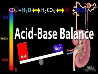
10. BASIC REGULATION OF ACID-BASE BALANCE
CO2 + H2O ↔ H2CO3 ↔ H+ + HCO3
The lungs help control acid-base balance by blowing off or
retaining CO2. The kidneys help regulate acid-base balance by
excreting or retaining HCO3
11. Buffer system
9/30/2023
11
Three Major Blood Buffer Systems:
Protein Buffer systems
Amino acids
Hemoglobin Buffer system
Phosphate Buffer system
Bicarbonate-carbonic acid Buffer system
12. Protein Buffer System
9/30/2023
12
Buffering capacity of plasma proteins is much
less than Hb (which operates only in
erythrocytes).
Buffering action of proteins:
In acidic medium: protein acts as a base, NH2
group takes up H+ ions forming NH3, proteins
become +vely charged.
In alkaline medium: protein acts as an acid.
Acidic COOH group dissociates and gives H+,
forming COO-. H+ combines with OH- to
produce a molecule of water, proteins become –
vely charged.
13. Hemoglobin as a Buffering agent
9/30/2023
13
Hemoglobin is a buffer for both CO2 and H+
CO2 diffuses across RBC membrane from
tissues.
The CO2 can bind directly with hemoglobin and
be released in the lungs. (20%)
The CO2 that reacts with water forms carbonic
acid that then dissociates into bicarbonate (70%)
in RBC.
Bicarbonate ions diffuse into plasma in exchange
for chloride ions.
H+ binds to hemoglobin and released in RBCs in
lungs to combine with bicarbonate & reform CO2
14. Phosphate Buffer System
9/30/2023
14
It is the major intracellular buffer. Its pK’ value of
6.86 is near the intracellular pH of 7.0.
Therefore, this buffer is very effective
intracellularly.
It consists of the following components:
1. H2PO4
- as the proton donor (i.e. the acid
component).
2. HPO4
- as the proton acceptor (i.e. the base
component).
H2PO4
- HPO4
- + H+
Phosphate buffer system works in conjunction
with the kidneys. A normal healthy kidney is
15. Bicarbonate/carbonic acid buffer
system
9/30/2023
15
The bicarbonate buffer system is the most
predominant extracellular buffer.
Mechanism of action of bicarbonate buffer.
When a strong acid, such as HCl is added to the
bicarbonate buffer solution, the increased hydrogen
ions are buffered by HCO3
-
HCl--------> H+ +Cl-
(strong acid)
HCO3
- + H+ ----------> H2CO3
(weak acid)
Thus, hydrogen ions from strong acid HCl react
with HCO3
- to form very weak acid H2CO3.
16. Bicarbonate/carbonic acid buffer
system
9/30/2023
16
The opposite reaction takes place when a strong
base, such as NaOH is added to the bicarbonate
buffer solution.
NaOH + H2CO3 ---------->NaHCO3 + H2O
(strong base) (weak base)
In this case hydroxyl ion (OH-) from NaOH
combines with H2CO3 to form weak base. Thus
strong base NaOH is replaced by a weak base
NaHCO3.
17. Bicarbonate/carbonic acid buffer
system
9/30/2023
17
At a pH 7.4, the ratio of bicarbonate to carbonic acid (HCO3
-
/ H2CO3) is 20:1.
Thus, the bicarbonate concentration is much higher (20
times) than carbonic acid in blood.
This is referred to as alkali reserve and is responsible for
the effective buffering of H+ ions, generated in the body.
Any alteration produced in the ratio between HCO3
- / H2CO3
leads to alkalosis or acidosis.
19. Respiratory mechanism in acid-base
balance
9/30/2023
19
The second line of defence against acid-
bases disturbances is by regulating the
concentration of carbonic acid (H2CO3) in
the blood and other body fluids by the
lungs.
The large volume of CO2 produced during
cellular metabolic activity endanger the
acid-base equilibrium of the body. But in
normal circumstances, all of this CO2 is
eliminated from the body in the expired air
via lungs.
20. Respiratory mechanism in acid-base
balance
9/30/2023
20
lungs function by maintaining one component
carbonic acid (H2CO3) of the bicarbonate buffer as
follows:
An increase in (H+) or (H2CO3) stimulates the
respiratory centre to increase the rate of
respiratory ventilation. When the ventilation rate
increases, more CO2 is released from the blood and
pH increases.
An increase in (OH-) or (HCO3-) depresses
respiratory ventilation. A decrease in ventilation rate
will cause a decrease in release of CO2 from the
blood. The increased blood CO2 will result in the
formation of more H2CO3. Thus there will be decrease
in pH.
21. Renal mechanism for pH
regulation
9/30/2023
21
Kidneys regulate the blood pH by maintaining the
alkali reserve, besides excreting or reabsorbing the
acidic or basic substances, as the situation
demands.
Urine pH is normally acidic ̴6 because the H+ ions
generated in the body in the normal circumstances,
are eliminated by acidified urine.
However it might vary between range 4.5-8
depending on the concentration of H+ ions.
22. Renal mechanism for pH
regulation
9/30/2023
22
Enzyme carbonic anhydrase is of central
importance in the renal regulation of pH which
occurs by the following mechanisms- :
1.Excretion of H+ ions
2.Reabsorption of bicarbonate
3.Excretion of titratable acid
4.Excretion of ammonium ions
25. Estimating blood pH
9/30/2023
25
The value of the extracellular [HCO3-/[CO2]
ratio (both in mmol/L) is 20 : 1. Taking pK’
value of carbonic acid to be 6.1, it can be
calculated from the Henderson–Hasselbalch
equation that this ratio represents a pH of
about 7.4.
pH = pKaH2CO3 + log [HCO3
-]
[H2CO3]
pH = 6.1+ log [25 mmol/L] = 6.1+1.3 =
7.4
[1.2 mmol/L]
26. ACID-Base disorders
9/30/2023
26
The acid-base disorders are
mainly classified as :
1.Acidosis: a decline in blood pH (<
7.35)
(a)Metabolic acidosis – due to
decrease in bicarbonate
(b)Respiratory acidosis -- due to an
increase in carbonic acid
2.Alkalosis: a rise in blood pH
(>7.45)
(a)Metabolic alkalosis – due to an
increase in bicarbonate
(b)Respiratory alkalosis – due to a
decrease in carbonic acid
27. 9/30/2023
27
If underlying problem is metabolic,
hyperventilation or hypoventilation can help :
respiratory compensation.
If problem is respiratory, renal mechanisms can
bring about metabolic compensation
28. Metabolic Acidosis: Bicarbonate Deficit
Increased acid production, uncontrolled diabetes mellitus,
alcoholism, starvation, renal acidosis, lactic acidosis, increased
acid ingestion, ethanol, salicylates, loss of bicarbonate, severe
diarrhea, intestinal fistulas, adrenal insufficiency,
hypoparathyroidism
Excess organic acids are added to body fluids or
bicarbonate is lost
Decrease in bicarbonate concentration
METABOLIC ACIDOSIS
Causes
29. Metabolic Acidosis
9/30/2023
29
Compensatory mechanism
1.Increasing rate of respiration to wash out CO2
(hence H2CO3) faster. Consequently, the ratio
HCO3-:H2CO3 is elevated.
2.Increasing excretion of H+ ions as NH4+ ions.
3.Increasing elimination of acid (H2PO4-) in the
urine.
30. Anion gap
9/30/2023
30
Knowledge of anion gap serves as an additional tool in
delineating the cause.
Anion gap is estimated by measuring the difference
between the sums of the concentrations of principal
cations (Na and K) and principal anions (Cl and
HCO3-).
31. Anion gap (metabolic acidosis)
9/30/2023
31
Increased anion gap: When excessive production of acids
is the cause of metabolic acidosis, concentration of
HCO3- decreases but that of Cl- remains unaffected.
Consequently, anion gap is increased.
Normal anion gap: In renal tubular acidosis, fall of
bicarbonate is accompanied by increase in chloride ion
concentration. Hence, anion gap does not change in
these conditions, which are, therefore, called normal
anion gap acidosis or hyperchloraemic acidosis.
32. Respiratory Acidosis: Carbonic Acid
Excess
Damage to the respiratory center in the medulla, drug or narcotic use,
obstruction of respiratory passages, respiratory and respiratory muscle
disorders
Decrease in the rate of pulmonary ventilation
Increase in the concentration of CO2, carbonic
acid, and hydrogen ions
RESPIRATORY ACIDOSIS
Potassium moves out of the cells
HYPERKALEMIA
VENTRICULAR FIBRILLATION
34. Respiratory Alkalosis: Carbonic Acid
Deficit
Anxiety, hysteria, fever, hypoxia, pain, pulmonary disorders,
lesions affecting the respiratory center in the medulla, brain
tumor, encephalitis, meningitis, hyperthyroidism, gram-
negative sepsis
Hyperventilation: Excessive pulmonary
ventilation
Decrease in hydrogen ion concentration
RESPIRATORY ALKALOSIS
Compensatory mechanism
1. Reduction in urinary ammonia formation
2.Increased excretion of bicarbonate.
35. Metabolic Alkalosis: Bicarbonate
Excess
Loss of stomach acid, gastric suctioning, persistent vomiting,
excess alkali intake, intestinal fistulas, hypokalemia,
Cushing’s syndrome or aldosteronism, potassium-diuretic
therapy
Excessive amounts of acid substance and hydrogen
ions are lost from the body or large amounts of
bicarbonate or lactate are added orally or IV
Excess of base elements
METABOLIC ALKALOSIS
40. Assessment of
oxygenation
9/30/2023
40
*Arterial partial pressure of oxygen (PaO2):
• The PaO2 level is a measurement of the amount of
oxygen dissolved in the blood.
• Normal values on room air are 80–100 mmHg.
• Analysis of PaO2 will identify hypoxaemia. A PaO2
level less than 60 mm Hg results in tissue hypoxia.
**Arterial oxygen saturation (SaO2):
• SaO2 (Oxyhemoglobin saturation) refers to the
number of hemoglobin binding sites that have
oxygen attached to them.
• How easily oxygen attaches to hemoglobin can be
affected by body temperature, pH, 2,3-
diphosphoglycerate levels, and CO2 levels.
41. Assessment of
ventilation
9/30/2023
41
Examination of the PaCO2 allows an assessment
of alveolar ventilation.
Alveolar ventilation is best assessed by
measuring the PaCO2, normal range (35-45 mm
Hg).
Increased ventilation will lower the PaCO2 and
lead to a respiratory alkalosis.
Decreased ventilation will raise the PaCO2 and
lead to a respiratory acidosis.
42. Components of Acid- Base
Balance
pH Measures the bloods acidity
Normal range 7.35- 7.45
Overall H+ from both respiratory and metabolic
factors.
pCO2
partial pressure of carbon dioxide in the blood
Normal range 35-45 mmHg
Presents the adequacy of alveolar ventilation
HCO3
The amount of bicarbonate in the blood
Normal range 22- 26 mEq/L
43. ABG sampling
Common sites includes:
1. Radial
2. Femoral
3. Branchial
4. Axillery artery
The radial artery is most
commonly used because of:
1. Accessible
2. Easily positioned
3. Comfortable to the patient
Heparinised blood is used for ABG but the correct
amount of heparin and blood is very important to
prevent coagulation of blood and to obtain accurate
test results.
44. Six Steps for ABG Analysis
9/30/2023
44
Steps
Step 1: Analyze the pH
pH < 7.35 = acidosis
pH > 7.45 = alkalosis
Step 2: Analyze the PaCO2
PaCO2 > 45 = acidosis
PaCO2 < 35 = alkalosis
Step 3: Analyze the HCO3
HCO3 - < 22 = acidosis
HCO3 - > 26 = alkalosis
Step 4: Match the PaCO2 or HCO3 -with pH
If pH and pCO2 match = respiratory
If pH and HCO3 match = metabolic
Step 5: Assess for compensation
Step 6: Analyze the PaO2 and SaO2
If PaO2 < 80 mm Hg or SaO2 < 95%, the patient has hypoxemia.
51. Question
Which of the following is
likely associated with her?
a. Metabolic alkalosis
b. Metabolic acidosis
c. Respiratory acidosis
d. Respiratory alkalosis
- pH = 7.21
- Na+= 130 mmol/L (RV: 135-145 mmol/L)
- Cl - = 80 mmol/L (RV: 96 -106 mmol/L)
- HCO3- = 10 mmol/L (RV: 22-26 mmol/L)
- pCO2= 25 mmHg (RV: 35-45 mmol/L)
- Anion gap= 40 mmo/L (RV: < 12)
*A40-year old female with type 1 DM, has the following
results for: