
Recommended
More Related Content
What's hot
What's hot (20)
Similar to Bone structure and type
Similar to Bone structure and type (20)
Recently uploaded
Recently uploaded (20)
Bone structure and type
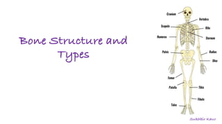
- 1. Bone Structure and Types Sukhbir Kaur
- 2. Introduction: • Bone: It is a rigid organ that constitutes part of the vertebrate skeleton. • Function: To support and protect the various organs of the body. • Bones come in a variety of shapes and sizes and have a complex internal and external structure. • They are lightweight yet strong and hard, and serve multiple functions. • In the human body at birth, there are over 270 bones , but many of these fuse together during development, leaving a total of 206 separate bones in the adult not counting numerous small sesamoid bones. • The largest bone in the body is the femur or thigh-bone, and the smallest is the stapes in the middle ear. • Bone tissue is a hard tissue, a type of dense connective tissue. It has a honeycomb-like matrix internally, which helps to give the bone rigidity.
- 3. Bone Structure Ø Matrix: It is made up of (90 and 95%) collagen fibers , and the ground substance. It is composed of organic and inorganic matter. v Inorganic component is made up of hydroxyapatite crystals (this is the bone mineral that gives bones their rigidity and tensile strength) and v Organic component collagen, an elastic protein which gives compressive strength. The collagen of bone is known as ossein. Ø Bone is formed by the hardening of this matrix around entrapped cells.
- 4. Bone Tissue and Composition • Bone tissue, or osseous tissue, is the major structural and supportive connective tissue of the body. • Bone tissue forms the rigid part of the bones that make up the skeleton. • Bone tissue refers specifically to the bone mineral matrix that forms the rigid sections of the organ, and the bone cells within it. • There are three types of bone tissue: cortical bone, cancellous bone and subchondral bone.
- 5. Types of Bone Tissue 1.Cortical Bone • The hard outer layer of bones composed of cortical bone also called COMPACT BONE. • Cortical referring to the outer (cortex) layer. The hard outer layer gives bone its smooth, white, and solid appearance, and accounts for 80% of the total bone mass of an adult human skeleton.
- 6. Also known as trabecular or spongy bone tissue, is the internal tissue of the skeletal bone and is open cell porous network. Thin formations of osteoblasts covered in endosteum create an irregular network of spaces, known as trabeculae. Within these spaces are bone marrow and stem cells that give rise to platelets, RBC’s and WBC’s. 2.Cancellous Bone
- 7. 3.Subchondral bone/tissue • Subchondral bone” is bone that sits underneath cartilage in a joint. Subchondral bone is found in large joints like the knees and hips, as well as in small joints like those of the hands and feet. • This is the smooth tissue at the ends of bones, which is covered with another type of tissue called cartilage. • The function of the subchondral bone is to attenuate forces generated through locomotion, with the compact subchondral bone plate providing firm support and the subchondral trabecular component providing elasticity for shock absorption during joint loading.
- 8. Osteon and Structure Of Cortical Bone • Cortical bone consists of multiple microscopic columns, each called an osteon. • It consists of concentric rings called lamellae which are formed by the osteoblasts that eventually differentiate into osteocytes. These osteocytes are located along the lamellae in spaces called Lacunae. • In the centre of the osteon is a canal called the Haversian canal. This contains the blood vessels, lymph vessels and neurons.
- 9. • Bone contains many of these individual osteons. The adjacent Haversian canals of these osteons are connected by Volkmann’s canals. • The columns are metabolically active, and as bone is reabsorbed and created, the nature and location of the cells within the osteon will change.
- 10. Bone marrow is the flexible tissue in the interior of bones. In humans, red blood cells are produced by cores of bone marrow in the heads of long bones in a process known as haematopoiesis. Bone marrow is also a key component of the lymphatic system, producing the lymphocytes that support the body’s immune system. • The two types of bone marrow are "red marrow“, which consists mainly of haematopoeitic tissue and "yellow marrow“, which is mainly made up of fat cells. RBC’s, platelets, and most white blood cells arise in red marrow. Both types of bone marrow contain numerous blood vessels and capillaries. Bone marrow
- 11. Bone Marrow • Also known as myeloid tissue, red bone marrow, can be found in almost any bone that holds cancellous tissue. • In new-borns, all such bones are filled exclusively with red marrow or haematopoietic marrow. • In adults, red marrow is found mainly in the flat bones, such as the pelvis, sternum, cranium, ribs, vertebrae and scapula, and in the cancellous material at the epiphyseal ends of long bones such as the femur and humerus.
- 12. At birth, all bone marrows are red. With age, more and more of it is converted to the yellow type; only around half of adult bone marrow is red. Yellow marrow is found in the medullary cavity, the hollow interior of the middle portion of long bones. In cases of severe blood loss, the body can convert yellow marrow back to red marrow to increase blood cell production.
- 13. Function of the Bone cells is to work in harmony to maintain a balance between bone formation and resorption, ultimately to control bone structure and function. They are responsible for the growing, shaping, and maintenance of bones. 4 types: • Osteoblast: It is found in the growing portions of bone, including endosteum and periosteum and is responsible for forming new bone. • Osteoclast: Osteoclasts are the cells responsible for bone resorption, thus they break down bone. Osteoclasts are large cells with multiple nuclei located on bone surfaces. • Osteocyte: Maintain mineral concentration of matrix. • Osteoprogenitor cells: Develop into osteoblasts. Bone is constantly remodelled by the resorption of osteoclasts and created by osteoblasts. Types of Bone Cells and their functions
- 14. Bone Cells :^I • Bone is a metabolically active tissue composed of several types of cells. • Osteoblasts are mononucleate bone-forming cells. They are located on the surface of osteon seams and make a protein mixture known as osteoid, which mineralizes to become bone. • Osteocytes are mostly inactive osteoblast.They originate from osteoblasts that have migrated into and become trapped and surrounded by bone matrix that they themselves produced. The spaces they occupy are known as lacunae.
- 15. There are five types of bones in the human body: Long, Short, Flat, Irregular, and Sesamoid. TYPES OF BONES
- 16. Types of Bones 1.Long bones are much longer than they are wide. They consist of 3 different sections • Epiphysis- contains spongy bone or cancellous bone and also contain red bone marrow that synthesise RBC’s. • Metaphysis- contains an important section i.e. Epiphyseal plate responsible for lengthening and elongating long bone as organism actually grows. • Diaphysis- long, curved shaft that contains compact bone that is responsible for giving bone, its strongness.
- 17. They are capable of resisting very high tensile and compressive forces due to which long bones support majority of our body’s weight. We have many different types of long bones in our body. Example : i) Clavicle (collar bone) ii) Humerus, radius, ulna (arm) iii) Femur, tibia, fibula (leg) iv) Metacarpals Long Bone (cont...)
- 18. 2.Short bones are roughly cube-shaped, and have only a thin layer of compact bone surrounding a spongy interior. They are as long as wide. They function by providing support as well as stability to other bones. They are located at the wrist (carpals) and ankle (tarsals). 3.Flat bones are thin and generally curved, with two parallel layers of compact bones sandwiching a layer of spongy bone. They have a large surface area. They : a) protect our internal organs and tissue b) Serve as attachment point for muscle because of their large surface area. Example : i) Skull (cranium)- flat bones fuse here to protect our brain. ii) Rib cage and Sternum- this acts not only as attachment point of muscle but also protect heart, lungs and vascular tissue in this region. iii) Scapula (shoulder blade bone) iv) Pelvis (hip bone)
- 19. 4.Sesamoid bones are bones embedded in tendons. This bone is in shape of sesame seed. Example of sesamoid bone is Patella (knee cap). 5.Irregular bones do not fit into the above categories. They consist of thin layers of compact bone surrounding a spongy interior. As implied by the name, their shapes are irregular and complicated. Thus, they have a unique shape and function. They help in protection and support of body. Example : i) Maxilla (upper jaw bone) and Mandible (lower jaw bone)- they help to eat and ingest food. ii) Sacrum iii) Vertebrae (cervical and lumbar)- they are responsible for protecting our spinal cord.
- 20. Bones consist of living cells embedded in a mineralized organic matrix. This matrix consists of organic components, mainly Type I Collagen – "organic" referring to materials produced as a result of the human body – and inorganic components, primarily hydroxyapatite and other salts of calcium and phosphate. The collagen fibers give bone its tensile strength, and the interspersed crystals of hydroxyapatite give bone its compressive strength. Collagen consists of strands of repeating units, which give bone tensile strength, and are arranged in an overlapping fashion that prevents shear stress. The function of ground substance is not fully known. Two types of bone can be identified microscopically according to the arrangement of collagen: Woven and lamellar.
- 21. Woven Bone Woven bone, (also known as fibrous bone) which is characterized by a haphazard organization of collagen fibers that occurs initially in all fetal bones. It is mechanically weaker, with a smaller number of randomly oriented collagen fibers, but forms quickly; it is for this appearance of the fibrous matrix that the bone is termed woven. It is soon replaced by lamellar bone. After a fracture, woven bone forms initially and is gradually replaced by lamellar bone during a process known as "bony substitution." Compared to woven bone, lamellar bone formation takes place more slowly.
- 22. It has a regular parallel alignment of collagen into sheets ("lamellae") and is mechanically strong. It is highly organized in concentric sheets with a much lower proportion of osteocytes to surrounding tissue. It makes its first appearance in humans in the fetus during the third trimester, is stronger and filled with many collagen fibers parallel to other fibers in the same layer (these parallel columns are called osteons). Lamellar Bone
- 23. Thank you