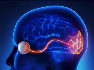
Optic nerve
•
1 like•1,062 views

second cranial nerve, origin of optic nerve, couurse and termination of optic nerve, clinical presentation incase of optic nerve damage,

Recommended
More Related Content
What's hot
What's hot (20)
Similar to Optic nerve
Similar to Optic nerve (20)
More from Dr. sana yaseen
More from Dr. sana yaseen (20)
Recently uploaded
Recently uploaded (20)
Optic nerve
- 2. OPTIC NERVE CN-II DR SANA YASEEN ANATOMY DEPARTMENT KIMS
- 3. LEARNING OBJECTIVE Introduction to cranial nerve II Origin, course and termination of optic nerve. Pupillary light reflex Accomodation reflex Clinical aspect of optic nerve
- 4. INTRODUCTION The anatomical course of the optic nerve describes the transmission of special sensory information from the retina of the eye to the primary visual cortex of the brain. It can be divided into extracranial (outside the cranial cavity) and intracranial components.
- 5. EXTRACRANIAL The optic nerve is formed by the convergence of axons from the retinal ganglion cells. These cells in turn receive impulses from the photoreceptors of the eye (the rods and cones). The nerve fibers arising from ganglionic cells makes up the optic nerve. These fibers converges towards optic disc hence piercing the three coats of an eyeball. (retina, choroid, sclera)
- 8. After its formation, the nerve leaves the bony orbit via the optic canal, a passageway through the sphenoid bone. It enters the cranial cavity, running along the surface of the middle cranial fossa (in close proximity to the pituitary gland).
- 11. INTRACRANIAL Within the middle cranial fossa, the optic nerves from each eye unite to form the optic chiasm. At the chiasm, fibres from the nasal (medial) half of each retina cross over to the contralateral optic tract, while fibres from the temporal (lateral) halves remain ipsilateral: Left optic tract – contains fibres from the left temporal (lateral) retina, and the right nasal (medial) retina. Right optic tract – contains fibres from the right temporal retina, and the left nasal retina.
- 15. Each optic tract travels to its corresponding cerebral hemisphere to reach the lateral geniculate nucleus (LGN), a relay system located in the thalamus; the fibres synapse here. Axons from the LGN then carry visual information via a pathway known as the optic radiation. Optic radiation pass through retrolenticular part of internal capsule and terminates in visual cortex. Brodmann area 17 This area occupies upper and lower lips of calcarine sulcus on medial surface of occipital lobe, where otic impulses recieves consciousness.
- 17. Association area 18 and 19 surrounding the brodmann are 17, recieves fiber from primary visual cortex. Visual association areas are responsible for interpretation of optic impressions as recognition of the object, perception of color, depth and other aspect of vision.
- 19. PUPILARY LIGHT REFLEX The pupillary light reflex involves adjustments in pupil size with changes in light levels. The reflex is consensual: Normally light that is directed in one eye produces pupil constriction in both eyes. The direct response is the change in pupil size in the eye to which the light is directed (e.g., if the light is shone in the right eye, the right pupil constricts). The consensual response is the change in pupil size in the eye opposite to the eye to which the light is directed (e.g., if the light is shone in the right eye, the left pupil also constricts consensually).
- 20. • Pupillary light reflex; direct and indirect
- 21. ACCOMODATION REFLEX The accommodation response is elicited when the viewer directs his eyes from a distant (greater than 30 ft. away) object to a nearby object. The stimulus is an “out-of-focus” image. The accommodation (near point) response is consensual (i.e., it involves the actions of the muscles of both eyes).
- 22. CLINICAL
- 24. SUMMARY Name: optic nerve Numerological order: II Funtion: NERVE OF VISION Functional component: SSA Origin: ganglionic layer of retina Ends: visual cortex in occipital lobe