3. Joseph Erlanger(1874-1965) and
Herbert Spencer Gasser (1888-
1963)
1944 Joseph Erlanger and Herbert Spencer Gasser "for their discoveries
relating to the highly differentiated functions of single nerve fibres
4. Joseph Erlanger and Herbert
Spencer Gasser
Joseph Erlanger was an American physiologist
Herbert Spencer Gasser was an American physiologist, and
recipient of the Nobel Prize for Physiology or Medicine in 1944 for
his work with action potentials in nerve fibers while on the faculty of
Washington University.
Erlanger and his student Gasser were interested in developing
tools that could measure impulses fired through nerve cells, and
they turned to the cathode-ray oscilloscope – an instrument that
allows electrical currents to be visualized as a moving two-
dimensional graph on a phosphorescent screen.
After its invention by Ferdinand Braun, the oscilloscope soon
became the most effective tool for detecting rapid changes in
electrical voltage, but still it was not sensitive enough to measure
the weak and rapid electrical impulses that are fired along nerve
cells
5. Eccles, Hodgkin and Huxley
The Nobel Prize in Physiology or Medicine 1963 was awarded jointly to Sir John Carew
Eccles, Alan Lloyd Hodgkin and Andrew Fielding Huxley "for their discoveries concerning the
ionic mechanisms involved in excitation and inhibition in the peripheral and central portions of
the nerve cell membrane".
6. Eccles, Hodgkin and Huxley
Sir Alan Lloyd Hodgkin British physiologist and biophysicist
Sir John Carew Eccles , Australian neurophysiologist
Sir Andrew Fielding Huxley English physiologist and biophysicist,
Huxley evidenced the existence of saltatory conduction in myelinated nerve fibres.
By showing how these impulses are generated and transmitted, the three scientists who received
an equal share of the 1963 Nobel Prize in Physiology or Medicine revealed the key triggers that
spark the nervous system's in-built electrical system into life.
Seeking ways of measuring electrical currents inside nerves, Alan Hodgkin and his student
Andrew Huxley turned to giant nerve fibres in the squid, which are almost a thousand times thicker
than their human counterparts.
Using tiny electrodes to record the electrical difference between the inside and outside of these
nerves, they were surprised to find that the polarity did not drop from negative to zero during the
transmission of an impulse as predicted, but in fact reversed, becoming electrically positive.
By carrying out a series of measurements and using complex mathematical models to interpret
the findings, Hodgkin and Huxley formulated a theory to propose how impulses are formed.
Changes in the permeability of the cell membrane allow charged atoms to flow in and out of a
nerve fibre, creating waves of electric charge that constitute the nerve impulse
7. Erwin Neher, Bert Sakmann
The Nobel Prize in Physiology or Medicine 1991 was awarded jointly to
Erwin Neher and Bert Sakmann "for their discoveries concerning the
function of single ion channels in cells"
8. Erwin Neher, Bert Sakmann
The two German cell physologisists Erwin Neher and Bert Sakmann
have together developed a technique that allows the registration of
the incredibly small electrical current (amounting to a picoampere-
10-12A) that passes through a single ion channel.
The technique is unique in that it records how a single channel
molecule alters it's shape and in that way controls the flow of current
within a time frame of a few millionths of a second.
They have demonstrated what happens during the opening or
closure of an ion channel with a diameter corresponding to that of a
single sodium or chloride ion.
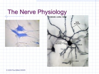
9. The neurone
Nodes of Ranvier
Dendrites
Schwann cell Nucleus of Schwann cell
Myelin sheath Axon Terminal dendrites
23. Characteristics of the Nerve
Impulse
An electrochemical event that occurs in nerve
cells following proper stimulation.
An all-or-none process which is fast acting
and quick to recover.
An event that is described by a voltage curve
that is called an action potential.
The nerve impulse can be conducted the
entire length of a nerve cell without
diminishment (“domino effect”).
24. Characteristics of a Nerve
Impulse Continued:
The nerve impulse serves as the primary
information signal used by the nervous system
to provide communication about stimuli, nerve
cell activity, neurotransmitter release and to
generate various output responses (motor
action, glandular secretion, etc.).
Typically initiated by graded or generator
potentials from a stimulus.
25. Graded potential
A change in potential that decreases with
distance
Localized depolarization or hyperpolarization
31. Nernst Equation
By the end of the 19th century, it was
known that the cytoplasm was high in K+
and that [Na+] was very low--and that this
relationship was reversed outside the cell.
The assumption was made that the cell
membrane was permiable to K+ but not to
Na+.
34. Resting Potential
Outside of cell
Sodium/Potassium pump
continuously and actively pumps
(3) Na+ out of the cell and (2) K+
into the cell.
Na+ channels are closed so Na+
are not able to move into the cell.
K+ channels are open so K+ can
diffuse out of the cell.
This generates a separation of
charges so that the inside of the
cell is relatively – and the outside
is relatively +.
The cell will remain in this state (at
rest) until it is stimulated.
Inside of cell
36. Action Potential
Appears when region of excitable
membrane depolarizes to threshold
Steps involved
Membrane depolarization and sodium
channel activation
Sodium channel inactivation
Potassium channel activation
Return to normal permeability
37. The Action Potential
Key Properties of the Action Potential
Threshold
Rising phase
Overshoot
Falling phase
Undershoot
Absolute refractory period
Relative refractory period
38. Introduction
Action Potential in the Nervous System
Conveys information over distances
Action potential
Spike
Nerve impulse
Discharge
39. AP Characteristics
Voltage-gated channels
All or none
Slow
Non-decremental
Self Propagated
41. Properties of the Action
Potential
The Ups and Downs of an Action
Potential
Oscilloscope to visualize an AP
Rising phase, overshoot, falling phase, and
undershoot
42. Properties of the Action
Potential
The Generation of an Action Potential
“All-or-none”: Cross threshold value for
action potential
Chain reaction
Opens Na+-permeable channels Na+ influx
depolarized membrane reaches threshold action
potential
43. Properties of the Action
Potential
Firing frequency reflects the magnitude of
the depolarizing current
45. Characteristics of action
potentials
Generation of action potential follows all-or-none
principle
Refractory period lasts from time action potential
begins until normal resting potential returns
Continuous propagation
spread of action potential across entire membrane in
series of small steps
Saltatory propagation
action potential spreads from node to node, skipping
internodal membrane
49. Action potential propagation
When the V-G Na+
channels open, they
cause a
depolarization of the
neighboring
membrane.
This causes the Na+
and K+ channels in
that piece of
membrane to be
activated
50. AP propagation cont.
The V_G chanels in
the neighboring
membrane then
open, causing that
membrane to
depolarize.
That depolarizes
the next piece of
membrane, etc.
It takes a while for
the Na+ channels to
return to their
voltage-sensitive
state. Until then,
they won’t respond
to a second
depolarization.
54. Action Potential Conduction
Propagation of the action potential
Down axon to the axon terminal
Orthodromic: Action potential travels in one
direction
Antidromic (experimental): Backward
propagation
Typical conduction velocity: 10 m/sec
Length of action potential: 2 msec
55. Action Potential Conduction
Factors Influencing Conduction Velocity
Spread of action potential along membrane
Dependent upon axon structure
Path of the positive charge
Inside of the axon (faster)
Across the axonal membrane (slower)
Axonal excitability
Axonal diameter (bigger = faster)
Number of voltage-gated channels
56. Action Potential Conduction
Factors Influencing Conduction Velocity
Myelin: Facilitates current flow
Layers of myelin sheath
Myelinating cells
Schwann cells in the PNS
Oligodendroglia in CNS
60. All-or-None Principle
Throughout depolarisation, the Na+ continues
to rush inside until the action potential
reaches its peak and the sodium gates close.
If the depolarisation is not great enough to
reach threshold, then an action potential
and hence an impulse are not produced.
This is called the All-or-None Principle.
66. Repolarization
1. The sodium/potassium
pumps return the cell to a
resting state by actively
pumping (3) Na+ out of
the cell and (2) K+ into
the cell.
2. The K+ continues to
diffuse out of the cell.
67.
68. Refractory Period
after AP
won’t fire again
relative & absolute
Relative
during after hyperpolarization
requires greater depolarization ~
69. Refractory Period
There are two types of
refractory period:
Absolute Refractory
Period – Na+ channels
are inactivated and no
matter what stimulus is
applied they will not re-
open to allow Na+ in &
depolarise the membrane to the threshold of an action potential.
Relative Refractory Period - Some of the Na+ channels have re-opened but the
threshold is higher than normal making it more difficult for the activated Na+
channels to raise the membrane potential to the threshold of excitation.
77. Anti-seizure Medications
Seizures caused by hyperactive brain
areas
Multiple chemical classes of drugs
All have same approach
Decrease propagation of action potentials
⇓ Na+, Ca++ influx (delay depolarization/prolong
repolarization)
⇑ Cl- influx (hyperpolarize membrane)
79. Clinical Correlation
It is the rate of action potential propagation that
determines neurologic function.
Determined by frequency of action potentials.
What is a seizure?
What is a seizure?
What would be the
What would be the
effect on the membrane
effect on the membrane
of ⇑ Cl- -influx
of ⇑ Cl influx
during a seizure?
during a seizure? Hyperpolarization & …
⇓ seizure
activity!