SlideShare a Scribd company logo
1 of 78
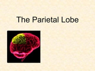
The Parietal Lobe
• ANATOMY

• FUNCTIONS

• CLINICAL EFFECTS
ANATOMY
BOUNDARIES OF PARIETAL LOBE

  – Anterior border - Central Fissure
  – Ventral border - Sylvan Fissure
  – Posterior border - Parieto-occipital sulcus
Subdivisions of the Parietal Lobes

          Post central sulcus
          Interparietal sulcus


1.   post central gyrus
2.   Somasthetic association area
3.   Superior parietal lobule
4.   Inferior parietal lobule –
                angular gyrus
                supra marginal gyrus
FUNCTIONS
PRIMARY SOMASTHETIC AREA
                     Body image representation
• Afferents – VP nucleus of thalamus
• Efferents - to SPL (area 5)
            - some to opp. Somatosensory cortex via
  corpus callosum.

SOMASTHETIC ASSOCIATION AREA
               Body in space
               Tactile discrimination
SUPERIOR PARIETAL LOBULE AND AREA 7
             Information processed in primary and
 association areas is transmitted to area 7, where
 polymodel association takes place.
 Area 7 is the highest level of somasthetic
 integration. (also have connections with all lobes).

     3 D analysis of body space interactions (body schema)
     Visual spatial properties
     Visual attention
     Motivation and grasping functions.
(parietal lobe lesions - there is ‘self grasping’ of forearm opp. the
  lesion )
• Areas 5 and 7
         have connections with premotor and motor
  cortex – so, lesions l/t disturbances in voluntary
  movement.
         also have connections with cingulate gyrus
  and prefrontal cortex . Therefore they mediate
  influence of emotion, attention and motivation
  on behavior - produced by somatosensory &
  visual stimuli.
INFERIOR PARIETAL LOBULE
        Last to mature anatomically and functionally.
 So, the functions are late, to develop b/w 5 and 8 yrs
  age. ( reading , calculations )
             Angular gyrus & Supra marginal gyrus -
  they have interconnections with visual, auditory,
  somasthetic, supr. colliculus, LGB and other lobes.
1. Multimodal assimilation – capacity for
   organizing , labelling and conceptualizing , using
   all senses.
   Ex : chair

2. Language capabilities
        angular gyrus - anomia
        supramarginal gyrus – conduction aphasia
        visual cortex to IPL connections – word
   blindness
3. Agraphia – lt. lobe
          Engrams for production and perception
   of written language are stored in IPL .
 So, misspellings, distorsions, and inversions occur.

4. Temporal sequential functions
            IPL is the main track of input and output.
Therefore, information is organized appropriately
    into a sequence here.

5. Caluculation (Lt.) and computation (Rt.)
IPL lesions l/t disruption of visual spatial
 functioning and temporal sequencing ability
 (apraxia).
    i.e either spatial sequential tasks lost - OXOXOX
or sequential grammar relations are lost.
Cerebral laterality
Either hemisphere
1. Cortical sensations.
2. Integration of sensory , motor and attention signals
   (i.e disengage attention - do other activity -immediately
   reengage correctly)
3. Optic radiation passes through.
4. Constructional ability – capacity to construct or draw
   3D/2D figures or shapes.
      Lt. – programming of movements necessary for
  constructional activity. (simplification of complex
  diagrams)
      Rt. – related to spatial relationships or imagery.
  (rotation of diagrams)
5. Short term memory
     Lt. – immediate recall for digits and words
     Rt. – immediate recall for geometric patterns

Left hemisphere
1. Language – comprehension
               reading
               writing
2. Calculations – verbal rote calculations and
   recognition of signs.
3. Non verbal symbolization (pantomime)
Right hemisphere
1. Constructional skills
2. Dressing apraxia
3. Calculations –
      arithmetic concepts of carrying and borrowing
      spatial alignment of written calculations.
   (computational difficulty – inability to manipulate
   no.s in spatial relation, like using decimals,etc –
   but he is able to do problems in his head )
4. Perceptual functions (inattention/neglect of lt.
   hemispace)
CLINICAL EFFECTS OF
PARIETAL LOBE LESIONS
• CORTICAL SENSORY SYNDROMES

• ASOMATAGNOSIAS

• APRAXIAS

• VISUAL DISORDERS

• AUDITORY NEGLECT
CORTICAL SENSORY SYNDROMES
Cortical defect is essentially one of sensory
 discrimination i.e impaired ability to integrate and
 localize stimuli.
        1. Loss of position sense and passive
 movement.
        2. Topagnosia – loss of localization of tactile,
 thermal and noxious stimuli.
        3. Astereognosis.
        4. Agraphesthesia.
        5. Loss of ‘two point’ discrimination.
• This type of sensory defect is sometimes referred
  to as “cortical” , although it can be produced as
  well by lesions of the subcortical connections.
• The perception of pain, touch, pressure, vibratory
  stimuli, and thermal stimuli is relatively intact in
  parietal lobe lesions.

Pseudothalamic pain syndrome : burning or
 constrictive pain, identical to the thalamic pain
 syndrome, resulted from vascular lesions restricted
 to the cortex.
Other parietal sensory defects :
• easy fatigability of sensory perceptions
• inconsistency of responses to painful and tactile
  stimuli
• Pain sensations outlast the stimuli & hyperpathia
• Tactile hallucinations

Motor deficits: mild hemiparesis +
                 hypotonia +
                 atrophy (unexplained by inactivity).
Optic ataxia : clumsiness in reaching for and
 grasping an object.

Pseudocerebellar syndrome : incoordination and
 intention tremor of contralateral limbs.

Fixed dystonic postures
Tests for cortical sensations
• Basic sensations must be intact.

A. Two point discrimination :
     Calipers or compass used.
     Sites – palms (8-15 mm) , dorsum (2-3cm)
   , shins (3-4 cm).

 Ask pt. to close eyes – respond as ‘one’ or ‘two’.
 Single and double points to be varied
 Compare with other side
B. Graphesthesia :
     done with pencil or swab stick.
     sites – palms, fingers and face.
 Digits like 1-9 , or shapes/symbols used.
 Stand beside the pt and face the area to be
   tested (so that he will be familiar).

C.   Stereognosis :
•    no preliminary visual demo given.
•    ex: key, pen or coin
•    abnormal side done first and then normal side.
D. Double simultaneous stimulation :
• Pin prick used (both must be equally        Sensory extinction
  sharp).                                     Sensory inattention
                                              Sensory suppression
 Eyes to be closed                           Sensory eclipse
 Pt is told to expect sensation either one   Tactile inattention
  side or both sides.                         Perceptual rivalry

 After stimuli, ask to indicate site of
  stimulus and their nature.

POSITIVE test – stimuli on involved half is
  ignored or sharp stimulus interpreted as
  dull.
• CORTICAL SENSORY SYNDROMES

• ASOMATAGNOSIAS

• APRAXIAS

• VISUAL DISORDERS

• AUDITORY NEGLECT
ASOMATAGNOSIAS
• The term asomatognosia denotes the inability to
  recognize part of one’s body.

• Visual and tactile sensory information is synthesized
  during development into a body schema
  (perception of one’s body and the relations of
  bodily parts to one another)
 ANOSAGNOSIA :          Unilateral asomatagnosia.
   (Denial of illness)   Anton–Babinski syndrome.
• Denial is more implict than explict, in many pts.
  (i.e they may not actively deny that they are ill)
  And some may act as if nothing were the matter.
• 7 times more frequent with Rt. sided lesions than
  left.
• Ass. with blunted emotionality – pts look
  dull, inattentive and apathetic. And also
  confused.
• Ass. with hallucinations of movement and
  allocheiria (one sided stimuli are felt on other
HEMI NEGLECT : neglect on one side of body in
 dressing and grooming.
Shave only one side or use only one sleeve of shirt.
Deviation of head and eyes to side of lesion.
Torsion of body to the side of lesion.
Fail to use one side of body, even though paralysis
 is not present
Finds impossible to wear eye glasses.
Sensory extinction - is subtle form of neglect.
• Basic disturbance in these cases is an inability to
  summate a series of ‘spatial impressions’ -
  tactile, kinesthetic, visual, or auditory — a defect
  referred to as amorphosynthesis.
• Denial and neglect - non-dominant parietal lesions.
• These disorders of spatial summation are strictly
  contralateral to the damaged parietal lobe (Rt > Lt)
       It must be distinguished from a true
  agnosia, which is a conceptual disorder and
  involves both sides of the body and extrapersonal
  space as a result of damage to the dominant
  hemisphere.
GERSTMANN SYNDROME
• An example of bilateral asomatognosia and is due
  to a left dominant parietal lesion.

1.   Finger agnosia
2.   Right-left confusion
3.   Acalculia
4.   Dysgraphia
• May be ass. with dyslexia or homonymous
  hemianopia / quadrantanopia.

Lesion – left inferior parietal lobule (angular
 gyrus).
Parietal lobe is – “ Lobe of hand ”.
 Hand is extensively represented
 Parietal lobe gathers information regarding
  various objects through hand
 Parietal lobe lesions (Gerstmann syndrome) –
  tetrad is related to
  functions of hand.
Tests for calculations
Components – Rote tables (add, multiply, etc)
         Recognition of signs (+ , - , * )
         Basic arithmetic(carrying, borrowing)
         Spatial alignment of written calculations

• Verbal rote examples : what is 4 plus 6 ?

• Verbal complex examples : what is 21 * 5 ?
• Written complex examples :

                          • Pt with rt. hemisphere
                            lesion & left neglect.


                          • Pt with rt. parietal
                            hematoma – showing
                            poor alignment and
                            calculation errors.
• Lt parietal lesions – inability to understand
  and carry out numericals.
  Severe acalculia = Anarithmetria.

• Rt parietal lesions – inability to align numbers
  and to do complex computations (
  borrowing, carrying, etc).
  But, pt can do problems in his head.
Tests for right – left confusion
 Identification on self
       ex : show your left foot.
 Crossed commands on self
       ex : with your right hand touch your left ear
 Identification on examiner
       ex : point my right elbow
 Crossed commands on examiner
       ex : with your left hand point my right foot.
Tests for finger agnosia
• Inability to name , point or recognize fingers on
  oneself or others.
1. Non verbal finger recognition :
          with pt eyes closed, touch one of his fingers.
   Ask him to touch the same finger of examiner, with
   eyes open.

2. Identifying named fingers on examiner’s hand :
         examiner places hand in some irregular
   position and asks pt – “ point to my middle finger”
3. Verbal identification (naming) of fingers :
           either examiner’s or pt’s hand kept in an
   irregular position. Examiner points to a finger and
   asks him – “name this finger?”
• CORTICAL SENSORY SYNDROMES

• ASOMATAGNOSIAS

• APRAXIAS

• VISUAL DISORDERS

• AUDITORY NEGLECT
APRAXIA AND PARIETAL LOBE

• An inability to carry out a commanded task despite
  the retention of motor and sensory function
• Sensory guidance of movement is lost
• Either spatial sequences lost (0VOOVOOOV)
  or sequential grammar relations lost
  (ex: father’s brother’s son).
• Defect - unable to show, but uses the object.
          - unable to do whole task, but does
  individual tasks.
• Failure to conceive or formulate an action , either
  spontaneously or on command.

• Sensory areas 5 and 7 in dominant parietal
  lobe, supplementary and premotor cortex of both
  cerebral hemispheres and their integral
  connections - are involved to accomplish these
  actions.
1.   Ideomotor apraxia *
2.   Ideational apraxia *
3.   Buccofacial apraxia *
4.   Constructional apraxia
5.   Dressing apraxia

•    Constructional apraxia – visuospatial orientation
•    Dressing apraxia – form of sensory extinction and loss of
     extra personal space.


* according to Liepmann
IDEOMOTOR APRAXIA (“how to do”)
• Most common type of apraxia
i. Buccofacial apraxia ( blowing a match )
ii. Limb apraxia ( flip a coin , comb hair )
iii. Whole body apraxia ( stand like boxer )

•   Commands to be alternated b/w right and left
    limbs
IDEATIONAL APRAXIA            (“what to do”)
• Disturbance of complex motor planning of a higher
  order .
• Pt able to do individual tasks, but cannot integrate
  them as a whole.
• ‘Conceptual apraxia’ – there is apparent inability to
  recognise the use of objects (object agnosia).
 ex: pt attempts to light a candle by striking it on matchbox
Praxis testing (done in an order)
1. Observe the actions – shaving ,dressing,eating.
2. Carry out familiar acts – blow a kiss, wave gudbye.
3. Imitate the examiner (‘do this after me’)
4. How to use objects (pantomime)
      simple acts – hammer nail, comb hair .
      complex acts – light and smoke cigar; open soda
   bottle, pour in glass and drink.
5. Demonstrate use of actual items

(both limbs and orofacial commands to be asked)
CONSTRUCTIONAL APRAXIA
• Constructional ability/praxis = visuoconstructive
  ability - high level non verbal cognitive function.
• Perceptual motor ability involving integration of
  occipito – parieto – frontal connections.
• Non dominant parietal lobe is imp. for this.
• Area 17       IPL (kinesthetic analysis of visual patterns
                     done here)


                    Premotor area
• Connections with frontal and occipital lobes provide
  necessary proprioceptive and visual information –
  for movement of body and manipulation of objects
  or constructional activities.
         Parietal lobes are principle areas of visual –
  motor integration.
Tests of constructional ability
 Reproduction drawings :
    given in order of complexity.
Drawings to command :
   1. draw a clock with 10:20 time
   2. draw a 2D figure - daisy in a pot
   3. draw a house – in way you can see two sides
 and the roof.

Block designs


 Lt. sided lesions – simplification of complex diagrams
 Rt. sided lesions – rotation of diagrams .
DRESSING APRAXIA

• Not a true apraxia.
• Combination of spatial disorientation and
  visuospatial inattention.
• CORTICAL SENSORY SYNDROMES

• ASOMATAGNOSIAS

• APRAXIAS

• VISUAL DISORDERS

• AUDITORY NEGLECT
VISUAL DISORDERS
• Inferior part of the parietal lobe – incongruous
  homonymous hemianopia or an inferior
  quadrantanopia. (in practice, the defect is complete
  or almost complete and congruous).
• Deep lesions - abolition of optokinetic nystagmus
  with target moving toward side of the lesion.
• Rt. angular gyrus - Left sided visual neglect.
• Topographagnosia - visual disorientation and loss
  of spatial (topographic) localization. Pts ar unable to
  orient themselves in an abstract spatial setting.
• Others –
    Deficits in localization of visual stimuli.
    Inability to compare the sizes of objects.
    Failure to avoid objects when walking.
    Inability to count objects.
    Disturbances in smooth-pursuit eye movement.s
    Loss of stereoscopic vision.
• “Spasticity of conjugate gaze” : eyes may deviate
  away from the lesion on forced lid closure.
• Optic ataxia : in reaching for a target, movement is
  misdirected and dysmetric. (distance to target is misjudged)
Tests for visual disorders

• Visual field testing


• Visual neglect :
     - casual observation of pt’s behaviour.
     - drawings made by the pt.
• Visual inattention


• Topographagnosia : tests for geographic
  disorientation.
Tests for geographic disorientation
• Geographic orientation is function of parietal lobe
  and its multimodal association area.
• Combination of processes – spatial
  orientation, right-left orientation ,visual perception
  and its memory.
1. History from relatives :
       Does he becomes lost in work?
       Does he have difficulty in orienting to new
   environment?
2. Localizing places in maps :
      Adequate literacy level and historical knowledge
   is necessary.
     ex : to locate cities or states on maps.

3. Ability to orient self in hospital :
    By observing the pt’s capacity to find their bed,
   ward and bathroom.
• CORTICAL SENSORY SYNDROMES

• ASOMATAGNOSIAS

• APRAXIAS

• VISUAL DISORDERS

• AUDITORY NEGLECT
AUDITORY NEGLECT
• This defect in appreciation of the left side of the
  environment is less apparent than is visual
  neglect.
• Many patients with acute right parietal lesions
  are initially unresponsive to voices or noises on
  the left side.
• Main lesion usually lies in the right superior
  lobule.
SUMMARY
EITHER PARIETAL LOBES (Rt. or Lt.)

1.   Loss of cortical sensations.
2.   Mild hemiparesis, hypotonia and hemiatrophy.
3.   Hemianopia / quadrantanopia .
4.   Visual inattention.
5.   Abolition of optokinetic nystagmus.
6.   Hemineglect ( more with Rt. parietal lobe lesions ).
DOMINANT PARIETAL LOBE

Additional phenomenon include,

1. Disorders of language ( anomia, aphasia, alexia,
   agraphia ).
2. Gerstmann syndrome
3. Tactile agnosia (bimanual astereognosis)
4. Bilateral ideomotor and ideational apraxia.
NON DOMINANT PARIETAL LOBE
Additional phenomenon include,

1.   Visuospatial disorders
2.   Topographic memory loss
3.   Anosagnosia
4.   Dressing and constructional apraxias.
5.   Confusion
6.   Tendency to keep eyes closed, resist lid opening
     and blepharospasm.
The Parietal Lobe: Functions, Anatomy, and Clinical Effects

More Related Content

What's hot

Occipital lobe
Occipital lobeOccipital lobe
Occipital lobesrimantp
 
Frontal lobe and its functions
Frontal lobe and its functionsFrontal lobe and its functions
Frontal lobe and its functionsdrnaveent
 
Disconnection syndrome
Disconnection syndromeDisconnection syndrome
Disconnection syndromegulabsoni
 
The Parietal Lobe
The Parietal LobeThe Parietal Lobe
The Parietal Lobeguest0e3a5d
 
Corpus callosum with disconnection syndromes
Corpus callosum with disconnection syndromes Corpus callosum with disconnection syndromes
Corpus callosum with disconnection syndromes Amruta Rajamanya
 
frontal lobe anatomy and clinical relevance
frontal lobe anatomy and clinical relevancefrontal lobe anatomy and clinical relevance
frontal lobe anatomy and clinical relevanceImran Rizvi
 
Anatomy & Function of Temporal lobe
Anatomy & Function of Temporal lobeAnatomy & Function of Temporal lobe
Anatomy & Function of Temporal lobeNeurologyKota
 
Frontallobe dr prashant mishra
Frontallobe dr prashant mishra Frontallobe dr prashant mishra
Frontallobe dr prashant mishra Prashant Mishra
 
Functional anatomy of Frontal lobe
Functional anatomy of Frontal lobeFunctional anatomy of Frontal lobe
Functional anatomy of Frontal lobeRooban Thavarajah
 
Cerebellum clinical
Cerebellum clinicalCerebellum clinical
Cerebellum clinicalmunnam37
 
Frontal lobe
Frontal lobeFrontal lobe
Frontal lobesai nath
 
Temporal lobe and its role in psychiatry
Temporal  lobe  and  its  role  in  psychiatryTemporal  lobe  and  its  role  in  psychiatry
Temporal lobe and its role in psychiatryDr Kaushik Nandy
 
Temporal lobe epilepsy
Temporal lobe epilepsyTemporal lobe epilepsy
Temporal lobe epilepsySiva Pesala
 
Frontal lobe & subcortical circuits
Frontal lobe & subcortical circuitsFrontal lobe & subcortical circuits
Frontal lobe & subcortical circuitsNeurologyKota
 

What's hot (20)

Occipital lobe
Occipital lobeOccipital lobe
Occipital lobe
 
Frontal lobe and its functions
Frontal lobe and its functionsFrontal lobe and its functions
Frontal lobe and its functions
 
Higher Mental Function
Higher  Mental FunctionHigher  Mental Function
Higher Mental Function
 
frontal lobe
frontal lobefrontal lobe
frontal lobe
 
Temporal lobe ppt
Temporal lobe pptTemporal lobe ppt
Temporal lobe ppt
 
Disconnection syndrome
Disconnection syndromeDisconnection syndrome
Disconnection syndrome
 
The Parietal Lobe
The Parietal LobeThe Parietal Lobe
The Parietal Lobe
 
Corpus callosum with disconnection syndromes
Corpus callosum with disconnection syndromes Corpus callosum with disconnection syndromes
Corpus callosum with disconnection syndromes
 
frontal lobe anatomy and clinical relevance
frontal lobe anatomy and clinical relevancefrontal lobe anatomy and clinical relevance
frontal lobe anatomy and clinical relevance
 
Frontal lobe &psychiatry- ppt
Frontal lobe &psychiatry- pptFrontal lobe &psychiatry- ppt
Frontal lobe &psychiatry- ppt
 
Anatomy & Function of Temporal lobe
Anatomy & Function of Temporal lobeAnatomy & Function of Temporal lobe
Anatomy & Function of Temporal lobe
 
Frontallobe dr prashant mishra
Frontallobe dr prashant mishra Frontallobe dr prashant mishra
Frontallobe dr prashant mishra
 
Functional anatomy of Frontal lobe
Functional anatomy of Frontal lobeFunctional anatomy of Frontal lobe
Functional anatomy of Frontal lobe
 
Temporal lobe ppt
Temporal lobe pptTemporal lobe ppt
Temporal lobe ppt
 
Cerebellum clinical
Cerebellum clinicalCerebellum clinical
Cerebellum clinical
 
Frontal lobe
Frontal lobeFrontal lobe
Frontal lobe
 
Temporal lobe and its role in psychiatry
Temporal  lobe  and  its  role  in  psychiatryTemporal  lobe  and  its  role  in  psychiatry
Temporal lobe and its role in psychiatry
 
Anatomy of Frontal lobe
Anatomy of Frontal lobeAnatomy of Frontal lobe
Anatomy of Frontal lobe
 
Temporal lobe epilepsy
Temporal lobe epilepsyTemporal lobe epilepsy
Temporal lobe epilepsy
 
Frontal lobe & subcortical circuits
Frontal lobe & subcortical circuitsFrontal lobe & subcortical circuits
Frontal lobe & subcortical circuits
 

Viewers also liked

Occipital lobe and clinical effects of its dysfunction
Occipital lobe and clinical effects of its dysfunctionOccipital lobe and clinical effects of its dysfunction
Occipital lobe and clinical effects of its dysfunctiondrnaveent
 
Occipital lobe 2 syndromes nwnw ppt
Occipital lobe 2 syndromes nwnw pptOccipital lobe 2 syndromes nwnw ppt
Occipital lobe 2 syndromes nwnw pptlaxmikant joshi
 
Cerebral cortex (frontal and parietal lobe)
Cerebral  cortex (frontal and parietal lobe)Cerebral  cortex (frontal and parietal lobe)
Cerebral cortex (frontal and parietal lobe)Aakriti Dhakal
 
Frontal lobe dr. arpit
Frontal lobe dr. arpitFrontal lobe dr. arpit
Frontal lobe dr. arpitArpit Koolwal
 
Parietal lobe tumor
Parietal lobe tumorParietal lobe tumor
Parietal lobe tumorvinodksahu
 
Temporal lobe
Temporal lobeTemporal lobe
Temporal lobePS Deb
 
Approach to temporal lobe anatomy,function,epilepsy MRI finding
Approach to temporal lobe anatomy,function,epilepsy MRI findingApproach to temporal lobe anatomy,function,epilepsy MRI finding
Approach to temporal lobe anatomy,function,epilepsy MRI findingDr Surendra Khosya
 
Disorders of neural tube closure and neuronal migration
Disorders of neural tube closure and neuronal migrationDisorders of neural tube closure and neuronal migration
Disorders of neural tube closure and neuronal migrationdrnaveent
 
Transcranial direct current stimulation
Transcranial direct current stimulation Transcranial direct current stimulation
Transcranial direct current stimulation Andri Andri
 
Temporal lobe and limbic system
Temporal lobe and limbic systemTemporal lobe and limbic system
Temporal lobe and limbic systemdrnaveent
 
Limbic system and psychiatric disorders
Limbic system and psychiatric disordersLimbic system and psychiatric disorders
Limbic system and psychiatric disordersKarrar Husain
 
Brain ppt
Brain pptBrain ppt
Brain pptcnps
 

Viewers also liked (17)

Frontal Lobe
Frontal LobeFrontal Lobe
Frontal Lobe
 
Occipital lobe and clinical effects of its dysfunction
Occipital lobe and clinical effects of its dysfunctionOccipital lobe and clinical effects of its dysfunction
Occipital lobe and clinical effects of its dysfunction
 
Occipital lobe 2 syndromes nwnw ppt
Occipital lobe 2 syndromes nwnw pptOccipital lobe 2 syndromes nwnw ppt
Occipital lobe 2 syndromes nwnw ppt
 
Occipital Lobe
Occipital LobeOccipital Lobe
Occipital Lobe
 
Cerebral cortex (frontal and parietal lobe)
Cerebral  cortex (frontal and parietal lobe)Cerebral  cortex (frontal and parietal lobe)
Cerebral cortex (frontal and parietal lobe)
 
Frontal lobe
Frontal lobeFrontal lobe
Frontal lobe
 
Frontal lobe dr. arpit
Frontal lobe dr. arpitFrontal lobe dr. arpit
Frontal lobe dr. arpit
 
Parietal lobe tumor
Parietal lobe tumorParietal lobe tumor
Parietal lobe tumor
 
Temporal lobe
Temporal lobeTemporal lobe
Temporal lobe
 
Approach to temporal lobe anatomy,function,epilepsy MRI finding
Approach to temporal lobe anatomy,function,epilepsy MRI findingApproach to temporal lobe anatomy,function,epilepsy MRI finding
Approach to temporal lobe anatomy,function,epilepsy MRI finding
 
Temporal lobe
Temporal lobeTemporal lobe
Temporal lobe
 
Disorders of neural tube closure and neuronal migration
Disorders of neural tube closure and neuronal migrationDisorders of neural tube closure and neuronal migration
Disorders of neural tube closure and neuronal migration
 
Transcranial direct current stimulation
Transcranial direct current stimulation Transcranial direct current stimulation
Transcranial direct current stimulation
 
Temporal lobe and limbic system
Temporal lobe and limbic systemTemporal lobe and limbic system
Temporal lobe and limbic system
 
15. neuroplasticity
15. neuroplasticity15. neuroplasticity
15. neuroplasticity
 
Limbic system and psychiatric disorders
Limbic system and psychiatric disordersLimbic system and psychiatric disorders
Limbic system and psychiatric disorders
 
Brain ppt
Brain pptBrain ppt
Brain ppt
 

Similar to The Parietal Lobe: Functions, Anatomy, and Clinical Effects

Parietal & occipital lobes
Parietal & occipital lobesParietal & occipital lobes
Parietal & occipital lobesneiloforhussain
 
NEUROLOGICAL ASSESSMENT PHYSIOTHERAPYT.pptx
NEUROLOGICAL ASSESSMENT PHYSIOTHERAPYT.pptxNEUROLOGICAL ASSESSMENT PHYSIOTHERAPYT.pptx
NEUROLOGICAL ASSESSMENT PHYSIOTHERAPYT.pptxRaghu Nadh
 
distribution of body schema and spatial relationship
distribution of body schema and spatial relationshipdistribution of body schema and spatial relationship
distribution of body schema and spatial relationshipNeurologyKota
 
Posterior association cortex
Posterior association cortexPosterior association cortex
Posterior association cortexPS Deb
 
11e posterior association cortex
11e posterior association cortex11e posterior association cortex
11e posterior association cortexPS Deb
 
Disconnection_syndrome.pptx
Disconnection_syndrome.pptxDisconnection_syndrome.pptx
Disconnection_syndrome.pptxzub Qazi
 
07b posterior association cortex
07b posterior association cortex07b posterior association cortex
07b posterior association cortexPS Deb
 
NeuroLocalisation.ppt
NeuroLocalisation.pptNeuroLocalisation.ppt
NeuroLocalisation.pptssuserf5fc05
 
9. cerebral cortex-08-09
9. cerebral cortex-08-099. cerebral cortex-08-09
9. cerebral cortex-08-09Nasir Koko
 
CONSCIOUSNESS, COGNITION AND MEMORY.pptx
CONSCIOUSNESS, COGNITION AND MEMORY.pptxCONSCIOUSNESS, COGNITION AND MEMORY.pptx
CONSCIOUSNESS, COGNITION AND MEMORY.pptxmohamed elshafei
 
Frontal Lobe ppt Psychiatry ppt ppt.pptx
Frontal Lobe ppt Psychiatry ppt ppt.pptxFrontal Lobe ppt Psychiatry ppt ppt.pptx
Frontal Lobe ppt Psychiatry ppt ppt.pptxAmrutha Gudimetla
 
Seminar Cerebral neurolocalization
Seminar Cerebral neurolocalizationSeminar Cerebral neurolocalization
Seminar Cerebral neurolocalizationBlenMamo
 
CNS Assessment
CNS AssessmentCNS Assessment
CNS Assessmentthommy003
 
Parietal Lobe SIgns 24_7_18
Parietal Lobe SIgns 24_7_18Parietal Lobe SIgns 24_7_18
Parietal Lobe SIgns 24_7_18PGINeurosurgery
 
Cortical lobar functions and its implications in psychiatry.pptx
Cortical lobar functions and its implications in psychiatry.pptxCortical lobar functions and its implications in psychiatry.pptx
Cortical lobar functions and its implications in psychiatry.pptxAdityaAgrawal238
 
Frontal lobe relation to psychiatry
Frontal lobe  relation to psychiatryFrontal lobe  relation to psychiatry
Frontal lobe relation to psychiatryDr.SIVA ANOOP YELLA
 
Attention
AttentionAttention
AttentionTarun
 

Similar to The Parietal Lobe: Functions, Anatomy, and Clinical Effects (20)

Parietal & occipital lobes
Parietal & occipital lobesParietal & occipital lobes
Parietal & occipital lobes
 
Parietal lobe.ppt
Parietal lobe.pptParietal lobe.ppt
Parietal lobe.ppt
 
NEUROLOGICAL ASSESSMENT PHYSIOTHERAPYT.pptx
NEUROLOGICAL ASSESSMENT PHYSIOTHERAPYT.pptxNEUROLOGICAL ASSESSMENT PHYSIOTHERAPYT.pptx
NEUROLOGICAL ASSESSMENT PHYSIOTHERAPYT.pptx
 
distribution of body schema and spatial relationship
distribution of body schema and spatial relationshipdistribution of body schema and spatial relationship
distribution of body schema and spatial relationship
 
Posterior association cortex
Posterior association cortexPosterior association cortex
Posterior association cortex
 
11e posterior association cortex
11e posterior association cortex11e posterior association cortex
11e posterior association cortex
 
Disconnection_syndrome.pptx
Disconnection_syndrome.pptxDisconnection_syndrome.pptx
Disconnection_syndrome.pptx
 
07b posterior association cortex
07b posterior association cortex07b posterior association cortex
07b posterior association cortex
 
NeuroLocalisation.ppt
NeuroLocalisation.pptNeuroLocalisation.ppt
NeuroLocalisation.ppt
 
9. cerebral cortex-08-09
9. cerebral cortex-08-099. cerebral cortex-08-09
9. cerebral cortex-08-09
 
CONSCIOUSNESS, COGNITION AND MEMORY.pptx
CONSCIOUSNESS, COGNITION AND MEMORY.pptxCONSCIOUSNESS, COGNITION AND MEMORY.pptx
CONSCIOUSNESS, COGNITION AND MEMORY.pptx
 
Frontal Lobe ppt Psychiatry ppt ppt.pptx
Frontal Lobe ppt Psychiatry ppt ppt.pptxFrontal Lobe ppt Psychiatry ppt ppt.pptx
Frontal Lobe ppt Psychiatry ppt ppt.pptx
 
Seminar Cerebral neurolocalization
Seminar Cerebral neurolocalizationSeminar Cerebral neurolocalization
Seminar Cerebral neurolocalization
 
Memory and tl
Memory and tlMemory and tl
Memory and tl
 
CNS Assessment
CNS AssessmentCNS Assessment
CNS Assessment
 
Parietal Lobe SIgns 24_7_18
Parietal Lobe SIgns 24_7_18Parietal Lobe SIgns 24_7_18
Parietal Lobe SIgns 24_7_18
 
Cortical lobar functions and its implications in psychiatry.pptx
Cortical lobar functions and its implications in psychiatry.pptxCortical lobar functions and its implications in psychiatry.pptx
Cortical lobar functions and its implications in psychiatry.pptx
 
Frontal lobe relation to psychiatry
Frontal lobe  relation to psychiatryFrontal lobe  relation to psychiatry
Frontal lobe relation to psychiatry
 
Pinel basics ch14
Pinel basics ch14Pinel basics ch14
Pinel basics ch14
 
Attention
AttentionAttention
Attention
 

More from drnaveent

neurological manifestations of scorpion sting
neurological manifestations of scorpion stingneurological manifestations of scorpion sting
neurological manifestations of scorpion stingdrnaveent
 
Tracts of the spinalcord
Tracts of the spinalcordTracts of the spinalcord
Tracts of the spinalcorddrnaveent
 
Cerebral oedema
Cerebral oedema Cerebral oedema
Cerebral oedema drnaveent
 
Neurocutaneous syndromes
Neurocutaneous syndromesNeurocutaneous syndromes
Neurocutaneous syndromesdrnaveent
 
Imaging sectional anatomy of brain part 2
Imaging sectional anatomy of brain   part 2Imaging sectional anatomy of brain   part 2
Imaging sectional anatomy of brain part 2drnaveent
 
Imaging sectional anatomy of brain part 1
Imaging sectional  anatomy  of  brain  part 1Imaging sectional  anatomy  of  brain  part 1
Imaging sectional anatomy of brain part 1drnaveent
 
Basal ganglia
Basal gangliaBasal ganglia
Basal gangliadrnaveent
 
Nerve physiology
Nerve physiologyNerve physiology
Nerve physiologydrnaveent
 
Disorders of neural tube closure and neuronal migration
Disorders of neural tube closure and neuronal migrationDisorders of neural tube closure and neuronal migration
Disorders of neural tube closure and neuronal migrationdrnaveent
 
bladder and its dysfunction
 bladder and its dysfunction bladder and its dysfunction
bladder and its dysfunctiondrnaveent
 
Supranuclear eye movement control (1)
Supranuclear eye movement control (1)Supranuclear eye movement control (1)
Supranuclear eye movement control (1)drnaveent
 
Higher cognitive __functions
Higher cognitive __functionsHigher cognitive __functions
Higher cognitive __functionsdrnaveent
 
Embryology of nervous system
Embryology of nervous systemEmbryology of nervous system
Embryology of nervous systemdrnaveent
 

More from drnaveent (16)

neurological manifestations of scorpion sting
neurological manifestations of scorpion stingneurological manifestations of scorpion sting
neurological manifestations of scorpion sting
 
Tracts of the spinalcord
Tracts of the spinalcordTracts of the spinalcord
Tracts of the spinalcord
 
Cerebral oedema
Cerebral oedema Cerebral oedema
Cerebral oedema
 
Neurocutaneous syndromes
Neurocutaneous syndromesNeurocutaneous syndromes
Neurocutaneous syndromes
 
Imaging sectional anatomy of brain part 2
Imaging sectional anatomy of brain   part 2Imaging sectional anatomy of brain   part 2
Imaging sectional anatomy of brain part 2
 
Imaging sectional anatomy of brain part 1
Imaging sectional  anatomy  of  brain  part 1Imaging sectional  anatomy  of  brain  part 1
Imaging sectional anatomy of brain part 1
 
Thalamus
ThalamusThalamus
Thalamus
 
Basal ganglia
Basal gangliaBasal ganglia
Basal ganglia
 
Nerve physiology
Nerve physiologyNerve physiology
Nerve physiology
 
Memory
MemoryMemory
Memory
 
Reflexes
Reflexes Reflexes
Reflexes
 
Disorders of neural tube closure and neuronal migration
Disorders of neural tube closure and neuronal migrationDisorders of neural tube closure and neuronal migration
Disorders of neural tube closure and neuronal migration
 
bladder and its dysfunction
 bladder and its dysfunction bladder and its dysfunction
bladder and its dysfunction
 
Supranuclear eye movement control (1)
Supranuclear eye movement control (1)Supranuclear eye movement control (1)
Supranuclear eye movement control (1)
 
Higher cognitive __functions
Higher cognitive __functionsHigher cognitive __functions
Higher cognitive __functions
 
Embryology of nervous system
Embryology of nervous systemEmbryology of nervous system
Embryology of nervous system
 

The Parietal Lobe: Functions, Anatomy, and Clinical Effects

  • 2. • ANATOMY • FUNCTIONS • CLINICAL EFFECTS
  • 4. BOUNDARIES OF PARIETAL LOBE – Anterior border - Central Fissure – Ventral border - Sylvan Fissure – Posterior border - Parieto-occipital sulcus
  • 5.
  • 6. Subdivisions of the Parietal Lobes Post central sulcus Interparietal sulcus 1. post central gyrus 2. Somasthetic association area 3. Superior parietal lobule 4. Inferior parietal lobule – angular gyrus supra marginal gyrus
  • 8. PRIMARY SOMASTHETIC AREA Body image representation • Afferents – VP nucleus of thalamus • Efferents - to SPL (area 5) - some to opp. Somatosensory cortex via corpus callosum. SOMASTHETIC ASSOCIATION AREA Body in space Tactile discrimination
  • 9.
  • 10. PRIMARY SOMASTHETIC AREA Body image representation • Afferents – VP nucleus of thalamus • Efferents - to SPL (area 5) - some to opp. Somatosensory cortex via corpus callosum. SOMASTHETIC ASSOCIATION AREA Body in space Tactile discrimination
  • 11. SUPERIOR PARIETAL LOBULE AND AREA 7 Information processed in primary and association areas is transmitted to area 7, where polymodel association takes place. Area 7 is the highest level of somasthetic integration. (also have connections with all lobes). 3 D analysis of body space interactions (body schema) Visual spatial properties Visual attention Motivation and grasping functions. (parietal lobe lesions - there is ‘self grasping’ of forearm opp. the lesion )
  • 12. • Areas 5 and 7 have connections with premotor and motor cortex – so, lesions l/t disturbances in voluntary movement. also have connections with cingulate gyrus and prefrontal cortex . Therefore they mediate influence of emotion, attention and motivation on behavior - produced by somatosensory & visual stimuli.
  • 13.
  • 14. INFERIOR PARIETAL LOBULE Last to mature anatomically and functionally. So, the functions are late, to develop b/w 5 and 8 yrs age. ( reading , calculations ) Angular gyrus & Supra marginal gyrus - they have interconnections with visual, auditory, somasthetic, supr. colliculus, LGB and other lobes.
  • 15. 1. Multimodal assimilation – capacity for organizing , labelling and conceptualizing , using all senses. Ex : chair 2. Language capabilities angular gyrus - anomia supramarginal gyrus – conduction aphasia visual cortex to IPL connections – word blindness
  • 16. 3. Agraphia – lt. lobe Engrams for production and perception of written language are stored in IPL . So, misspellings, distorsions, and inversions occur. 4. Temporal sequential functions IPL is the main track of input and output. Therefore, information is organized appropriately into a sequence here. 5. Caluculation (Lt.) and computation (Rt.)
  • 17. IPL lesions l/t disruption of visual spatial functioning and temporal sequencing ability (apraxia). i.e either spatial sequential tasks lost - OXOXOX or sequential grammar relations are lost.
  • 19. Either hemisphere 1. Cortical sensations. 2. Integration of sensory , motor and attention signals (i.e disengage attention - do other activity -immediately reengage correctly) 3. Optic radiation passes through. 4. Constructional ability – capacity to construct or draw 3D/2D figures or shapes. Lt. – programming of movements necessary for constructional activity. (simplification of complex diagrams) Rt. – related to spatial relationships or imagery. (rotation of diagrams)
  • 20. 5. Short term memory Lt. – immediate recall for digits and words Rt. – immediate recall for geometric patterns Left hemisphere 1. Language – comprehension reading writing 2. Calculations – verbal rote calculations and recognition of signs. 3. Non verbal symbolization (pantomime)
  • 21. Right hemisphere 1. Constructional skills 2. Dressing apraxia 3. Calculations – arithmetic concepts of carrying and borrowing spatial alignment of written calculations. (computational difficulty – inability to manipulate no.s in spatial relation, like using decimals,etc – but he is able to do problems in his head ) 4. Perceptual functions (inattention/neglect of lt. hemispace)
  • 22.
  • 24. • CORTICAL SENSORY SYNDROMES • ASOMATAGNOSIAS • APRAXIAS • VISUAL DISORDERS • AUDITORY NEGLECT
  • 25. CORTICAL SENSORY SYNDROMES Cortical defect is essentially one of sensory discrimination i.e impaired ability to integrate and localize stimuli. 1. Loss of position sense and passive movement. 2. Topagnosia – loss of localization of tactile, thermal and noxious stimuli. 3. Astereognosis. 4. Agraphesthesia. 5. Loss of ‘two point’ discrimination.
  • 26. • This type of sensory defect is sometimes referred to as “cortical” , although it can be produced as well by lesions of the subcortical connections. • The perception of pain, touch, pressure, vibratory stimuli, and thermal stimuli is relatively intact in parietal lobe lesions. Pseudothalamic pain syndrome : burning or constrictive pain, identical to the thalamic pain syndrome, resulted from vascular lesions restricted to the cortex.
  • 27. Other parietal sensory defects : • easy fatigability of sensory perceptions • inconsistency of responses to painful and tactile stimuli • Pain sensations outlast the stimuli & hyperpathia • Tactile hallucinations Motor deficits: mild hemiparesis + hypotonia + atrophy (unexplained by inactivity).
  • 28. Optic ataxia : clumsiness in reaching for and grasping an object. Pseudocerebellar syndrome : incoordination and intention tremor of contralateral limbs. Fixed dystonic postures
  • 29. Tests for cortical sensations • Basic sensations must be intact. A. Two point discrimination : Calipers or compass used. Sites – palms (8-15 mm) , dorsum (2-3cm) , shins (3-4 cm).  Ask pt. to close eyes – respond as ‘one’ or ‘two’.  Single and double points to be varied  Compare with other side
  • 30. B. Graphesthesia : done with pencil or swab stick. sites – palms, fingers and face.  Digits like 1-9 , or shapes/symbols used.  Stand beside the pt and face the area to be tested (so that he will be familiar). C. Stereognosis : • no preliminary visual demo given. • ex: key, pen or coin • abnormal side done first and then normal side.
  • 31.
  • 32. D. Double simultaneous stimulation : • Pin prick used (both must be equally Sensory extinction sharp). Sensory inattention Sensory suppression  Eyes to be closed Sensory eclipse  Pt is told to expect sensation either one Tactile inattention side or both sides. Perceptual rivalry  After stimuli, ask to indicate site of stimulus and their nature. POSITIVE test – stimuli on involved half is ignored or sharp stimulus interpreted as dull.
  • 33. • CORTICAL SENSORY SYNDROMES • ASOMATAGNOSIAS • APRAXIAS • VISUAL DISORDERS • AUDITORY NEGLECT
  • 34. ASOMATAGNOSIAS • The term asomatognosia denotes the inability to recognize part of one’s body. • Visual and tactile sensory information is synthesized during development into a body schema (perception of one’s body and the relations of bodily parts to one another)
  • 35.  ANOSAGNOSIA : Unilateral asomatagnosia. (Denial of illness) Anton–Babinski syndrome. • Denial is more implict than explict, in many pts. (i.e they may not actively deny that they are ill) And some may act as if nothing were the matter. • 7 times more frequent with Rt. sided lesions than left. • Ass. with blunted emotionality – pts look dull, inattentive and apathetic. And also confused. • Ass. with hallucinations of movement and allocheiria (one sided stimuli are felt on other
  • 36. HEMI NEGLECT : neglect on one side of body in dressing and grooming. Shave only one side or use only one sleeve of shirt. Deviation of head and eyes to side of lesion. Torsion of body to the side of lesion. Fail to use one side of body, even though paralysis is not present Finds impossible to wear eye glasses. Sensory extinction - is subtle form of neglect.
  • 37. • Basic disturbance in these cases is an inability to summate a series of ‘spatial impressions’ - tactile, kinesthetic, visual, or auditory — a defect referred to as amorphosynthesis. • Denial and neglect - non-dominant parietal lesions. • These disorders of spatial summation are strictly contralateral to the damaged parietal lobe (Rt > Lt) It must be distinguished from a true agnosia, which is a conceptual disorder and involves both sides of the body and extrapersonal space as a result of damage to the dominant hemisphere.
  • 38. GERSTMANN SYNDROME • An example of bilateral asomatognosia and is due to a left dominant parietal lesion. 1. Finger agnosia 2. Right-left confusion 3. Acalculia 4. Dysgraphia
  • 39. • May be ass. with dyslexia or homonymous hemianopia / quadrantanopia. Lesion – left inferior parietal lobule (angular gyrus).
  • 40. Parietal lobe is – “ Lobe of hand ”.  Hand is extensively represented  Parietal lobe gathers information regarding various objects through hand  Parietal lobe lesions (Gerstmann syndrome) – tetrad is related to functions of hand.
  • 41. Tests for calculations Components – Rote tables (add, multiply, etc) Recognition of signs (+ , - , * ) Basic arithmetic(carrying, borrowing) Spatial alignment of written calculations • Verbal rote examples : what is 4 plus 6 ? • Verbal complex examples : what is 21 * 5 ?
  • 42. • Written complex examples : • Pt with rt. hemisphere lesion & left neglect. • Pt with rt. parietal hematoma – showing poor alignment and calculation errors.
  • 43. • Lt parietal lesions – inability to understand and carry out numericals. Severe acalculia = Anarithmetria. • Rt parietal lesions – inability to align numbers and to do complex computations ( borrowing, carrying, etc). But, pt can do problems in his head.
  • 44. Tests for right – left confusion  Identification on self ex : show your left foot.  Crossed commands on self ex : with your right hand touch your left ear  Identification on examiner ex : point my right elbow  Crossed commands on examiner ex : with your left hand point my right foot.
  • 45. Tests for finger agnosia • Inability to name , point or recognize fingers on oneself or others. 1. Non verbal finger recognition : with pt eyes closed, touch one of his fingers. Ask him to touch the same finger of examiner, with eyes open. 2. Identifying named fingers on examiner’s hand : examiner places hand in some irregular position and asks pt – “ point to my middle finger”
  • 46. 3. Verbal identification (naming) of fingers : either examiner’s or pt’s hand kept in an irregular position. Examiner points to a finger and asks him – “name this finger?”
  • 47. • CORTICAL SENSORY SYNDROMES • ASOMATAGNOSIAS • APRAXIAS • VISUAL DISORDERS • AUDITORY NEGLECT
  • 48. APRAXIA AND PARIETAL LOBE • An inability to carry out a commanded task despite the retention of motor and sensory function • Sensory guidance of movement is lost • Either spatial sequences lost (0VOOVOOOV) or sequential grammar relations lost (ex: father’s brother’s son). • Defect - unable to show, but uses the object. - unable to do whole task, but does individual tasks.
  • 49. • Failure to conceive or formulate an action , either spontaneously or on command. • Sensory areas 5 and 7 in dominant parietal lobe, supplementary and premotor cortex of both cerebral hemispheres and their integral connections - are involved to accomplish these actions.
  • 50.
  • 51.
  • 52. 1. Ideomotor apraxia * 2. Ideational apraxia * 3. Buccofacial apraxia * 4. Constructional apraxia 5. Dressing apraxia • Constructional apraxia – visuospatial orientation • Dressing apraxia – form of sensory extinction and loss of extra personal space. * according to Liepmann
  • 53. IDEOMOTOR APRAXIA (“how to do”) • Most common type of apraxia i. Buccofacial apraxia ( blowing a match ) ii. Limb apraxia ( flip a coin , comb hair ) iii. Whole body apraxia ( stand like boxer ) • Commands to be alternated b/w right and left limbs
  • 54. IDEATIONAL APRAXIA (“what to do”) • Disturbance of complex motor planning of a higher order . • Pt able to do individual tasks, but cannot integrate them as a whole. • ‘Conceptual apraxia’ – there is apparent inability to recognise the use of objects (object agnosia). ex: pt attempts to light a candle by striking it on matchbox
  • 55.
  • 56. Praxis testing (done in an order) 1. Observe the actions – shaving ,dressing,eating. 2. Carry out familiar acts – blow a kiss, wave gudbye. 3. Imitate the examiner (‘do this after me’) 4. How to use objects (pantomime) simple acts – hammer nail, comb hair . complex acts – light and smoke cigar; open soda bottle, pour in glass and drink. 5. Demonstrate use of actual items (both limbs and orofacial commands to be asked)
  • 57. CONSTRUCTIONAL APRAXIA • Constructional ability/praxis = visuoconstructive ability - high level non verbal cognitive function. • Perceptual motor ability involving integration of occipito – parieto – frontal connections. • Non dominant parietal lobe is imp. for this. • Area 17 IPL (kinesthetic analysis of visual patterns done here) Premotor area
  • 58.
  • 59. • Connections with frontal and occipital lobes provide necessary proprioceptive and visual information – for movement of body and manipulation of objects or constructional activities. Parietal lobes are principle areas of visual – motor integration.
  • 60. Tests of constructional ability  Reproduction drawings : given in order of complexity.
  • 61. Drawings to command : 1. draw a clock with 10:20 time 2. draw a 2D figure - daisy in a pot 3. draw a house – in way you can see two sides and the roof. Block designs  Lt. sided lesions – simplification of complex diagrams  Rt. sided lesions – rotation of diagrams .
  • 62. DRESSING APRAXIA • Not a true apraxia. • Combination of spatial disorientation and visuospatial inattention.
  • 63.
  • 64. • CORTICAL SENSORY SYNDROMES • ASOMATAGNOSIAS • APRAXIAS • VISUAL DISORDERS • AUDITORY NEGLECT
  • 65. VISUAL DISORDERS • Inferior part of the parietal lobe – incongruous homonymous hemianopia or an inferior quadrantanopia. (in practice, the defect is complete or almost complete and congruous). • Deep lesions - abolition of optokinetic nystagmus with target moving toward side of the lesion. • Rt. angular gyrus - Left sided visual neglect. • Topographagnosia - visual disorientation and loss of spatial (topographic) localization. Pts ar unable to orient themselves in an abstract spatial setting.
  • 66. • Others – Deficits in localization of visual stimuli. Inability to compare the sizes of objects. Failure to avoid objects when walking. Inability to count objects. Disturbances in smooth-pursuit eye movement.s Loss of stereoscopic vision. • “Spasticity of conjugate gaze” : eyes may deviate away from the lesion on forced lid closure. • Optic ataxia : in reaching for a target, movement is misdirected and dysmetric. (distance to target is misjudged)
  • 67. Tests for visual disorders • Visual field testing • Visual neglect : - casual observation of pt’s behaviour. - drawings made by the pt.
  • 68. • Visual inattention • Topographagnosia : tests for geographic disorientation.
  • 69. Tests for geographic disorientation • Geographic orientation is function of parietal lobe and its multimodal association area. • Combination of processes – spatial orientation, right-left orientation ,visual perception and its memory. 1. History from relatives : Does he becomes lost in work? Does he have difficulty in orienting to new environment?
  • 70. 2. Localizing places in maps : Adequate literacy level and historical knowledge is necessary. ex : to locate cities or states on maps. 3. Ability to orient self in hospital : By observing the pt’s capacity to find their bed, ward and bathroom.
  • 71. • CORTICAL SENSORY SYNDROMES • ASOMATAGNOSIAS • APRAXIAS • VISUAL DISORDERS • AUDITORY NEGLECT
  • 72. AUDITORY NEGLECT • This defect in appreciation of the left side of the environment is less apparent than is visual neglect. • Many patients with acute right parietal lesions are initially unresponsive to voices or noises on the left side. • Main lesion usually lies in the right superior lobule.
  • 74. EITHER PARIETAL LOBES (Rt. or Lt.) 1. Loss of cortical sensations. 2. Mild hemiparesis, hypotonia and hemiatrophy. 3. Hemianopia / quadrantanopia . 4. Visual inattention. 5. Abolition of optokinetic nystagmus. 6. Hemineglect ( more with Rt. parietal lobe lesions ).
  • 75. DOMINANT PARIETAL LOBE Additional phenomenon include, 1. Disorders of language ( anomia, aphasia, alexia, agraphia ). 2. Gerstmann syndrome 3. Tactile agnosia (bimanual astereognosis) 4. Bilateral ideomotor and ideational apraxia.
  • 76. NON DOMINANT PARIETAL LOBE Additional phenomenon include, 1. Visuospatial disorders 2. Topographic memory loss 3. Anosagnosia 4. Dressing and constructional apraxias. 5. Confusion 6. Tendency to keep eyes closed, resist lid opening and blepharospasm.

Editor's Notes

  1. Tell that she came to hospital mfor routine check up. Or Some tell that they r in rest room , rather than hospitalSome makes excuses for the fatigue of paralysed limb , when asked to perform some action