SlideShare a Scribd company logo
1 of 40
Juliana Knocikova
Transport mechanisms
and biological signals –
fundamenals of origin
and propagation
The nervous system
The nervous system serves three functions
1. Senses changes both in and outside the body (the sensory function)
2. Interprets and explains the changes (the integrative function)
3. Responds to the interpretation by making muscles interact and glands
secrete hormones or other chemicals into the bloodstream (the motor
function). How do nerve impulses start?
Several types of receptors initiate nerve impulses in
senzory neurons as a result of:
• touch
• pressure
• stretching
• sound waves
• motion
Excitable cells – neurons and muscle cells
• dendrites – collect and
gather all informations
•Soma – cell body
(contains own DNA) -
decision for action
potential (AP) happend
there, neurotransmitters
are producing there
• axon hillock – point at
which all information are
cumulated and AP is
initiated
• axons – carries all
information down a
neuron and is controlled
by the soma (coated by
myelin layer)
Basic constitution of the nerve cell - neuron
The connection
between neurons –
synapse
•Every cell has a resting membrane potential (RMP)
•Intracellular space is negatively charged under comparison with
extracellular space (typically - 40-90 mV)
Resting membrane potential and its level is a result of:
1. Unbalanced distribution of positive and negative ions inside
and outside the cell
2. Semipermeable membrane (different permeability
for different ions)
K : Cl : Na = 100 : 45 : 4
3. PrĂ­tomnosti tzv. Sodium-potassium exchange pump
in cell membrane
Basic resting membrane potential (RMP)
Neurones send messages electrochemically; this means that chemicals
(ions) transported by the ion channels in the membrane cause an
electrical impulse.
Resting membrane potential
Ion Concentration
inside cell
[mmol.dm-3
]
Concentration
outside cell
[mmol. dm-3
]
K+ 150 2.5
Na+ 15 145
Cl- 9 101
Rest – condition under which the neurone does not send a signal
These ions do not move down
their concentration gradient
K+ ions do not move out of the
neurone due to positive charges
outside the membrane. This
repels the movement of any more
K+ ions out of the cell.
Cl- ions do not move into the cell
because of the negatively
charged protein molecules (that
cannot cross the surface
membrane) repel them.
Ion pumps generates
concentration difference of ions
Resting membrane potential
[ ]
[ ]
[ ]
[ ]e
ie
Cl
Cl
iK
K
−
−
+
+
=
Donnan’s balance – conjuction of concentrations related to the anions
and cations on the one side of the membrane is the same as on the other
side of the membrane
[ ]eK +
[ ]eCl−
[ ]iK +
[ ]iCl−
=⇒
[ ]
[ ]i
e
K
K
K
F
TR
U +
+
=+ ln.
.
Nernst’s equation – magnitude of the resting membrane potential
equals approximately to the equilibrium point of the pottasium
iones
Equilibrium point for pottasium iones depends on:
R - universal gas constant
T - thermodynamic temperature
F - Faraday’ constant
- concentration of pottasium iones
in extracellulary space
- concentration of pottasium iones
in intracellulary space
[ ]eK +
[ ]iK +
Resting membrane potential
[ ] [ ] [ ]
[ ] [ ] [ ]eCliNaiK
iCleNaeK
K
ClPNaPKP
ClPNaPKP
F
TR
U −++
−++
−++
−++
+
++
++
= ln.
.
Goldman’s equation – rather expression of the resting mebrane
potential magnitude because of sodium, pottasium and chloride
iones
P K+ , P Na+ , P Cl- - coefficients of membrane permeability
for pre K+, Na+, Cl-
[K+
], [Na+
], [Cl-
] - concentrations
+
•Ion pump, kind of active transport
(energy feed is needed)
•The Na+K+ATPase pump uses energy to
move 3Na+ out and 2K+ into neuron
The Sodium – Potassium Pump (Na+K+ATPase)
Ion pumps
And a result of Na+K+ATPase pump and the leak channels cause a stable
imbalance of Na+ and K+ ions across the membrane. The imbalance in
voltage causes a potential difference across the cell membrane - called the
resting potential. Resting membrane potential is always negative
(-70 mV in humans).
• (Na+, K+)-ATPase, in plasma membranes of most animal cells, is
an antiport ion pump. It catalyzes ATP-dependent transport of Na+
out of a cell in exchange for K+ entering the cell.
• (H+, K+)-ATPase, involved in acid secretion in the stomach, is an
antiport pump. It catalyzes ATP-dependent transport of H+ out of the
gastric parietal cell (toward the stomach lumen) in exchange for K+
entering the cell.
• Ca++-ATPases, in endoplasmic reticulum (ER) and plasma
membranes of many cells, catalyze ATP-dependent transport of Ca+
+ away from the cytosol, into the ER lumen or out of the cell. Some
evidence indicates that these pumps
are antiporters, transporting protons
in the opposite direction. Ca++-ATPase
pumps function to keep cytosolic [Ca++]
low, allowing Ca++ to serve as a signal.
Ion pumps
Active transport
Active transport is the transport of biochemicals, and other
atomic/molecular substances, across the membranes. Unlike passive
transport, this process requires the expenditure of cellular energy to move
molecules against their concentration gradient.
Primary transport - energy from hydrolysis of ATP is directly coupled to
the movement of a specific substance across a membrane independent of
any other species.
Secondary active transport, the required energy is derived from energy
stored in the form of concentration differences in a second solute.
Typically, the concentration
gradient of the second solute
was created by primary active
transport, and the diffusion
of the second solute across
the membrane drives secondary
active transport of the first solute.
Active transport
The action potential (AP)
Occures when neurone send information down the axon, reversion of the
membrane potential is caused because of stimulus application.
AP has two main phases:
depolarization - a stimulus cause the
membrane potential to change. This
change is detected by the voltage-gated
(sodium, potassium) ion channels. The
sodium channels open for 0.5ms when
the potential reaches threshold level as
an effect of the voltage change. This
results in sodium ions entering inside
and making the inside of the cell more
positive.
The normal voltage polarity (negative inside) is reversed (becomes
positive inside the cell).
Repolarization - at a certain
point, the depolarisation of the
membrane causes the sodium
channels to close. As a result the
potassium channels open for
0.5ms, causing potassium ions to
rush out, making the inside
more negative again
The action potential (AP)
A slight ‘overshoot’ in the movement of potassium ions (called
hyperpolarisation) became marked. The resting membrane potential is
restored by the Na+K+ATPase pump
The original polarity is restored
The action potential (AP)
“All or nothing” low – only if the stimulus causes enough sodium ions
to enter inside the cell to change the membrane potential to a certain
threshold level, AP is created. It is necessary to achieve this threshold.
Absolute refractory period
• during AP, the second stimulus will not cause a new AP
• Na+ channels are open or recovering
Relative refractory period
• second AP may be created only if the stimulus has to be great, it is
more difficult to generate it
• occurs when the membrane is hyperpolarised (-80mV), where the K+
channels are open
The action potential (AP)
Channel2.swf
Propagation of the action potential
3 processes of the AP propagation are possible:
• mechanism of local current – AP is moved along an axon automatically.
The local reversal of the membrane potential is detected by the
surrounding voltage-gated ion channels, which open when the potential
changes enough.
• saltatory propagation – myelin sheath is interruped by nodes of Ranvier
and behave between them as a electrical insulator. AP jump from one node
to another. This increases the speed of propagation (from 1m/s
in unmyelinated neurons up to 100 m/s in myelinated neurons)
• synaptic propagation – see the next chapter
propagation of the impulses – comparison
betwen healthy and damaged nerve
Propagation of the action potential
Factrors which may cause affection of the
AP velocity:
• temperature
higher temperature = faster velocity
• axon diameter
larger diameter = faster velocity
• myelin sheath – saltatory propagation
of AP is faster
The synapse – active connection between neurons
Electrical - direct connection between neurons
Chemical – there is a space between neurons. Neurons communicate
by neurotransmitters (chemical molecules)
Excitatory - increase probability of impulse generation
Inhibitory - decrease probability of impulse propagation
Synaptic propagation
Propagation of the action potential
1. The action potential activates a
calcium channel and Ca++ diffuses
into the neuron.
2. This Ca++ causes vesicles to fuse
with the cell membrane. Through
exocytosis, neurotransmitters
(chemicals) are released into the
synapse.
3. These neurotransmitters diffuse
across the synapse and bind to
receptors on another neuron. This
causes special Na+ channels to
open and an action potential is
initiated in the next neuron.
Synaptic propagation of AP between two neurons
The synapse
Excitatory and inhibitory synapse
How many synapses
are in one neuron?
1,000 to 10,000
Strength duration curve
Chronaxie - The minimum interval of time necessary to electrically
stimulate a muscle or nerve fiber, using twice the minimum current
needed to elicit a threshold response.
Reobase - The minimal strength of an electrical stimulus that is able to
cause excitation of a tissue.
bireflexarc.swf
The action potential (AP)
Single unit activity
Multipotential
Electrical activity of the muscle
Human body contains over 400
skeletal muscles, 40-50% of total
body weight
Functions of skeletal muscle
• force production for locomotion
and breathing
• force production for postural
support
• heat production during cold stress
Microstructure:
Sarcolemma - muscle cell membrane
Myofibrils - threadlike strands within muscle fibers
• Actin (thin filament)
Troponin
Tropomyosin
• Myosin (thick filament)
Electrical activity of the muscle
The light areas are called the I-BANDS and the darker areas the A-
BANDS. Near the center of each I-BAND is a thin dark line called the
Z-LINE (or Z-membrane in the drawing below). The Z-LINE is where
adjacent sarcomeres come together and the thin myofilaments of
adjacent sarcomeres overlap slightly. A sarcomere can be defined as the
area between Z-lines.
Electrical activity of the muscle
1. The impulse is transferred from a neuron to the SARCOLEMMA of a
muscle cell.
2. The impulse travels along the SARCOLEMMA and T-TUBULES. From
the T-TUBULES, the impulse passes to the SARCOPLASMIC
RETICULUM (SR).
3. The calcium gates in the membrane of the SR open. As a result,
CALCIUM diffuses out of the SR and among the myofilaments.
4. Calcium fills the sites in the TROPONIN molecules. This alters the shape
and position of the TROPONIN which causes movement of the attached
TROPOMYOSIN molecule.
5. Movement of TROPOMYOSIN permits the myosin head to contact
ACTIN.
6. Contact with ACTIN causes the
myosin head to swivel.
Electrical activity of the muscle
7. During the swivel, the myosin head is attached to ACTIN.
When the HEAD swivels it pulls the ACTIN (and, therefore, the
entire thin myofilament) forward. Many MYOSIN HEADS are
swivelling simultaneously, or nearly so, and their collective
efforts are enough to pull the entire thin myofilament).
8.At the end of the swivel, ATP fits into the binding site on the
cross-bridge and this breaks the bond between the cross-bridge
(myosin) and actin. The myosin head then swivels back. As it
swivels back, the ATP breaks down to ADP and P and the cross-
bridge again binds to an actin molecule.
9. As a result, the HEAD is once again bound firmly to ACTIN.
Because the HEAD was not attached to actin when it swivelled
back, the HEAD will bind to a different ACTIN molecule (i.e.,
one further back on the thin myofilament). Once the HEAD is
attached to ACTIN, the cross-bridge again swivels, so step 7 is
repeated
Electrical activity of the muscle
Types of contractions:
1 - isotonic - tension or force generated by the muscle is greater than the
load and the muscle shortens
2 - isometric - load is greater than the tension or force generated by the
muscle & the muscle does not shorten
Twitch - the response of a skeletal
muscle to a single stimulation:
latent period - no change in length;
time during which impulse is traveling
along sarcolemma to sarcoplasmic
reticulum, calcium is being released,
and so on (in other words, muscle
cannot contract instantaneously!)
contraction period - tension increases
(cross-bridges are swivelling)
relaxation period - muscle relaxes
(tension decreases) and tends to return
to its original length
Electrical activity of the heart muscle
electrical activity initiates the heart muscle to contract causing the
opening and closing of the valves to deliver the blood around the body
and back to the heart and lungs.
Myocard – spetial type of
muscle – two muscle cells
are conneted by intercalar
discs. The mechanical
connection enables
transport of the impulses
Plateau phase – result of permeability
for Ca ++ ions increasing, AP in
ventricle (0.3s)
Electrical activity of the heart
1. sinoatrial node - an area of nerve cells
known as the pacemaker situated in the
upper wall of the right atrium. Impulese
are initiated there
2. electrical impulse is picked up by a
further electrical node called the
atrioventricular node, which is situated
in the lower part of the right atrium
close to the valves between the upper and
lower chambers of the heart
3. impulses flow down the central wall of
the heart (called the septum), between
the two ventricles and into the left and
right bundle branches via the electrical
conductive tissue to carry the impulse
over each of the ventricles.
1 Sinoatrial node (Pacemaker)
2 Atrioventricular node
3 Atrioventricular Bundle
(Bundle of Hiss)
4 Left & Right Bundle branches
5 Bundle Branches (branches of
Purkyne)
Electrical activity of the heart
0078a.swf
ElektrickĂĄ aktivita srdca
Electrical activity of the heart
An electrocardiogram (ECG) represents the electrical current moving
through the heart during a heartbeat.
P wave – depolarization
of the atria
QRS complex –
depolarization of the
ventricles
T wave – repolarization
of the ventricles
Biological signal - properties
May be defined as a carrier of
informations, which are expressed
by its properties
Basic properties of biological signal:
• wavelength
• amplitude
• frequency
Biological signal - properties
Time and frequency domain
Biological signal - properties
Ventricle tachycardia (on the left) broken by the defibrilator charge,
normal rhythm on the right side
for attention
Š 2007 Juliana Knocikova

More Related Content

What's hot

Membrane transport(Passive mediated)
Membrane transport(Passive mediated)Membrane transport(Passive mediated)
Membrane transport(Passive mediated)Malar Malar
 
Transport system
Transport systemTransport system
Transport systemPraveen Garg
 
Transport across cell membrane i and ii
Transport across cell membrane i and iiTransport across cell membrane i and ii
Transport across cell membrane i and iiSanjogBam
 
Transport across cell membrane
Transport across cell membraneTransport across cell membrane
Transport across cell membranemuti ullah
 
Ch 3 Cell Physiology
Ch 3 Cell PhysiologyCh 3 Cell Physiology
Ch 3 Cell Physiologyguest970cb3
 
Membrane transport final
Membrane transport  finalMembrane transport  final
Membrane transport finalMubashir Iqbal
 
Sodium potassium pump
Sodium potassium pumpSodium potassium pump
Sodium potassium pumpAbhijeet2509
 
Transport across cell membrane
Transport across cell membraneTransport across cell membrane
Transport across cell membraneAnu Priya
 
11.10 (dr. husun bano)transport of substances across cell membrane
11.10 (dr. husun bano)transport of substances across cell membrane11.10 (dr. husun bano)transport of substances across cell membrane
11.10 (dr. husun bano)transport of substances across cell membraneFati Naqvi
 
Transport across the cell membrane
Transport across the cell membraneTransport across the cell membrane
Transport across the cell membraneAmina Rajah
 
Transport across cell membranes
Transport across cell membranesTransport across cell membranes
Transport across cell membranesOla Elgaddar
 
Transport through plasma membrane
Transport through plasma membraneTransport through plasma membrane
Transport through plasma membraneKomang Sumertawan
 
active and passive transport of plasma membrane
active and passive transport of plasma membraneactive and passive transport of plasma membrane
active and passive transport of plasma membraneAnand Raj
 
Transportation through cell membrane.
Transportation through cell membrane.Transportation through cell membrane.
Transportation through cell membrane.Reffat Yassa
 
Cell membrane transport
Cell membrane transportCell membrane transport
Cell membrane transportDinDin Horneja
 
Transport across cell membrane
Transport across cell membraneTransport across cell membrane
Transport across cell membraneSADDA_HAQ
 

What's hot (20)

Membrane transport(Passive mediated)
Membrane transport(Passive mediated)Membrane transport(Passive mediated)
Membrane transport(Passive mediated)
 
Transport system
Transport systemTransport system
Transport system
 
Transport across cell membrane i and ii
Transport across cell membrane i and iiTransport across cell membrane i and ii
Transport across cell membrane i and ii
 
Transport across cell membrane
Transport across cell membraneTransport across cell membrane
Transport across cell membrane
 
Ch 3 Cell Physiology
Ch 3 Cell PhysiologyCh 3 Cell Physiology
Ch 3 Cell Physiology
 
Membrane transport final
Membrane transport  finalMembrane transport  final
Membrane transport final
 
Sodium potassium pump
Sodium potassium pumpSodium potassium pump
Sodium potassium pump
 
TRANSPORT ACROSS CELL MEMBRANE
TRANSPORT ACROSS CELL MEMBRANETRANSPORT ACROSS CELL MEMBRANE
TRANSPORT ACROSS CELL MEMBRANE
 
Transport across cell membrane
Transport across cell membraneTransport across cell membrane
Transport across cell membrane
 
11.10 (dr. husun bano)transport of substances across cell membrane
11.10 (dr. husun bano)transport of substances across cell membrane11.10 (dr. husun bano)transport of substances across cell membrane
11.10 (dr. husun bano)transport of substances across cell membrane
 
Na k pump
Na k pumpNa k pump
Na k pump
 
Transport across the cell membrane
Transport across the cell membraneTransport across the cell membrane
Transport across the cell membrane
 
Transport across cell membranes
Transport across cell membranesTransport across cell membranes
Transport across cell membranes
 
Sodium potassium pump
Sodium potassium pumpSodium potassium pump
Sodium potassium pump
 
Transport through plasma membrane
Transport through plasma membraneTransport through plasma membrane
Transport through plasma membrane
 
active and passive transport of plasma membrane
active and passive transport of plasma membraneactive and passive transport of plasma membrane
active and passive transport of plasma membrane
 
Transportation through cell membrane.
Transportation through cell membrane.Transportation through cell membrane.
Transportation through cell membrane.
 
Cell membrane transport
Cell membrane transportCell membrane transport
Cell membrane transport
 
Transport across cell membrane
Transport across cell membraneTransport across cell membrane
Transport across cell membrane
 
Transport processes
Transport processesTransport processes
Transport processes
 

Similar to Active transport, biological signals

Nerve and Muscle 1.pdf
Nerve and Muscle 1.pdfNerve and Muscle 1.pdf
Nerve and Muscle 1.pdfssuser00317c1
 
1 Sem Chap-3 Neuromuscular phys..pptx
1 Sem Chap-3 Neuromuscular phys..pptx1 Sem Chap-3 Neuromuscular phys..pptx
1 Sem Chap-3 Neuromuscular phys..pptxMegersa4
 
Neural Transmission
Neural TransmissionNeural Transmission
Neural Transmissionvacagodx
 
Mechanism of Generation and Propagation of Nerve Impulse.docx
Mechanism of Generation and Propagation of Nerve Impulse.docxMechanism of Generation and Propagation of Nerve Impulse.docx
Mechanism of Generation and Propagation of Nerve Impulse.docxAbhinav Baranwal
 
BSC Lecture Action potential.pptx
BSC Lecture Action potential.pptxBSC Lecture Action potential.pptx
BSC Lecture Action potential.pptxWallerianDegenration
 
Presentation EXCITABLE TISSUES.pptx
Presentation EXCITABLE TISSUES.pptxPresentation EXCITABLE TISSUES.pptx
Presentation EXCITABLE TISSUES.pptxFranciKaySichu
 
Nerve Conduction and Synapse
Nerve Conduction and SynapseNerve Conduction and Synapse
Nerve Conduction and SynapseSado Anatomist
 
Synapses by Dr Pandian M .
Synapses by Dr Pandian M .Synapses by Dr Pandian M .
Synapses by Dr Pandian M .Pandian M
 
Cell membrane potential
Cell membrane potentialCell membrane potential
Cell membrane potentialPersonal
 
neurons and synapses
neurons and synapsesneurons and synapses
neurons and synapseslearneasy5
 
Fundamental Types of Neurons.ppt
Fundamental Types of Neurons.pptFundamental Types of Neurons.ppt
Fundamental Types of Neurons.pptMuhammadUmair472123
 
Muscloskeletal system.pdf
Muscloskeletal system.pdfMuscloskeletal system.pdf
Muscloskeletal system.pdfssuser7d3c1b
 
Neuron
NeuronNeuron
NeuronRGCL
 
BSC Lecture Action potential.pdf
BSC Lecture Action potential.pdfBSC Lecture Action potential.pdf
BSC Lecture Action potential.pdfWallerianDegenration
 
OVERVIEW OF THE EXCITABLE TISSUE- PART ONE
OVERVIEW OF THE EXCITABLE TISSUE- PART ONEOVERVIEW OF THE EXCITABLE TISSUE- PART ONE
OVERVIEW OF THE EXCITABLE TISSUE- PART ONEsahar sasi
 
Lecture4
Lecture4Lecture4
Lecture4vanilacoke
 
Neuron communication
Neuron communicationNeuron communication
Neuron communicationtahirchuhan786
 
NERVE AND MUSCLE VETERINARY PHYSIOLOGY .pdf
NERVE AND MUSCLE VETERINARY PHYSIOLOGY .pdfNERVE AND MUSCLE VETERINARY PHYSIOLOGY .pdf
NERVE AND MUSCLE VETERINARY PHYSIOLOGY .pdfTatendaMageja
 

Similar to Active transport, biological signals (20)

Nerve and Muscle 1.pdf
Nerve and Muscle 1.pdfNerve and Muscle 1.pdf
Nerve and Muscle 1.pdf
 
1 Sem Chap-3 Neuromuscular phys..pptx
1 Sem Chap-3 Neuromuscular phys..pptx1 Sem Chap-3 Neuromuscular phys..pptx
1 Sem Chap-3 Neuromuscular phys..pptx
 
Neural Transmission
Neural TransmissionNeural Transmission
Neural Transmission
 
Mechanism of Generation and Propagation of Nerve Impulse.docx
Mechanism of Generation and Propagation of Nerve Impulse.docxMechanism of Generation and Propagation of Nerve Impulse.docx
Mechanism of Generation and Propagation of Nerve Impulse.docx
 
BSC Lecture Action potential.pptx
BSC Lecture Action potential.pptxBSC Lecture Action potential.pptx
BSC Lecture Action potential.pptx
 
Presentation EXCITABLE TISSUES.pptx
Presentation EXCITABLE TISSUES.pptxPresentation EXCITABLE TISSUES.pptx
Presentation EXCITABLE TISSUES.pptx
 
Neurons
NeuronsNeurons
Neurons
 
Nerve Conduction and Synapse
Nerve Conduction and SynapseNerve Conduction and Synapse
Nerve Conduction and Synapse
 
Synapses by Dr Pandian M .
Synapses by Dr Pandian M .Synapses by Dr Pandian M .
Synapses by Dr Pandian M .
 
Cell membrane potential
Cell membrane potentialCell membrane potential
Cell membrane potential
 
neurons and synapses
neurons and synapsesneurons and synapses
neurons and synapses
 
Fundamental Types of Neurons.ppt
Fundamental Types of Neurons.pptFundamental Types of Neurons.ppt
Fundamental Types of Neurons.ppt
 
Nervous coordination
Nervous coordinationNervous coordination
Nervous coordination
 
Muscloskeletal system.pdf
Muscloskeletal system.pdfMuscloskeletal system.pdf
Muscloskeletal system.pdf
 
Neuron
NeuronNeuron
Neuron
 
BSC Lecture Action potential.pdf
BSC Lecture Action potential.pdfBSC Lecture Action potential.pdf
BSC Lecture Action potential.pdf
 
OVERVIEW OF THE EXCITABLE TISSUE- PART ONE
OVERVIEW OF THE EXCITABLE TISSUE- PART ONEOVERVIEW OF THE EXCITABLE TISSUE- PART ONE
OVERVIEW OF THE EXCITABLE TISSUE- PART ONE
 
Lecture4
Lecture4Lecture4
Lecture4
 
Neuron communication
Neuron communicationNeuron communication
Neuron communication
 
NERVE AND MUSCLE VETERINARY PHYSIOLOGY .pdf
NERVE AND MUSCLE VETERINARY PHYSIOLOGY .pdfNERVE AND MUSCLE VETERINARY PHYSIOLOGY .pdf
NERVE AND MUSCLE VETERINARY PHYSIOLOGY .pdf
 

Recently uploaded

Transposable elements in prokaryotes.ppt
Transposable elements in prokaryotes.pptTransposable elements in prokaryotes.ppt
Transposable elements in prokaryotes.pptArshadWarsi13
 
Grafana in space: Monitoring Japan's SLIM moon lander in real time
Grafana in space: Monitoring Japan's SLIM moon lander  in real timeGrafana in space: Monitoring Japan's SLIM moon lander  in real time
Grafana in space: Monitoring Japan's SLIM moon lander in real timeSatoshi NAKAHIRA
 
Analytical Profile of Coleus Forskohlii | Forskolin .pptx
Analytical Profile of Coleus Forskohlii | Forskolin .pptxAnalytical Profile of Coleus Forskohlii | Forskolin .pptx
Analytical Profile of Coleus Forskohlii | Forskolin .pptxSwapnil Therkar
 
Call Girls in Munirka Delhi 💯Call Us 🔝8264348440🔝
Call Girls in Munirka Delhi 💯Call Us 🔝8264348440🔝Call Girls in Munirka Delhi 💯Call Us 🔝8264348440🔝
Call Girls in Munirka Delhi 💯Call Us 🔝8264348440🔝soniya singh
 
Call Girls in Mayapuri Delhi 💯Call Us 🔝9953322196🔝 💯Escort.
Call Girls in Mayapuri Delhi 💯Call Us 🔝9953322196🔝 💯Escort.Call Girls in Mayapuri Delhi 💯Call Us 🔝9953322196🔝 💯Escort.
Call Girls in Mayapuri Delhi 💯Call Us 🔝9953322196🔝 💯Escort.aasikanpl
 
Evidences of Evolution General Biology 2
Evidences of Evolution General Biology 2Evidences of Evolution General Biology 2
Evidences of Evolution General Biology 2John Carlo Rollon
 
Harmful and Useful Microorganisms Presentation
Harmful and Useful Microorganisms PresentationHarmful and Useful Microorganisms Presentation
Harmful and Useful Microorganisms Presentationtahreemzahra82
 
‏‏VIRUS - 123455555555555555555555555555555555555555
‏‏VIRUS -  123455555555555555555555555555555555555555‏‏VIRUS -  123455555555555555555555555555555555555555
‏‏VIRUS - 123455555555555555555555555555555555555555kikilily0909
 
Recombinant DNA technology( Transgenic plant and animal)
Recombinant DNA technology( Transgenic plant and animal)Recombinant DNA technology( Transgenic plant and animal)
Recombinant DNA technology( Transgenic plant and animal)DHURKADEVIBASKAR
 
Solution chemistry, Moral and Normal solutions
Solution chemistry, Moral and Normal solutionsSolution chemistry, Moral and Normal solutions
Solution chemistry, Moral and Normal solutionsHajira Mahmood
 
Scheme-of-Work-Science-Stage-4 cambridge science.docx
Scheme-of-Work-Science-Stage-4 cambridge science.docxScheme-of-Work-Science-Stage-4 cambridge science.docx
Scheme-of-Work-Science-Stage-4 cambridge science.docxyaramohamed343013
 
The dark energy paradox leads to a new structure of spacetime.pptx
The dark energy paradox leads to a new structure of spacetime.pptxThe dark energy paradox leads to a new structure of spacetime.pptx
The dark energy paradox leads to a new structure of spacetime.pptxEran Akiva Sinbar
 
LIGHT-PHENOMENA-BY-CABUALDIONALDOPANOGANCADIENTE-CONDEZA (1).pptx
LIGHT-PHENOMENA-BY-CABUALDIONALDOPANOGANCADIENTE-CONDEZA (1).pptxLIGHT-PHENOMENA-BY-CABUALDIONALDOPANOGANCADIENTE-CONDEZA (1).pptx
LIGHT-PHENOMENA-BY-CABUALDIONALDOPANOGANCADIENTE-CONDEZA (1).pptxmalonesandreagweneth
 
Pests of safflower_Binomics_Identification_Dr.UPR.pdf
Pests of safflower_Binomics_Identification_Dr.UPR.pdfPests of safflower_Binomics_Identification_Dr.UPR.pdf
Pests of safflower_Binomics_Identification_Dr.UPR.pdfPirithiRaju
 
Pests of castor_Binomics_Identification_Dr.UPR.pdf
Pests of castor_Binomics_Identification_Dr.UPR.pdfPests of castor_Binomics_Identification_Dr.UPR.pdf
Pests of castor_Binomics_Identification_Dr.UPR.pdfPirithiRaju
 
Speech, hearing, noise, intelligibility.pptx
Speech, hearing, noise, intelligibility.pptxSpeech, hearing, noise, intelligibility.pptx
Speech, hearing, noise, intelligibility.pptxpriyankatabhane
 
BIOETHICS IN RECOMBINANT DNA TECHNOLOGY.
BIOETHICS IN RECOMBINANT DNA TECHNOLOGY.BIOETHICS IN RECOMBINANT DNA TECHNOLOGY.
BIOETHICS IN RECOMBINANT DNA TECHNOLOGY.PraveenaKalaiselvan1
 
GenBio2 - Lesson 1 - Introduction to Genetics.pptx
GenBio2 - Lesson 1 - Introduction to Genetics.pptxGenBio2 - Lesson 1 - Introduction to Genetics.pptx
GenBio2 - Lesson 1 - Introduction to Genetics.pptxBerniceCayabyab1
 
SOLUBLE PATTERN RECOGNITION RECEPTORS.pptx
SOLUBLE PATTERN RECOGNITION RECEPTORS.pptxSOLUBLE PATTERN RECOGNITION RECEPTORS.pptx
SOLUBLE PATTERN RECOGNITION RECEPTORS.pptxkessiyaTpeter
 

Recently uploaded (20)

Transposable elements in prokaryotes.ppt
Transposable elements in prokaryotes.pptTransposable elements in prokaryotes.ppt
Transposable elements in prokaryotes.ppt
 
Grafana in space: Monitoring Japan's SLIM moon lander in real time
Grafana in space: Monitoring Japan's SLIM moon lander  in real timeGrafana in space: Monitoring Japan's SLIM moon lander  in real time
Grafana in space: Monitoring Japan's SLIM moon lander in real time
 
Analytical Profile of Coleus Forskohlii | Forskolin .pptx
Analytical Profile of Coleus Forskohlii | Forskolin .pptxAnalytical Profile of Coleus Forskohlii | Forskolin .pptx
Analytical Profile of Coleus Forskohlii | Forskolin .pptx
 
Call Girls in Munirka Delhi 💯Call Us 🔝8264348440🔝
Call Girls in Munirka Delhi 💯Call Us 🔝8264348440🔝Call Girls in Munirka Delhi 💯Call Us 🔝8264348440🔝
Call Girls in Munirka Delhi 💯Call Us 🔝8264348440🔝
 
Call Girls in Mayapuri Delhi 💯Call Us 🔝9953322196🔝 💯Escort.
Call Girls in Mayapuri Delhi 💯Call Us 🔝9953322196🔝 💯Escort.Call Girls in Mayapuri Delhi 💯Call Us 🔝9953322196🔝 💯Escort.
Call Girls in Mayapuri Delhi 💯Call Us 🔝9953322196🔝 💯Escort.
 
Evidences of Evolution General Biology 2
Evidences of Evolution General Biology 2Evidences of Evolution General Biology 2
Evidences of Evolution General Biology 2
 
Harmful and Useful Microorganisms Presentation
Harmful and Useful Microorganisms PresentationHarmful and Useful Microorganisms Presentation
Harmful and Useful Microorganisms Presentation
 
‏‏VIRUS - 123455555555555555555555555555555555555555
‏‏VIRUS -  123455555555555555555555555555555555555555‏‏VIRUS -  123455555555555555555555555555555555555555
‏‏VIRUS - 123455555555555555555555555555555555555555
 
Recombinant DNA technology( Transgenic plant and animal)
Recombinant DNA technology( Transgenic plant and animal)Recombinant DNA technology( Transgenic plant and animal)
Recombinant DNA technology( Transgenic plant and animal)
 
Solution chemistry, Moral and Normal solutions
Solution chemistry, Moral and Normal solutionsSolution chemistry, Moral and Normal solutions
Solution chemistry, Moral and Normal solutions
 
Scheme-of-Work-Science-Stage-4 cambridge science.docx
Scheme-of-Work-Science-Stage-4 cambridge science.docxScheme-of-Work-Science-Stage-4 cambridge science.docx
Scheme-of-Work-Science-Stage-4 cambridge science.docx
 
The dark energy paradox leads to a new structure of spacetime.pptx
The dark energy paradox leads to a new structure of spacetime.pptxThe dark energy paradox leads to a new structure of spacetime.pptx
The dark energy paradox leads to a new structure of spacetime.pptx
 
LIGHT-PHENOMENA-BY-CABUALDIONALDOPANOGANCADIENTE-CONDEZA (1).pptx
LIGHT-PHENOMENA-BY-CABUALDIONALDOPANOGANCADIENTE-CONDEZA (1).pptxLIGHT-PHENOMENA-BY-CABUALDIONALDOPANOGANCADIENTE-CONDEZA (1).pptx
LIGHT-PHENOMENA-BY-CABUALDIONALDOPANOGANCADIENTE-CONDEZA (1).pptx
 
Pests of safflower_Binomics_Identification_Dr.UPR.pdf
Pests of safflower_Binomics_Identification_Dr.UPR.pdfPests of safflower_Binomics_Identification_Dr.UPR.pdf
Pests of safflower_Binomics_Identification_Dr.UPR.pdf
 
Hot Sexy call girls in Moti Nagar,🔝 9953056974 🔝 escort Service
Hot Sexy call girls in  Moti Nagar,🔝 9953056974 🔝 escort ServiceHot Sexy call girls in  Moti Nagar,🔝 9953056974 🔝 escort Service
Hot Sexy call girls in Moti Nagar,🔝 9953056974 🔝 escort Service
 
Pests of castor_Binomics_Identification_Dr.UPR.pdf
Pests of castor_Binomics_Identification_Dr.UPR.pdfPests of castor_Binomics_Identification_Dr.UPR.pdf
Pests of castor_Binomics_Identification_Dr.UPR.pdf
 
Speech, hearing, noise, intelligibility.pptx
Speech, hearing, noise, intelligibility.pptxSpeech, hearing, noise, intelligibility.pptx
Speech, hearing, noise, intelligibility.pptx
 
BIOETHICS IN RECOMBINANT DNA TECHNOLOGY.
BIOETHICS IN RECOMBINANT DNA TECHNOLOGY.BIOETHICS IN RECOMBINANT DNA TECHNOLOGY.
BIOETHICS IN RECOMBINANT DNA TECHNOLOGY.
 
GenBio2 - Lesson 1 - Introduction to Genetics.pptx
GenBio2 - Lesson 1 - Introduction to Genetics.pptxGenBio2 - Lesson 1 - Introduction to Genetics.pptx
GenBio2 - Lesson 1 - Introduction to Genetics.pptx
 
SOLUBLE PATTERN RECOGNITION RECEPTORS.pptx
SOLUBLE PATTERN RECOGNITION RECEPTORS.pptxSOLUBLE PATTERN RECOGNITION RECEPTORS.pptx
SOLUBLE PATTERN RECOGNITION RECEPTORS.pptx
 

Active transport, biological signals

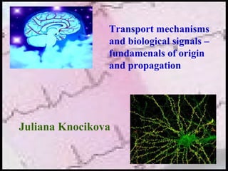
  • 1. Juliana Knocikova Transport mechanisms and biological signals – fundamenals of origin and propagation
  • 2. The nervous system The nervous system serves three functions 1. Senses changes both in and outside the body (the sensory function) 2. Interprets and explains the changes (the integrative function) 3. Responds to the interpretation by making muscles interact and glands secrete hormones or other chemicals into the bloodstream (the motor function). How do nerve impulses start? Several types of receptors initiate nerve impulses in senzory neurons as a result of: • touch • pressure • stretching • sound waves • motion Excitable cells – neurons and muscle cells
  • 3. • dendrites – collect and gather all informations •Soma – cell body (contains own DNA) - decision for action potential (AP) happend there, neurotransmitters are producing there • axon hillock – point at which all information are cumulated and AP is initiated • axons – carries all information down a neuron and is controlled by the soma (coated by myelin layer) Basic constitution of the nerve cell - neuron The connection between neurons – synapse
  • 4. •Every cell has a resting membrane potential (RMP) •Intracellular space is negatively charged under comparison with extracellular space (typically - 40-90 mV) Resting membrane potential and its level is a result of: 1. Unbalanced distribution of positive and negative ions inside and outside the cell 2. Semipermeable membrane (different permeability for different ions) K : Cl : Na = 100 : 45 : 4 3. PrĂ­tomnosti tzv. Sodium-potassium exchange pump in cell membrane Basic resting membrane potential (RMP)
  • 5. Neurones send messages electrochemically; this means that chemicals (ions) transported by the ion channels in the membrane cause an electrical impulse. Resting membrane potential Ion Concentration inside cell [mmol.dm-3 ] Concentration outside cell [mmol. dm-3 ] K+ 150 2.5 Na+ 15 145 Cl- 9 101 Rest – condition under which the neurone does not send a signal These ions do not move down their concentration gradient K+ ions do not move out of the neurone due to positive charges outside the membrane. This repels the movement of any more K+ ions out of the cell. Cl- ions do not move into the cell because of the negatively charged protein molecules (that cannot cross the surface membrane) repel them. Ion pumps generates concentration difference of ions
  • 6. Resting membrane potential [ ] [ ] [ ] [ ]e ie Cl Cl iK K − − + + = Donnan’s balance – conjuction of concentrations related to the anions and cations on the one side of the membrane is the same as on the other side of the membrane [ ]eK + [ ]eCl− [ ]iK + [ ]iCl− =⇒ [ ] [ ]i e K K K F TR U + + =+ ln. . Nernst’s equation – magnitude of the resting membrane potential equals approximately to the equilibrium point of the pottasium iones Equilibrium point for pottasium iones depends on: R - universal gas constant T - thermodynamic temperature F - Faraday’ constant - concentration of pottasium iones in extracellulary space - concentration of pottasium iones in intracellulary space [ ]eK + [ ]iK +
  • 7. Resting membrane potential [ ] [ ] [ ] [ ] [ ] [ ]eCliNaiK iCleNaeK K ClPNaPKP ClPNaPKP F TR U −++ −++ −++ −++ + ++ ++ = ln. . Goldman’s equation – rather expression of the resting mebrane potential magnitude because of sodium, pottasium and chloride iones P K+ , P Na+ , P Cl- - coefficients of membrane permeability for pre K+, Na+, Cl- [K+ ], [Na+ ], [Cl- ] - concentrations +
  • 8. •Ion pump, kind of active transport (energy feed is needed) •The Na+K+ATPase pump uses energy to move 3Na+ out and 2K+ into neuron The Sodium – Potassium Pump (Na+K+ATPase) Ion pumps And a result of Na+K+ATPase pump and the leak channels cause a stable imbalance of Na+ and K+ ions across the membrane. The imbalance in voltage causes a potential difference across the cell membrane - called the resting potential. Resting membrane potential is always negative (-70 mV in humans).
  • 9. • (Na+, K+)-ATPase, in plasma membranes of most animal cells, is an antiport ion pump. It catalyzes ATP-dependent transport of Na+ out of a cell in exchange for K+ entering the cell. • (H+, K+)-ATPase, involved in acid secretion in the stomach, is an antiport pump. It catalyzes ATP-dependent transport of H+ out of the gastric parietal cell (toward the stomach lumen) in exchange for K+ entering the cell. • Ca++-ATPases, in endoplasmic reticulum (ER) and plasma membranes of many cells, catalyze ATP-dependent transport of Ca+ + away from the cytosol, into the ER lumen or out of the cell. Some evidence indicates that these pumps are antiporters, transporting protons in the opposite direction. Ca++-ATPase pumps function to keep cytosolic [Ca++] low, allowing Ca++ to serve as a signal. Ion pumps
  • 10. Active transport Active transport is the transport of biochemicals, and other atomic/molecular substances, across the membranes. Unlike passive transport, this process requires the expenditure of cellular energy to move molecules against their concentration gradient. Primary transport - energy from hydrolysis of ATP is directly coupled to the movement of a specific substance across a membrane independent of any other species. Secondary active transport, the required energy is derived from energy stored in the form of concentration differences in a second solute. Typically, the concentration gradient of the second solute was created by primary active transport, and the diffusion of the second solute across the membrane drives secondary active transport of the first solute.
  • 12. The action potential (AP) Occures when neurone send information down the axon, reversion of the membrane potential is caused because of stimulus application. AP has two main phases: depolarization - a stimulus cause the membrane potential to change. This change is detected by the voltage-gated (sodium, potassium) ion channels. The sodium channels open for 0.5ms when the potential reaches threshold level as an effect of the voltage change. This results in sodium ions entering inside and making the inside of the cell more positive. The normal voltage polarity (negative inside) is reversed (becomes positive inside the cell).
  • 13. Repolarization - at a certain point, the depolarisation of the membrane causes the sodium channels to close. As a result the potassium channels open for 0.5ms, causing potassium ions to rush out, making the inside more negative again The action potential (AP) A slight ‘overshoot’ in the movement of potassium ions (called hyperpolarisation) became marked. The resting membrane potential is restored by the Na+K+ATPase pump The original polarity is restored
  • 14.
  • 15. The action potential (AP) “All or nothing” low – only if the stimulus causes enough sodium ions to enter inside the cell to change the membrane potential to a certain threshold level, AP is created. It is necessary to achieve this threshold. Absolute refractory period • during AP, the second stimulus will not cause a new AP • Na+ channels are open or recovering Relative refractory period • second AP may be created only if the stimulus has to be great, it is more difficult to generate it • occurs when the membrane is hyperpolarised (-80mV), where the K+ channels are open
  • 16. The action potential (AP) Channel2.swf
  • 17. Propagation of the action potential 3 processes of the AP propagation are possible: • mechanism of local current – AP is moved along an axon automatically. The local reversal of the membrane potential is detected by the surrounding voltage-gated ion channels, which open when the potential changes enough. • saltatory propagation – myelin sheath is interruped by nodes of Ranvier and behave between them as a electrical insulator. AP jump from one node to another. This increases the speed of propagation (from 1m/s in unmyelinated neurons up to 100 m/s in myelinated neurons) • synaptic propagation – see the next chapter
  • 18. propagation of the impulses – comparison betwen healthy and damaged nerve Propagation of the action potential Factrors which may cause affection of the AP velocity: • temperature higher temperature = faster velocity • axon diameter larger diameter = faster velocity • myelin sheath – saltatory propagation of AP is faster
  • 19. The synapse – active connection between neurons Electrical - direct connection between neurons Chemical – there is a space between neurons. Neurons communicate by neurotransmitters (chemical molecules) Excitatory - increase probability of impulse generation Inhibitory - decrease probability of impulse propagation Synaptic propagation Propagation of the action potential
  • 20. 1. The action potential activates a calcium channel and Ca++ diffuses into the neuron. 2. This Ca++ causes vesicles to fuse with the cell membrane. Through exocytosis, neurotransmitters (chemicals) are released into the synapse. 3. These neurotransmitters diffuse across the synapse and bind to receptors on another neuron. This causes special Na+ channels to open and an action potential is initiated in the next neuron. Synaptic propagation of AP between two neurons
  • 21. The synapse Excitatory and inhibitory synapse
  • 22. How many synapses are in one neuron? 1,000 to 10,000
  • 23. Strength duration curve Chronaxie - The minimum interval of time necessary to electrically stimulate a muscle or nerve fiber, using twice the minimum current needed to elicit a threshold response. Reobase - The minimal strength of an electrical stimulus that is able to cause excitation of a tissue. bireflexarc.swf
  • 24. The action potential (AP) Single unit activity Multipotential
  • 25. Electrical activity of the muscle Human body contains over 400 skeletal muscles, 40-50% of total body weight Functions of skeletal muscle • force production for locomotion and breathing • force production for postural support • heat production during cold stress Microstructure: Sarcolemma - muscle cell membrane Myofibrils - threadlike strands within muscle fibers • Actin (thin filament) Troponin Tropomyosin • Myosin (thick filament)
  • 27. The light areas are called the I-BANDS and the darker areas the A- BANDS. Near the center of each I-BAND is a thin dark line called the Z-LINE (or Z-membrane in the drawing below). The Z-LINE is where adjacent sarcomeres come together and the thin myofilaments of adjacent sarcomeres overlap slightly. A sarcomere can be defined as the area between Z-lines. Electrical activity of the muscle
  • 29. Electrical activity of the muscle 1. The impulse is transferred from a neuron to the SARCOLEMMA of a muscle cell. 2. The impulse travels along the SARCOLEMMA and T-TUBULES. From the T-TUBULES, the impulse passes to the SARCOPLASMIC RETICULUM (SR). 3. The calcium gates in the membrane of the SR open. As a result, CALCIUM diffuses out of the SR and among the myofilaments. 4. Calcium fills the sites in the TROPONIN molecules. This alters the shape and position of the TROPONIN which causes movement of the attached TROPOMYOSIN molecule. 5. Movement of TROPOMYOSIN permits the myosin head to contact ACTIN. 6. Contact with ACTIN causes the myosin head to swivel.
  • 30. Electrical activity of the muscle 7. During the swivel, the myosin head is attached to ACTIN. When the HEAD swivels it pulls the ACTIN (and, therefore, the entire thin myofilament) forward. Many MYOSIN HEADS are swivelling simultaneously, or nearly so, and their collective efforts are enough to pull the entire thin myofilament). 8.At the end of the swivel, ATP fits into the binding site on the cross-bridge and this breaks the bond between the cross-bridge (myosin) and actin. The myosin head then swivels back. As it swivels back, the ATP breaks down to ADP and P and the cross- bridge again binds to an actin molecule. 9. As a result, the HEAD is once again bound firmly to ACTIN. Because the HEAD was not attached to actin when it swivelled back, the HEAD will bind to a different ACTIN molecule (i.e., one further back on the thin myofilament). Once the HEAD is attached to ACTIN, the cross-bridge again swivels, so step 7 is repeated
  • 31. Electrical activity of the muscle Types of contractions: 1 - isotonic - tension or force generated by the muscle is greater than the load and the muscle shortens 2 - isometric - load is greater than the tension or force generated by the muscle & the muscle does not shorten Twitch - the response of a skeletal muscle to a single stimulation: latent period - no change in length; time during which impulse is traveling along sarcolemma to sarcoplasmic reticulum, calcium is being released, and so on (in other words, muscle cannot contract instantaneously!) contraction period - tension increases (cross-bridges are swivelling) relaxation period - muscle relaxes (tension decreases) and tends to return to its original length
  • 32. Electrical activity of the heart muscle electrical activity initiates the heart muscle to contract causing the opening and closing of the valves to deliver the blood around the body and back to the heart and lungs. Myocard – spetial type of muscle – two muscle cells are conneted by intercalar discs. The mechanical connection enables transport of the impulses Plateau phase – result of permeability for Ca ++ ions increasing, AP in ventricle (0.3s)
  • 33. Electrical activity of the heart 1. sinoatrial node - an area of nerve cells known as the pacemaker situated in the upper wall of the right atrium. Impulese are initiated there 2. electrical impulse is picked up by a further electrical node called the atrioventricular node, which is situated in the lower part of the right atrium close to the valves between the upper and lower chambers of the heart 3. impulses flow down the central wall of the heart (called the septum), between the two ventricles and into the left and right bundle branches via the electrical conductive tissue to carry the impulse over each of the ventricles. 1 Sinoatrial node (Pacemaker) 2 Atrioventricular node 3 Atrioventricular Bundle (Bundle of Hiss) 4 Left & Right Bundle branches 5 Bundle Branches (branches of Purkyne)
  • 34. Electrical activity of the heart 0078a.swf
  • 36. Electrical activity of the heart An electrocardiogram (ECG) represents the electrical current moving through the heart during a heartbeat. P wave – depolarization of the atria QRS complex – depolarization of the ventricles T wave – repolarization of the ventricles
  • 37. Biological signal - properties May be defined as a carrier of informations, which are expressed by its properties Basic properties of biological signal: • wavelength • amplitude • frequency
  • 38. Biological signal - properties Time and frequency domain
  • 39. Biological signal - properties Ventricle tachycardia (on the left) broken by the defibrilator charge, normal rhythm on the right side
  • 40. for attention Š 2007 Juliana Knocikova