Eyestar 900 brochure
•
0 likes•44 views

The Eyestar 900 features swept-source technology, enabling precise measurement, as well as topographic assessment of the front and back corneal surface and the anterior chamber, including the lens, as well as imaging of all these structures. It also includes cornea-to-retina biometry of the entire eye.

Recommended
More Related Content
Similar to Eyestar 900 brochure
Similar to Eyestar 900 brochure (20)
More from Haag-Streit UK (HS-UK)
More from Haag-Streit UK (HS-UK) (20)
Recently uploaded
Recently uploaded (20)
Eyestar 900 brochure
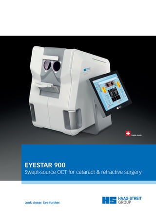
- 1. EYESTAR 900 Swept-source OCT for cataract & refractive surgery
- 2. 02 | 03 EYESTAR 900 One fully-automated device, for both cataract & refractive surgery The growing demand for improved outcomes, both in cataract and refractive surgery, has raised the bar for anterior chamber diagnostics. To meet the needs of the ambitious surgeon in a busy practice, the Eyestar 900 offers versatile diagnostic tools for both cataract and refractive surgery, in one fully-automated combination device. Its cutting-edge swept-source OCT technology enables precise measure- ment of the entire eye and offers comprehensive data and high-quality images of the anterior chamber. It allows for cornea-to-retina biometry, as well as topographic assessment of the anterior and posterior corneal surface and visualisation of the anterior chamber, including the lens. The Eyestar 900 features excellent cataract penetration. It boasts well- established dual zone reflective keratometry, specifically for cataract application, providing precise keratometry and astigmatism measurement compatible with existing IOL formulas. Equipped with this information, the eye care specialist may achieve excellent outcomes in cataract surgery, accurately diagnose diseases, and simply document the eye status. Furthermore, it also offers Class-A topography to 12 mm diameter of the anterior and posterior surface of the cornea, Keratoconus screening* and OCT imaging of the anterior segment, including the crystalline lens and the chamber angle. This allows the user to easily verify any measurement and to identify anatomical anomalies that may interfere with planned surgical procedures. Full automation enables fast data acquisition in typically under 40 seconds for both eyes, allowing for easy delegation, and thus improving workflow ef- ficiency in a busy practice. In sum, the Eyestar 900 is a swept-source OCT device designed specifically for the ambitious surgeon wanting to measure, diagnose, plan and image the eye for improved outcomes, and more confidence. * Available in a future EyeSuite software release
- 3. Identify anatomical anomalies with confidence Imaging of the entire anterior chamber, topograph- ic maps of the anterior and posterior cornea and pachymetry maps enable users to improve their surgical plan and review patients’ suitability for specific interventions, such as toric, multifocal IOL or refractive surgery. Precise data, for excellent surgical outcomes The Eyestar 900’s swept-source OCT technology provides precise measurements of the entire eye, from the cornea to the retina and imaging of the anterior chamber including the lens. This provides the basis for reliable diagnosis and accurate surgical planning. Fully-automated, for easy delegation The fully-automated measurement process allows for easy delegation, optimises workflow and enables the user to measure both eyes in typically under 40 seconds.
- 4. 04 | 05 EYESTAR 900 Diagnose, plan, predict & control The Eyestar 900 is based on future-proof technology: swept- source OCT. It provides the user with precise measurements, comprehensive topography and pachymetry maps, complete cornea-to-retina biometry and high quality, detailed cross- sectional eye images. Acquiring all this data simultaneously, in a fast, fully-automated measurement process results in excellent data quality, usability and patient comfort. The Eyestar 900 also uses unique, patent-protected Mandala scan technology, which is designed for highly precise data acquisition. Unlike classic radial or line scans, that scan any point only once (besides the apex), trajectories of the Mandala scan are aligned in an interwoven and highly dense pattern, both in the centre and in the periphery. This, combined with the OCT inherent motion compensation, results in a detailed and highly precise three- dimensional data set. This comprehensive information set supports the eye care specialist to accurately diagnose a patient, plan surgical proce- dures, predict outcomes and control the intervention efficacy of cataract, refractive and anterior chamber surgeries. TYPICAL RADIAL SCAN UNIQUE MANDALA SCAN
- 5. TOPOGRAPHICAL DATA Tear film independent topography… Eyestar 900 provides corneal topography in compliance with Class A- topographer standards. The maps of the Cataract Suite cover 7.5 mm and the maps of the Anterior Chamber Suite up to 12 mm in diameter, and provide comprehensive information of the anterior and posterior surface of the cornea, and pachymetry. The Anterior Chamber Suite offers additional tools like trend/progression and difference views for more detailed analysis of the topographic data collected. MANDALA SCAN TECHNOLOGY Makes rescanning obsolete Due to the highly dense Mandala scan pattern, the user can create virtual radial scans or line scan stacks, as well as individual B-scans, any time after the data acquisition, and at any location of the 18mm diameter OCT scan volume previously acquired. This unique feature eliminates the need for time-consuming rescans, if a new cross-section is required. SWEPT-SOURCE OCT TECHNOLOGY Quantify what you see… The refraction-corrected B-scan OCT imaging of the anterior chamber allows visual anatomy assessment. In addition, the software also determines the three-dimensional lens orientation and location and displays the respective information and numerical data in an intuitive detailed results screen, featuring the B-scan cross-section in the direction of the maximum lens tilt. TOPOGRAPHICAL DATA SWEPT-SOURCE OCT TECHNOLOGY MANDALA SCAN TECHNOLOGY
- 6. COMPREHENSIVE OVERVIEW OF DATA FULLY-AUTOMATED MEASUREMENT PROCESS GRAPHIC IOL PLANNING & LATEST GENERATION IOL CALCULATION VISION SIMULATION & ZERNIKE WAVE FRONT ANALYSIS 06 | 07 Cataract Suite Optimized workflow, fewer surprises The Eyestar 900’s Cataract Suite enables acquisition of all mea- surement data necessary for state-of-the-art cataract planning in an optimized, fully-automated measurement workflow. The binocular measurement is typically completed in under 40 seconds, from the time the patient is asked to look into the device to the finalization of the measurements. In this short time, all data useful for planning of spherical, toric, multifocal and phakic IOL is collected. The result overview presents all data from axial measurements to topography maps and 16 B-scans of the anterior chamber in an intuitive display. All data can be reviewed on detail screens. Measurements taken include axial measurements of all eye compartments, corneal front and back topography as well as keratometry, B-scan imaging of the anterior chamber, including the lens, and assessment of lens tilt and decentration, as well as Zernike analysis and vision simulation.
- 7. SWEPT-SOURCE OCT TECHNOLOGY Detailed information, excellent outcomes Biometry based on swept-source OCT provides the user with much more than just axial length measurements and keratometry. Detailed information of the cornea front and back surface have the potential to significant- ly improve cataract planning for astigmatic and post-refractive patients. The topography maps allow the surgeon to screen for signs of corneal pathol- ogies, that may limit the patient’s post-cataract surgery visual potential. In toric candidates, the symmetry and regularity of the astigmatism on the cornea front and back are readily available, allowing a thorough judgement of the patient’s eligibility for a premium IOL. MORE DATA FOR EXCELLENT DIAGNOSIS & OUTCOMES Measure, visualize & understand Anterior chamber B-scan imaging, including the lens, and identification of lens tilt and decentration are beneficial for patient education, particularly when it is a question of premium toric or multifocal IOL. Vision simulation and Zernike analysis support the surgeon to set the patients expactaion right and helps to choos the optimum procedure. FULLY-AUTOMATED ACQUISITION PROCESS Precise & efficient Fast and reliable measurement acquisition is the key to efficiently achieving excellent outcomes. Providing a fully-automated and quick acquisition pro- cess enables easy delegation, improved patient comfort and compliance. Built-in tear film quality assessment leads to highly precise keratometry, complemented by swept-source OCT based laser precision biometry, topo- graphy, pachymetry and tomography of the entire eye. MORE DATA FOR EXCELLENT DIAGNOSIS & OUTCOMES FULLY-AUTOMATED ACQUISITION PROCESS SWEPT-SOURCE OCT TECHNOLOGY
- 8. 08 | 09 EyeSuite IOL The ultimate planning platform for any IOL EyeSuite IOL features a comprehensive set of state-of-the-art IOL calculation formulas for any IOL-type or corneal condition in cataract surgery. It includes the latest generation calculation methods – Hill-RBF, Barrett and Olsen – for spherical, as well as toric, IOL calculations. One of the key features of these methods is the use of biometry data beyond axial length (AL) and keratometry (K). Central corneal thickness (CCT), anterior chamber depth (ACD), lens thickness (LT), posterior keratometry (simPK) and white-to-white (WtW) are additional parameters that improve prediction accuracy. Extreme and unusual eyes, in particular, will benefit from the additional information. Toric calculations incorporate the front and back corneal surface for increased accuracy in calculating the IOL spherical equivalent, cylinder power and orientation. This information is then displayed in an intuitive graphic planning tool, enabling accurate transfer of the plan to the operating room. For post-refractive cases, EyeSuite IOL again features a complete set of state-of-the-art calculation methods, such as Barrett’s True K and True K Toric, which both incorporate measurement of the posterior cornea, the Masket formula or Shammas No-History method.
- 9. HILL-RBF METHOD Certainty Hill-RBF is a purely data-driven IOL calculation technique incorporating pattern recognition and sophisticated data interpolation. It features a boundary model, informing the user of the calculation’s reliability. Hill-RBF performs equally well on long, normal and short eyes. It clearly outperforms second- and third-generation formulas. Paired with the Abulafia-Koch method, Hill-RBF is available for spherical and toric IOL calculations. Unlike static theoretical formulas, the Hill-RBF method is an ongoing project and is continuously updated for even better overall depth of accuracy. FEWER REFRACTIVE SURPRISES Identify the unusual Having B-scan swept-source OCT images of the entire anterior chamber available at the time of measurement allows visual identification of unusual tilt and decentration of the crystalline lens. Furthermore, it facilitates easy monitoring of automated gate positions for the biometry measurements. All this additional information enables further minimization of refractive surprises. HILL-RBF METHOD FEWER REFRACTIVE SURPRISES EYESUITE SOFTWARE Flexible integration The EyeSuite software is designed for optimum patient flow in busy prac- tices. The easy-to-use Eyestar 900 is fully networkable with Haag-Streit devices and your own practice network. The EyeSuite script language or command line interface works fluently with almost any EMR system, and supports standard interfaces like DICOM for excellent compatibility. EYESUITE SOFTWARE
- 10. 10 | 11 Anterior Chamber Suite Comprehensive data for thorough diagnosis, clear visualization for more understanding The Eyestar 900 Anterior Chamber Suite offers precise measure- ments, comprehensive data analysis, and excellent images of the anterior chamber. Thanks to its cutting-edge swept source OCT technology, the Anterior Chamber Suite provides class A-topography up to 12 mm diameter of the anterior and posterior surface of the cornea, and OCT imaging of the anterior segment, including the lens and the chamber angle, with up to 18mm diameter coverage. Its corneal topography feature set includes difference and trend views for maps and indices, as well as sophisticated screening aids for corneal ectasia. The integrated Belin ABCD grading system provides the user with intuitive data for efficient keratoconus classification. A user definable progression display enables intuitive follow-up on any measurement parameter available with the Eyestar. Other tools include Zernike wavefront analysis of the cornea and simu- lation of visual acuity for patient education. Due to image acquisition using the cutting-edge, patent-protected Mandala scan technology, virtual B-scans may be created any time and anywhere in the already acquired volume, minimizing the need for the rescanning of patients to visualize details. FOUR-IN-ONE DIFFERENCE MAP – THREE TIME POINTS CORNEAL ECTASIA DISPLAY INCLUDING BELIN ABCD GRADING SYSTEM CROSS-SECTIONS AT INDIVIDUAL LOCATIONS
- 11. HIGH QUALITY IMAGES DENSE SCANS Visualize key structures The Eyestar 900 Anterior Chamber Suite offers high quality 18 mm diame- ter images for visual inspection of key structures, such as lens position, ICL vault* or chamber angle*. The basic measurement tool kit supports manual point-to-point distance, angle and area measurements in the refraction- corrected radial scans. Due to the highly dense Mandala scan pattern, the user can create virtual, radial and line scan stacks, as well as individual B-scans, any time after the data acquisition and at any location of the 18 mm OCT scan volume previously acquired. FUTURE PROOF Enhanced features, increased functionality An expandable device, the Eyestar 900 will boast increased functionality in the future. Recent EyeSuite software releases include integrated corneal ectasia screening aids with dedicated summary displays, including the Belin ABCD grading system. Other planned software releases include ICL vault analysis* and chamber angle assessment*. CONFIDENCE THROUGH OCT TECHNOLOGY Comprehensive analysis of the anterior chamber The swept source OCT-based topography of the Eyestar 900 covers up to 12 mm diameter on the anterior cornea and provides highly accurate assessment of the cornea front and back, as well as data on its thickness. Intuitive displays, such as difference, trend and corneal ectasia displays, support the user in the diagnostic process. FUTURE PROOF Platzhalter-Bild CONFIDENCE THROUGH OCT TECHNOLOGY *New future modules and suites are currently being developed. HIGH QUALITY IMAGES DENSE SCANS
- 12. 12 | 13 Intuitive efficient Ergonomics for patient comfort precision Precise measurement data, intuitive map information and OCT imaging of the anterior chamber is essential in efficiently diag- nosing and treating patients. The combination of swept-source OCT, reflective keratometry, high-resolution imaging and the fully- automated measurement allow efficient, patient-friendly, simultaneous acquisition of all this information in a single device. Patient comfort and short examination time are key contributors to optimized data quality. Moreover, data collection delegation is crucial to efficiently running a busy practice. Taking this into account, Haag-Streit developed a unique fully-automated measurement process for the Eyestar 900, minimizing the user’s learning curve and optimizing patient comfort. Platzhalter-Bild
- 13. INTUITIVE USER INTERFACE Optimized workflow The familiar look-and-feel of the touch-screen optimized EyeSuite software, used in all Haag-Streit devices, enables efficient interaction and improved adaptation. Like any Haag-Streit device, the Eyestar 900 is easy to integrate into almost any practice management system. FLEXIBLE SPACE-SAVING Fits any room The Eyestar 900 touch screen can be mounted on either side of the device, or even on its back. In combination with the all-in-one device’s small footprint, this makes it a space-saver that will fit in any examination room. FULLY-AUTOMATED ACQUISITION PROCESS Patient compliance efficacy Patients, especially the elderly, tend to tire quickly during an eye exam, impairing optimal data collection. The fully-automated data acquisition process and its fast measurement with simultaneous data recording lead to excellent patient comfort and thus improved cooperation, which has a positive effect on measurement quality. FULLY-AUTOMATED ACQUISITION PROCESS INTUITIVE USER INTERFACE FLEXIBLE SPACE-SAVING
- 14. Swept-source OCT visualizes details in high definition INTRA CORNEAL RING SEGMENTS (INTACS) CORNEAL TRANSPLANT DMEK PRE-OP IOL DMEK POST-OP IOL
- 15. Technical specifications Eyestar 900 Technology Measurement variables modes Swept-source OCT Wave length 1060nm Scan Speed 30kHz Topography, Imaging, Measurement Dual Zone Keratometry Infrared LED 850nm Measurement points 32 Anterior Keratometry High resolution imaging Resolution Full HD 1080p Color and Infrared Enface eye imaging, Measurement Laser safety Class 1 laser product Supported EMR interfaces DICOM EyeSuite script language GDT Eyesuite command line interface The above-mentioned measurement ranges are based on the standard settings of the device for automatic measurement and analysis. Contraindication: There are no known contraindications. Intended purpose The Eyestar 900 is a non-invasive, non-contact biometer used for obtaining the following information: � Corneal shapes � Anterior chamber dimensions � Axial eye length � Front- and cross-sectional images � Lens dimension and position Corneal Thickness CCT Measurement range 300–800μm Display resolution 1μm Anterior chamber depth ACD Measurement range 1.8–6.3mm Display resolution 0.01mm Lens thickness LT Measurement range 0.5–6.5mm Display resolution 0.01mm Axial length AL Measurement range 14–38mm Display resolution 0.01mm Topography Topography system Type A Map display 7.5mm Anterior Keratometry K Measurement range 32.1–67.5dpt Display resolution 0.01dpt Posterior simulated Keratometry SimPK Measurement range 3.9–9.5dpt Display resolution 0.01dpt White to white WTW Measurement range 7–16mm Display resolution 0.01mm Pupillometry PD Measurement range 2–13mm Onboard IOL Calculation methods Hill-RBF Hill-RBF/Abulafia-Koch for toric IOL Barrett Universal 2 Barrett Toric Calculator Barrett True K and True K Toric Olsen and Olsen Toric Haigis Hoffer Q Holladay 1 SRK/T and SRK II Masket and Modified Masket Shammas No-History IOL calculation data interfaces Holladay IOL Consultant PhacoOptics Measurements Dimensions: 480 × 560 × 460 mm Weight: 31.0 kg Topography Topography system Type A Map display Up to 12mm Maps/Displays Anterior corneal topography maps Posterior corneal topography maps Pachymetry maps Difference maps Progression (timeline) displays Corneal ectasia display Belin ABCD grading OCT Imaging Area/Volume Up to 18mm on anterior cornea converging to the retina Scans Patented Mandala scan technology Radial scans Line scan stacks Custom single line scan Included in future software release Total Keratometric Power ICL vault analysis Chamber angle Cataract Suite Anteror Chamber Suite
- 16. ©HAAG-STREIT AG, 3098 Koeniz, Switzerland 4. Edition / 2023 – 01 HS-Art. No. 1511.7220695.02040 0297 HAAG-STREIT AG Gartenstadtstrasse 10 3098 Koeniz Switzerland Phone +41 31 978 01 11 Fax +41 31 978 02 82 info@haag-streit.com www.haag-streit.com