1. Home Exam Answers:
1. How do you expect the charge distribution of a protein to change between pH 0 and 14? That is,
describe qualitatively how (and why) the charge varies as a function of pH.
Answer (1).
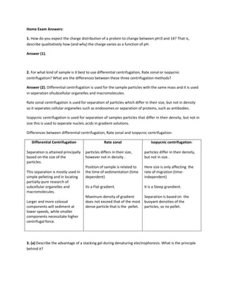
2. For what kind of sample is it best to use differential centrifugation, Rate zonal or isopycnic
centrifugation? What are the differences between these three centrifugation methods?
Answer (2). Differential centrifugation is used for the sample particles with the same mass and it is used
in seperation ofsubcellular organelles and macromolecules.
Rate zonal centrifugation is used for separation of particles which differ in their size, but not in density
so it seperates cellular organelles such as endosomes or separation of proteins, such as antibodies.
Isopycnic centrifugation is used for separation of samples particles that differ in their density, but not in
size this is used to seperate nucleic acids in gradient solutions.
Differences between differential centrifugation, Rate zonal and Isopycnic centrifugation:
Differential Centrifugation Rate zonal Isopycnic centrifugation:
Separation is attained principally particles differs in their size, particles differ in their density,
based on the size of the however not in density . but not in size .
particles.
Position of sample is related to Here size is only affecting the
This separation is mostly used in the time of sedimentation (time rate of migration (time-
simple pelleting and in locating dependent) independent)
partially-pure research of
subcellular organelles and Its a Flat gradient. It is a Steep grandient.
macromolecules.
Maximum density of gradient Separation is based on the
Larger and more colossal does not exceed that of the most buoyant densities of the
components will sediment at dense particle that is the pellet. particles, so no pellet.
lower speeds, while smaller
components necessitate higher
centrifugal force.
3. (a) Describe the advantage of a stacking gel during denaturing electrophoresis. What is the principle
behind it?
2. Answer 3. (a)The main advantage is that the proteins electrophorese quickly through the stacking gel
and "pile" at the border between the two gels, before they move in the gels. This upturns the firmness
of the proteins before they enter the running gel and increases resolution. So when the electrophoresis
is started, ions from the higher reservoir enter the stacking gel since at that pH they have a middling
fractional negative charge. The stacking gel buffer ions continue moving in the stacking gel, but when
the ions enters the pH of the stacking gel, they are converted into zwitterions with a net charge of zero,
and therefore stop motion toward the anode. The electrical resistance in the stacking gel then increases
since the number of ions moving through the stacking gel decreases. To maintain constant current
throughout the circuit, there will be a confined increase in the voltage in the stacking gel (from Ohms
Law, V=iR). This will cause the proteins to travel quickly and all stack in a single, very thin disc right
behind the ions in the stacking gel (which are in front because they have the highest charge density and
electrophoretic mobility of any ion in the stacking gel). The proteins will not pass the ions since if they
did, they would straightway slow down since they would no longer be in an area of reduced charged
carriers and higher voltage. At the stacking gel/running gel interface, all the proteins can’t migrate at the
same speed, owing to sieving effects of the more concentrated gel, and hence will be separated in the
running gel.
(b) Describe the principle of a 2D-gel and discuss various staining methods for gels (sensitivity,
advantages and disadvantages).
Answer 3. (b) The main principle of this technique is that it separates the proteins according to two
properties in two separate steps: the first-dimension step, isoelectric focusing (IEF), separates proteins
according to their isoelectric points (pI); the second-dimension step, SDS-polyacrylamide gel
electrophoresis (SDS-PAGE), separates proteins according to their molecular weights (Mr, relative
molecular weight). Every spot on the consequential two-dimensional array resembles to a single protein
species in the sample. Many different proteins can be separated, and info such as the protein pI, the
apparent molecular weight, and the amount of each protein is achieved.
Various Staining methods for Gels:
1. Coomassie Brilliant Blue (CBB): It is a quick and strong way of envisaging proteins in a gel,
however they generally lack in sensitivity.CBB staining procedure is relatively quick and very
easy.Since there is presence of alcohol some of the proteins release the dye during the
background destaining process.
2. Silver staining is the most sensitive non-radioactive method (below 1 ng). Silver staining detects
the proteins mainly on the gel surface .Silver staining is a complex, multi-step process using
several reagents for which quality is critical.For the reason that it is not an endpoint procedure,
the staining intensities can differ from gel to gel.
3. Fluorescent stains uses fluorescence as a detection method and the staining is an endpoint
staining and is thus highly reproducible. Fluorescent protein gel stains are usually well-suited
with consequent protein analysis.
4. (a) Mention two important special features of ESI.
Answer 4. (a) 1. The ability to analyze the compounds from aqueous or organic solutions by coupling
with Liquid chromatography.
3. 2. It can induce the fragmentation of small peptides typically below 3000Da thus allowing the
sequencing information to be obtained.
(b) Make a simple drawing of a MALDI-TOF mass spectrometer.
Answer 4. (b)
4. (c) What simple relation is used to determine molecular masses in MALDI-TOF mass spectrometry?
Answer 4. (c) MALDI-TOF mass spectrometers operate on the principle relation
K.E. = [mv2]/2 = zeEs
Where K.E. = kinetic energy; m = the mass of the ion; v = velocity of the ion; z = number of charges; e =
the charge on an electron in coulombs; E = electric field gradient; and s = the distance of the ion source
region.
d) What is the lowest detection limit of peptides in MALDI-TOF mass spectrometry?
e) Using peptide mapping-How would you determine the identity of a protein present in a mix with
other proteins?
Answer 4. (e). To identify the unidentified protein of interest in a mix the protein is first cleaved into
smaller peptides, whose absolute masses can be accurately measured with a mass spectrometer such as
MALDI-TOF or ESI-TOF. These masses are then related to either a database comprising of known protein
sequences or even the genome databases.
f) How can you use MALDI-TOF mass spectrometry to determine if a protein is phosphorylated or not on
a serine or a threonine amino acid?
4. Answer 4. (f).
(i) Degradation of the phosphoprotein into small peptides by specific enzymatic or chemical
reactions.
(ii) Identification of the phosphopeptides by -80 (or multiples of -80)-Da mass shifts in the mass
spectra after dephosphorylation with alkaline phosphatase.
(iii) Location of the phosphorylation sites by mass mapping.
(ref: Liao PC, Leykam J, Andrews PC, Gage DA, Allison J., Anal Biochem. 1994 May 15;219(1):9-20.)
5. You are trying to purify a Very Interesting Protein (VIP) with the help of ion-exchange
chromatography. After the purification step you analyse the fractions by SDS-PAGE and Western
Blotting. The Antibody identifies a protein of 22 kDa as VIP, but in the SDS gel you see that the fraction
also contains three contaminating proteins with molecular masses 6 kDa, 28 kDa, and 60 kDa. How
would you continue to purify your protein using chromatography? Discuss the advantages and
disadvantages of the possible techniques.
Answer 5.We can use the gel filtration chromatography here, since it separates the molecules according
to their size and shape. Hence our protein of interest can be sieved from the contaminating proteins
with molecular masses 6 kDa, 28 kDa, and 60 kDa and thus can be purified. To perform this
separation,all the proteins with known molecular weights are run on the column and their elution
volumes noted. If the elution volumes are then plotted against the log molecular weight of the
matching proteins, a straight line is obtained for the separation range of the gel being used.
The main advantage of gel filtration is that it’s the best method for separation of molecules differing in
molecular weight because:
1. It doesn’t rely on temperature, pH, ionic strength and buffer composition. So separation can be
carried out under any conditions.
2. There is very slight adsorption
3. The elution volume is associated to the molecular weight.
The disadvantages of the gel filtration chromatography:
1. The column should be precisely prepared to get optimum separation.
2. Any cracks or cutoffs in the column will interfere.
3. The size of the sample and the rate of buffer flow must be strictly controlled.
6. (a) Describe the principles of CD-spectroscopy.
5. Answer 6. (a).The physical principle of CD-spectroscopy involved states that the Chiral or asymmetric
molecules produce a CD spectrum because they absorb left and right handed polarized light to different
extents and thus are considered to be "optically active"
The difference between the absorption of left and right handed circularly-polarized light and is
measured as a function of wavelength. CD is measured as a quantity called mean residue ellipticity,
whose units are degrees-cm2/dmol.
6. (b) What type of information can be obtained from a far- and near- UV CD spectrum, respectively?
Answer 6. (b).
i. The near-UV CD spectrum has very high sensitivity for the native state of a protein. It can be
used as a fingerprint of the correctly folded conformation.
ii. The far –UV CD spectrum detects the occurrence of ordered secondary structure and can be
used to guess the secondary structure .CD spectra of natively unfolded proteins, being
measured in the far ultraviolet region, These values are discrete enough to differentiate
them from ordered proteins.
7. Interactions between biomolecules can be studied using several different techniques mentioned
during this course. Suppose you would like to study the interaction between a DNA-binding protein and
DNA. How would you characterize this interaction in detail? Describe the experiment(s) you have in
mind.
Answer 7.
1. Chromatin Immunoprecipitation (ChIP) Assays: The ChIP method can be used to screen
transcriptional regulation through histone modification (epigenetics) or transcription factor-DNA
binding interactions. The ChIP assay method permits study of DNA-Protein interactions in living
cells by considering the cells with formaldehyde or other crosslinking reagents in order to
stabilize the interactions for downstream purification and detection. It can be used to do
quantitative study when coupled with qPCR investigation.
2. DNA Electrophoretic Mobility Shift Assay (EMSA): It is used to investigate the proteins binding
to known DNA oligonucleotide probes and can be used to evaluate the ability of affinity or
specificity of the interface. The method is built on the statement that protein-DNA complexes
migrate more gently than free DNA molecules when exposed to non-denaturing polyacrylamide
or agarose gel electrophoresis.
3. Reporter Assays: It delivers a real-time in vivo read-out of translational movement for a
promoter of interest. Reporter genes are fusions of a target promoter DNA sequence and a
reporter gene DNA sequence. Here we can incorporate the reporters such as firefly luciferase,
Renilla luciferase or alkaline phosphatase. This technique detects real-time data and it’s a
potent tool for mutational analysis of promoters which is adaptable to high-throughput
screening.