
Recommended
More Related Content
What's hot
What's hot (20)
Similar to Lymphatic system
Similar to Lymphatic system (20)
Recently uploaded
Recently uploaded (20)
Lymphatic system
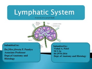
- 1. Dr.(Mrs.)Sweta P. Pandya Associate Professor Dept.of Anatomy and Histology Submitted to:
- 2. All body tissues are bathed in tissue fluid, consisting of the diffusible constituent of blood & waste material from cell. Some tissue fluid returnes to capillaries at their venous end the reminder defuses through the more permeable wall of the lymph capillaries, forming lymph. Introduction:- 2
- 3. The lymphatic system and its organs are widespread and scattered throughout the body. It functions to service almost every region of the body. Because the vessels of the lymphatic system span the entire body it becomes an easy portal for the spread of cancer and other diseases, which is why disorders and diseases of this system can be so devastating. 3
- 4. Cardiovascular & lymphatic system both are supply fluid flow in to the body. but bothe are deferent type of fluid. Lymphatic system does not having closed circuit & central pump like heart. 4
- 5. Lymph Lymph vessels Lymph node Lymph organ eg. Spleen, Thymus Diffuse lymphoid tissue eg. Tonsil It consist of... 5
- 6. 6
- 7. 7
- 8. The cardiovascular system pumps blood through its system but it cannot return all the fluid from the body cells. The lymph system picks up 60% of the fluid dropped off at the cellular level, at this point we are talking about interstial fluid, the IF picks up plasma and becomes tissue fluid. The tissue fluid is then picked up by lymph capillaries. The tissue fluid is called lymph The origin on lymph :- 8
- 9. 9
- 10. Selected lymph nodes of cattle 10
- 11. 11
- 12. The thoracic duct is the chief collecting trunk of the lymphatic system.It drains lymph from all parts of the body except the right forelimb, right side of head neck and thorax. It extends from about the level of the first lumbar vertebra forwards a little beyond the thoracic inlet.Its beginning is in the form of a reservoir, the cisterna or receptaculum chyli. This reservoir is formed by the meeting of the two lymphatic trunks, The lumbar trunk and gastro intestinal trunk. The cisterna chyli is in the form of elongated dilatation and lies in an oblique manner upwards and forwards a little above and in front of the level of the hiatus aorticus, between the first and second lumbar arteries and on the right face of the right crus of diaphragm. 12
- 13. It may also be found lying a little lower down or behind the right crus of the diaphragm when its position will be to the left of the posterior vena cava. The duct proceeds from the anterior aspect of the cisterna chyli is the thoracic duct, which passes forwards and slightly upwards, enters the thoracic cavity through the hiatus aorticus and crosses the right face of the terminal part of the thoracic aorta to gain its dorsal face. The thoracic duct from the cisterna chyli passes forwards on the right part of the dorsal face of the aorta covered by pleura and fat. The thoracic duct may remain single in some cases. But generally divide into two branches At the 6th or 7th dorsal vertebra it inclines ventrally crosses obliquely over the left face of the oesophagus, passes forward on the left face of the trachea to the thoracic inlet. The extra thoracic part is on the deep face of the left scalenus muscle. 13
- 14. It curves backwards and inwards under the bicarotid trunk and opens on the dorsal face of the left jugular confluence or anterior vena cava. In its course it receives efferents of the intercostal, mediastinal and bronchial glands and at thoracic inlet it receives the duct from the left posterior cervical and left tracheal duct. The chief tributaries are gastro intestinal trunks, efferents of the mediastinal lymph glands and in its termination, the efferents of the posterior cervical, left costocervical, prescapular and sternal lymph glands and left tracheal duct. 14
- 15. The lymph conveyed by the lymphatics from the hind limb, pelvic walls and organs, inguinal structures and the abdominal walls, finally reaches either the lumbar lymph glands placed in the sublumbar region or the efferents of these glands. The efferents leaving these lumbar glands from two lumbar trunks, which unite to form single lumbar trunk. This in its turn joins the gastrointestinal trunk to form the cisterna chyli. The lumbar trunk is formed by the union of the efferents from the iliac lymph glands. It also receives efferents from the lumbar and renal glands. 15 Lumbar trunk:-
- 16. The gastro-intestinal trunk is formed at the ventral face of the posterior vena cava just behind the dorsal border of the liver by the confluence of the radicles gastric and intestinal branches. The gastric trunk is on the left of the coeliac artery. The intestinal trunk follows on the anterior mesenteric artery. The trunk formed by the union of these two trunks, bends dorsally between the aorta and posterior vena cava and unites with the lumbar trunk. 16
- 17. Right lymphatic duct:- It is a short (usually absent) vessel, which drains the lymph from the right side of head, neck, right forelimb and thorax. When absent, several ducts open directly into thoracic duct or jugular confluence. It is formed by the union of right tracheal lymph duct and the efferents of right posterior cervical and right axillary glands. It lies on the deep face of the scalenus above terminal part of the right jugular vein. It opens into the anterior vena cava. 17
- 18. Tracheal duct:- These are right and left and are formed essentially by the confluence of efferent vessels from the atlantal lymph gland. They receive efferents of cervical, costocervical and prescapular glands. They pass along each side of the trachea and oesophagus in relation to the carotid arteries. The right duct joins the efferents of the posterior cervical and costocervical glands to form the right lymphatic duct or it may join the right common jugular vein. The left duct opens into the thoracic duct. 18
- 19. The secondary lymphatic organs are ◦ the spleen, ◦ the lymph nodes and ◦ other organs, such as the tonsils, Peyer patches, and the appendix. All the secondary organs are the places where lymphocytes encounter and bind with antigens, after which they proliferate and become actively engaged cells. 19
- 20. Pterygoid lymph node Parotid lymph node Mandibular lymph node Suprapharyngeal lymph node Peripharyngeal lymph node Atlantal lymph node 20
- 21. 21
- 22. Pterygoid lymph node:- It is a very small gland (absent in horse and dog) situated on the upper part of the lateral face of the pterygoid near the maxillary tuberosity. Afferents: Hard palate, gums etc. Efferents: To mandibular node. Protid lymph node:- It is situated superficially on the lateral face of the posterior portion of the masseter muscle partly under cover of the parotid salivary gland. Afferents: Muscles of the head, eye, eyelids, external ear, parotid salivary glands, lips, cheeks, anterior part of turbinates, frontal, nasal, malar and premaxillary bones. Efferents: To atlantal node. 22
- 23. Suprapharyngeal lymph node These are situated close together on the median line of the dorsal wall of the pharynx medial to the great cornu of hyoid bone. Afferents: Tongue, hard palate, pharynx, soft palate, sublingual, mandibular salivary glands, larynx, maxillary and palatine sinuses. Efferents: To tracheal lymph ducts. 23
- 24. Peripharyngeal lymph node:- It is small and is situated on the lateral aspect of the pharynx inseparably blended with the tonsil under the cover of mandibular salivary gland ventral to the carotid artery and atlantal node. Afferents: Salivary glands, hyoid muscles and efferents of parotid, mandibular and suprapharyngeal lymph nodes. Efferents: To the tracheal ducts. Atlantal lymph node:- It is situated below the wing of the atlas under cover of the mandibular salivary gland. Afferents: Hyoid and cervical muscles, salivary glands, tongue, efferents of parotid mandibular and suprapharyngeal nodes. Efferents: To tracheal duct. 24
- 25. 25 Intercostal lymph node Medastinal lymph node Bronchial lymph node Pulmonary lymph node Pericardial lymph node Diaphragmatic lymph node Sternal lymph node LYMPH NODES IN THE THORACIC CAVITY
- 26. INTERCOSTAL LYMPH NODE These are small glands situated in the upper part of the intercostal spaces (absent in dog) on the course of intercostal vessels. Afferents: Vertebrae, ribs, pleura, intercostal and spinal muscles. Efferents: To mediastinal lymph nodes 26
- 27. Anterior mediastinal lymph node:- These are situated in the anterior mediastinum along the oesophagus trachea anterior vena cava and common brachiocephalic trunk. Two or four are found ventral to the trachea and oesophagus. The largest often lies along the origin of the internal thoracic artery. Afferents: Pleura, trachea, oesophagus, thymus, pericardium, heart and lungs. Efferents: On the left to the thoracic duct on the right to the right lymphatic duct or costocervical nodes. 27
- 28. Middle mediastinal lymph node:- They are situated on the superior part of middle mediastinum over the oesophagus on the right side. Afferents: Pleura, pericardium, longus colli, vertebrae, etc. Efferents: To the efferent of the posterior mediastinal glands. Posterior mediastinal lymph node These lie along the oesophagus in the posterior mediastinum Afferents: Oesophagus, lungs, pericardium, mediastinum, diaphragm, liver and spleen. Efferents: To thoracic duct. 28
- 29. Dorsal mediastinal lymph node:- They are situated on either side of the aorta and between it and vertebrae. Afferents: Similar to intercostal and also from diaphragm pericardium mediastinum. Efferents: To the efferent from posterior mediastinal or to the thoracic duct. Ventral mediastinal lymph node:- These lie on the transversus thoracic muscle at the apex of the pericardium. Afferents: Pleura diaphragm pericardium and sternum. Efferents: To the anterior sternal node. 29
- 30. Bronchial lymph node:- They are right, left, middle and apical bronchial nodes. The right bronchial is situated on the right face of the trachea. The left bronchial is situated between the aortic arch and pulmonary artery. The middle bronchial at the bifurcation of the trachea. The apical bronchial is at the origin of the apical bronchus. Afferents: Lungs, oesophagus bronchi and heart. Efferents: On the left to the efferents of the posterior mediastinal gland on the right to the middle mediastinal and anterior mediastinal nodes. 30
- 31. Pulmonary lymph node:- They are inconstant and found in the chief bronchi in the lungs. Afferents: Lungs. Efferents: To bronchial and mediastinal nodes. 31 Pericardial lymph node:- They are small and inconstant and are found between the aortic arch and vena hemiazygos. Afferents: Pericardium. Efferents: To left bronchial node.
- 32. Diaphragmatic lymph node:- It is a small gland close to the foramen vena cava of diaphragm. Afferents: Diaphragm. Efferents: To posterior mediastinal nodes. Sternal lymph node:- These nodes are situated along the course of internal thoracic vessels. The largest of these is the anterior sternal nodes. Afferents: Intercostal muscles, pericardium, pectoral and abdominal muscles. Efferents: To the anterior mediastinal nodes. 32
- 33. Cervical lymph node:- These are placed along the course of the common carotid artery in the upper, middle and lower third of the neck as anterior, middle and posterior cervical. Afferents: The larynx, trachea, oesophagus, thyroid, thymus and muscles and bones of the neck. The posterior cervicals receive in addition the efferents from the prescapular and axillary nodes. Efferents: Anterior and middle cervicals go to the tracheal ducts and those from the posterior cervical on the left join the left tracheal duct or thoracic duct and on the right lymphatic duct. 33
- 34. Axillary lymph node:- It is placed on the medial face of teres major. Afferents: Muscles of shoulder, arm, forearm, latissimus dorsi, shoulder, elbow and carpal joints etc. Efferents: To posterior cervical lymph nodes. 34
- 35. 35 Costocervical lymph node:- It is situated in front of the first rib under the scalenus muscle (absent in the horse) dorsal to the carotid artery. Afferents: Trachea, oesophagus cervical, shoulder muscles and those of intercostal and anterior mediastinal nodes. Efferents: On the left to the thoracic duct and on the right to the right lymphatic duct. Prescapular lymph node:- It is situated at the anterior border of the supraspinatus about 10 to 13 cm, above the shoulder joint under cover of the omotransversarius and brachiocephalicus. Afferents: Skin and muscles of neck forelimb and thorax. Efferents: Similar to the preceding.
- 36. These lymph nodes are parietal and visceral in the abdomen and pelvis. The former lodges in the abdominal walls while the latter are situated on the viscera contained in these cavities. Visceral lymph nodes Gastric lymph nodes Mesenteric lymph nodes Parietal lymph nodes Precrural lymph nodes Sacral lymph node Ischiatic lymph node Superficial inguinal lymph node (in the male) Supramammary lymph node (in the female) Popliteal lymph node 36
- 37. These are (i) ruminal (ii) reticular (iii) omasal and (iv) abomasal nodes. Three or four glands situated on the right face of the atrium are the atrial nodes. They receive afferents from spleen, reticulum, rumen and the efferents of the other gastric nodes. Their efferents unite to form a common efferent, which in turn unites with efferents of the mesenteric nodes to form the mesenteric trunk. One or two nodes situated in the left longitudinal groove of the rumen are the left ruminal nodes.The efferents go to anterior ruminal nodes. A number of nodes in the right longitudinal groove of the rumen are the right ruminal nodes. The efferents go to the gastric trunk and anterior ruminal nodes. 37
- 38. A number of nodes situated in the anterior transverse groove are the anterior ruminal nodes. The efferents go to the atrial nodes. The nodes situated on the reticulo-omasal junction are the reticular glands. Their efferents go to atrial nodes. Nodes situated along the greater curvature of the omasum are omasal nodes. Their efferent goes to atrial nodes. Nodes situated along the lesser curvature of omasum and greater curvature of abomasum is abomasal nodes. Their efferents pass to the hepatic nodes. Nodes situated at the portal fissure of the liver are hepatic nodes. Their efferents go to the gastric trunk. The pancreatic nodes lie on the inferior face of the pancreas. The efferent joins the intestinal trunk. 38
- 39. The duodenal lymph nodes are in the mesoduodenum and the efferents go to hepatic or abomasal lymph nodes. Afferents: Efferents of popliteal, superficial inguinal, precrural, abdominal muscles, urinary organs and the vesiculae seminalis. Efferents: To internal iliac nodes and lumbar trunk. 39
- 40. Precrural lymph nodes:- It is situated superficially in front of the tensor fasciae latae about a handbreadth above the patella. Afferents: Skin of leg, thigh, hip, abdomen and muscles of these regions. Efferents: To deep inguinal and internal iliac nodes. Sacral lymph node It consists of medial and lateral groups. 40
- 41. Ischiatic lymph node It is a small node on the lateral face of the sacrosciatic ligament at the lesser sciatic foramen under cover of biceps femoris. A second one often occurs at the medial side of tuber ischii. Afferents: Gluteal muscles, hip joint, rectum, anus, vulva, root of penis, prostate and bulbourethral glands. Efferents: To internal iliac nodes. Superficial inguinal lymph node (in the male) It is situated above the neck of the scrotum. Afferents: Scrotum, prepuce and skin. Efferents: To deep inguinal lymph node. 41
- 42. Supramammary lymph node (in the female):- It is situated above the posterior border of the mammary gland. Afferents: Mammary gland Efferents: To deep inguinal node. Popliteal lymph node:- It is small node situated between the two heads of the gastrocnemius muscle and covered by the biceps femoris and semitendinosus muscles about the level of the stifle joint. Afferents: Muscles of hind limp and skin Efferents: To deep inguinal node. 42
- 43. Fowl:- The lymphatics of the chicken consist of lymph vessels and lymph glands. The lymph nodes are very small and are in the form of lymph nodules, which are very numerous in the walls of the alimentary tract and a few are found in the cervical region. The lymph vessels from the abdomen and posterior part of the body join together to form a plexus about the level of the coeliac artery. From this lymphatic plexus a right and left thoracic duct leads forward to the jugular vein of its respective side. Before entering the jugular vein they receive the duct from the head, neck, thoracic limbs and anterior part of the body. 43
- 44. The suprapharyngeal and internal iliac lymph nodes are comparatively very large. Posterior mediastinal lymph nodes are absent. Intercostal lymph nodes are absent. Portal lymph nodes occur in the course of portal vein. Mesenteric lymph nodes are two elongated nodes occurring at the root of mesentery. Supramammary lymph node is single or sometimes two in number related in the inguinal mammary glands. Popliteal lymph node is on the gastrocnemius between biceps femoris and semitendinosus. It is more superficial than in other animals and hence is commonly palpable. 44
- 45. The lymph nodes are more numerous, smaller and occur in groups unlike in the ox in which they are less numerous, compact and larger. The mesenteric lymph nodes are situated in the great mesentery chiefly near its root. The cubital lymph nodes are present behind the biceps brachii on the brachial vessels and median nerve. Their efferents pass to axillary and prescapular glands. Deep inguinal lymph nodes are situated in the proximal part of the femoral canal between the pectineus and sartorius. 45