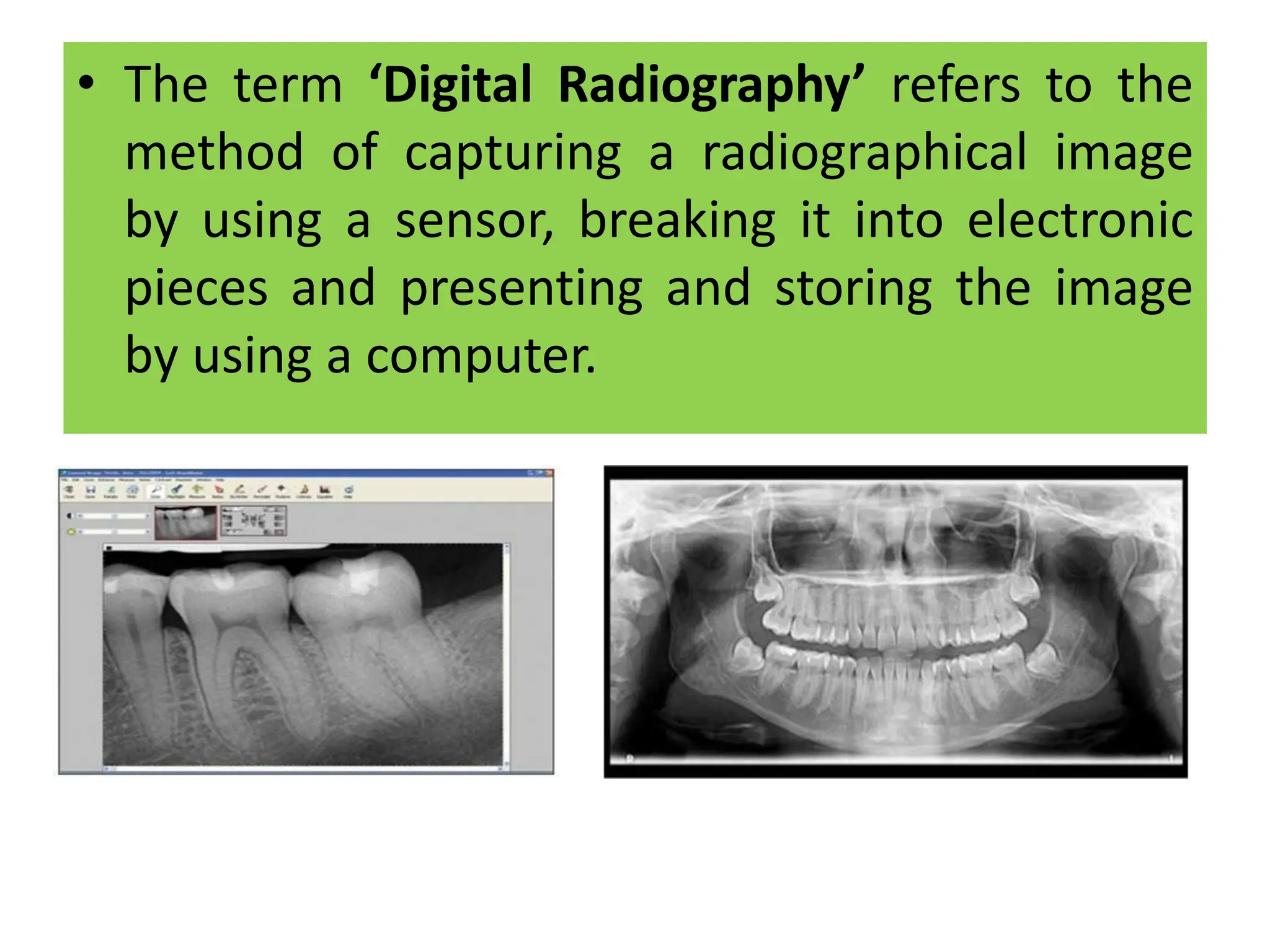
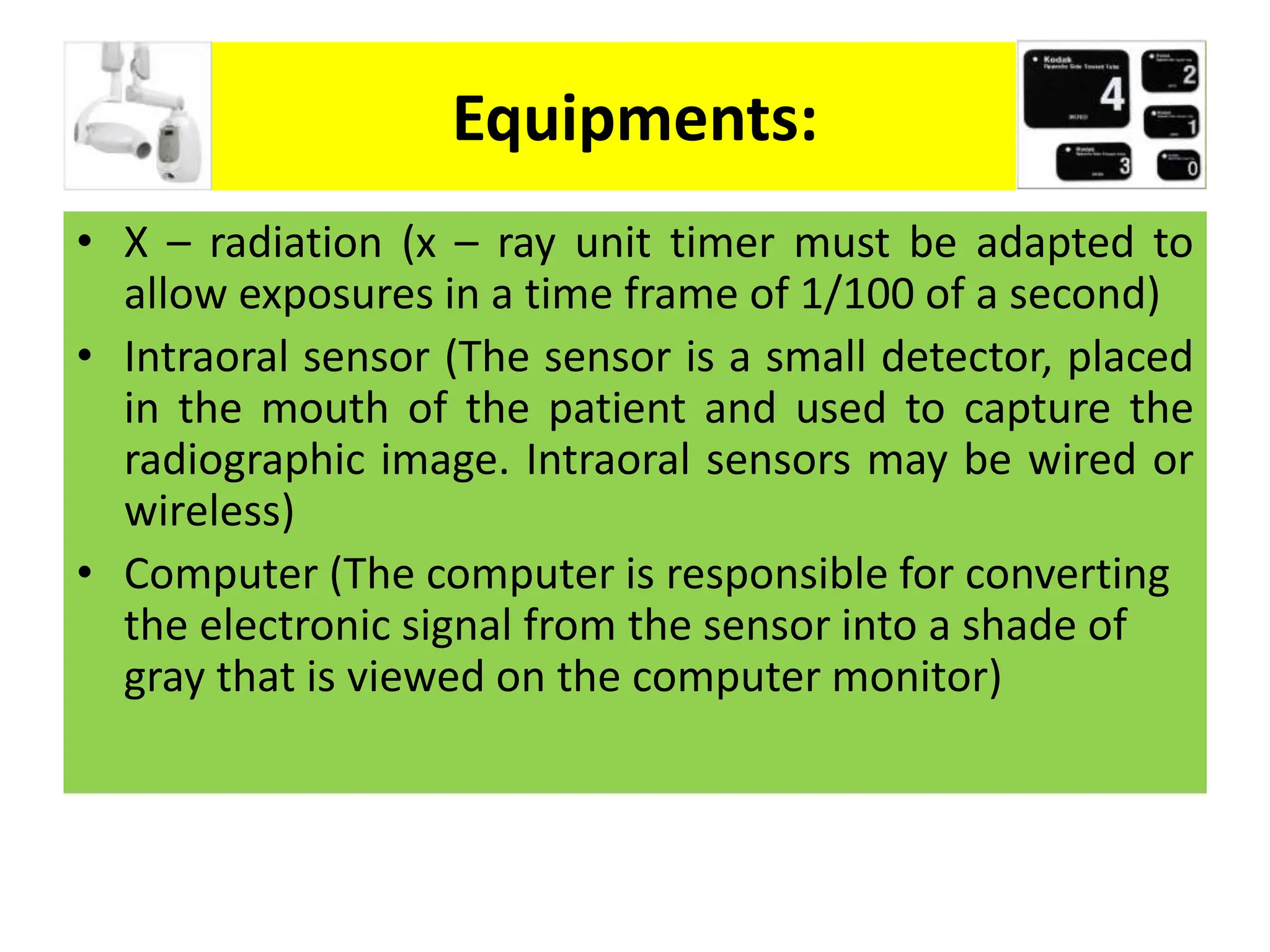
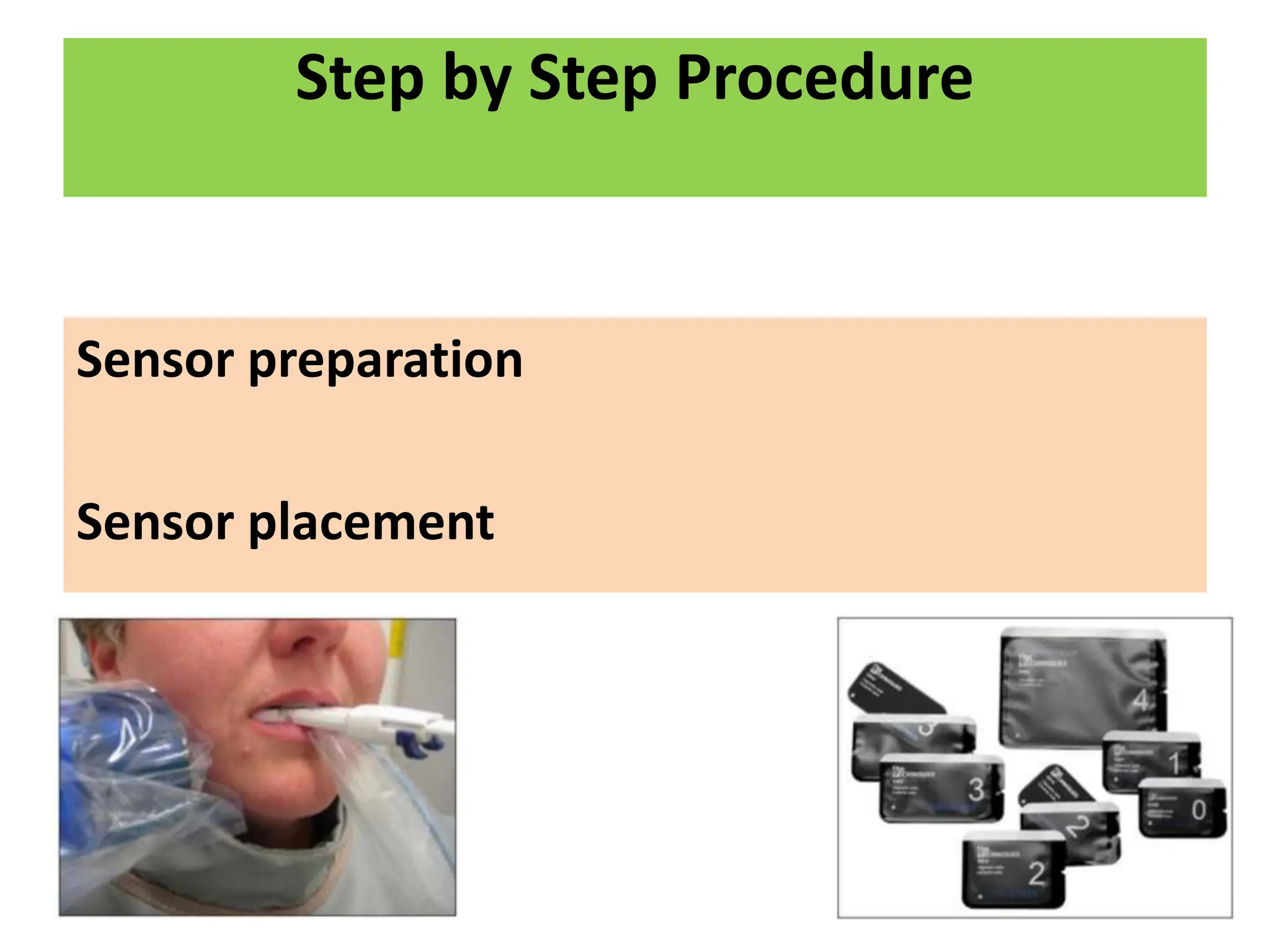
- Digital radiography involves capturing a radiographic image using an intraoral sensor, converting it to electronic data, and storing/viewing it on a computer.
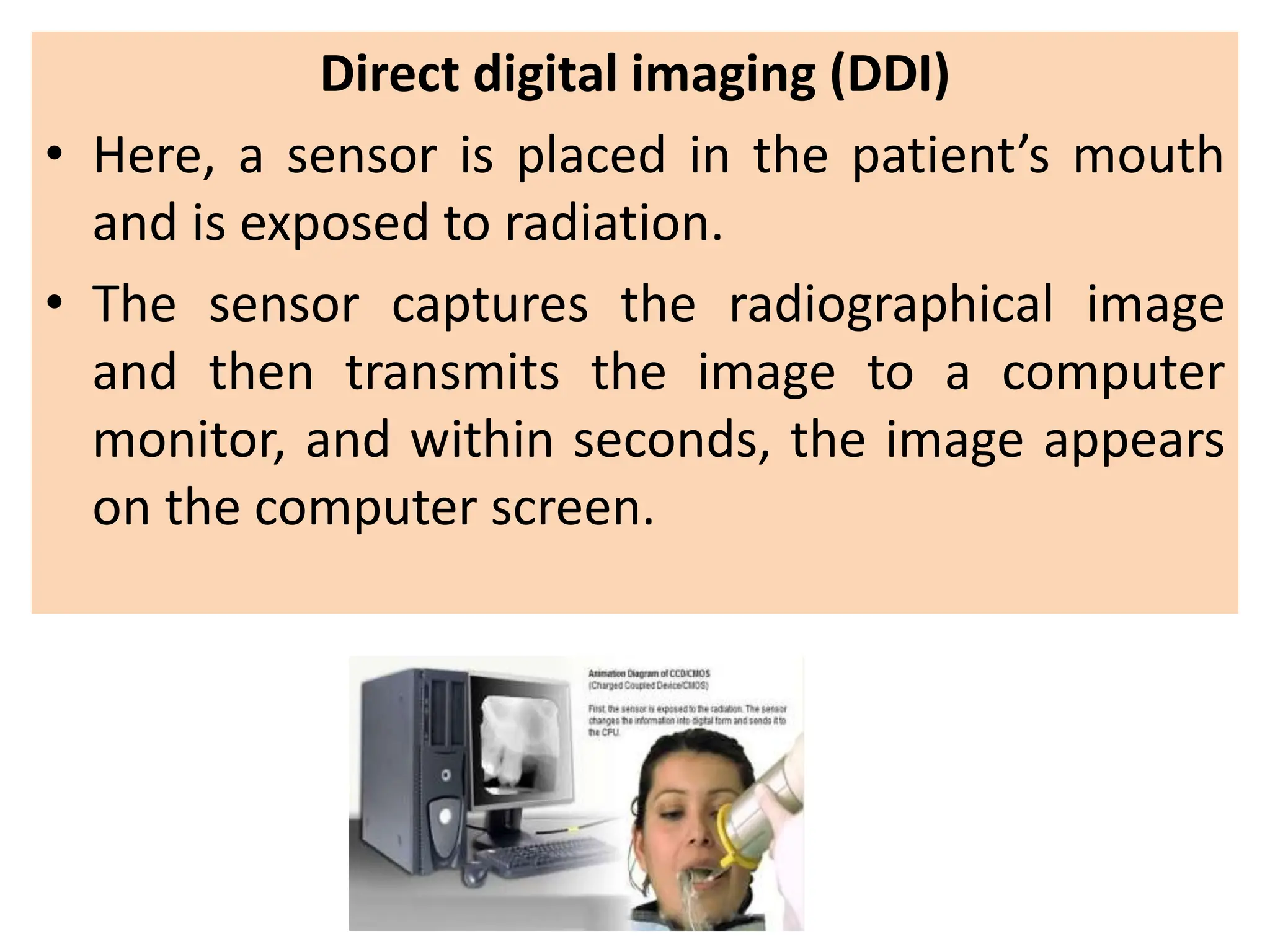
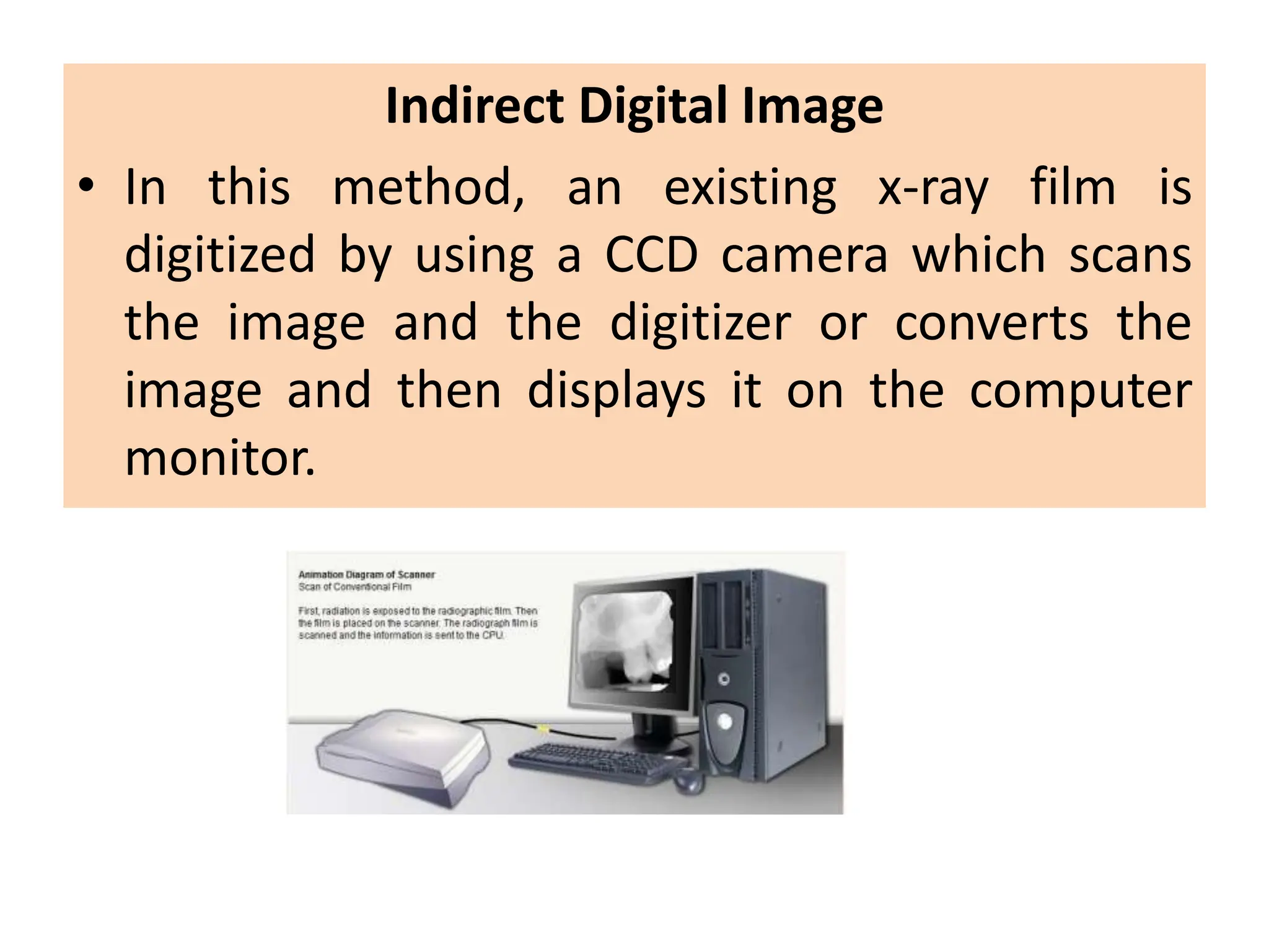
- There are three main methods of digital imaging: direct digital imaging using an intraoral sensor, indirect using digitization of films, and storage phosphor imaging using reusable plates.
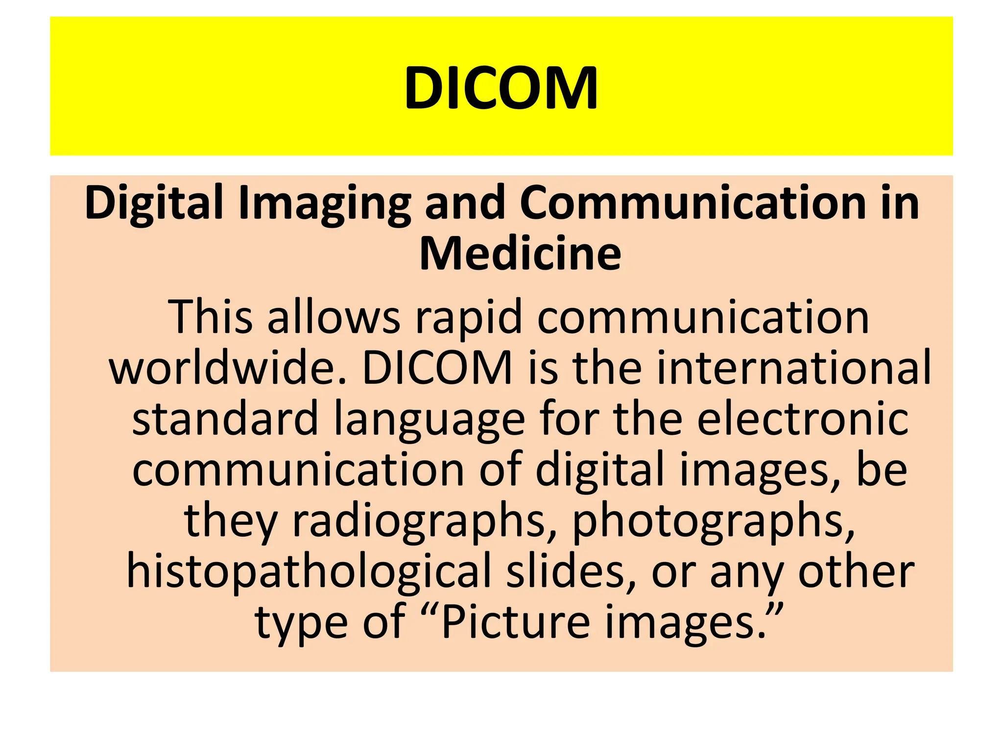
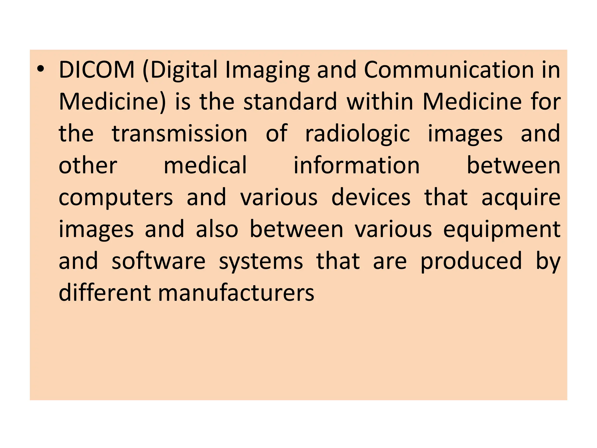
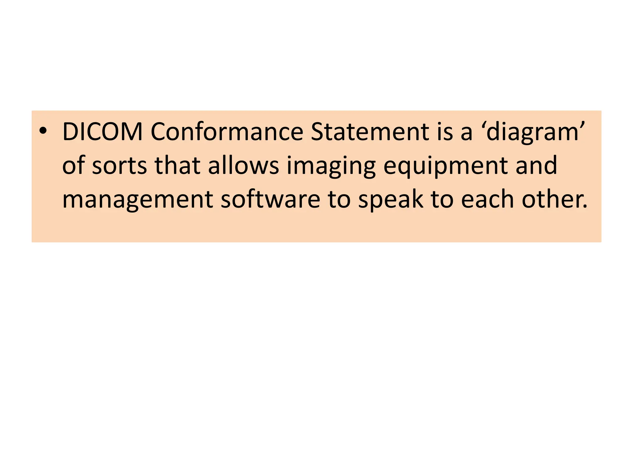
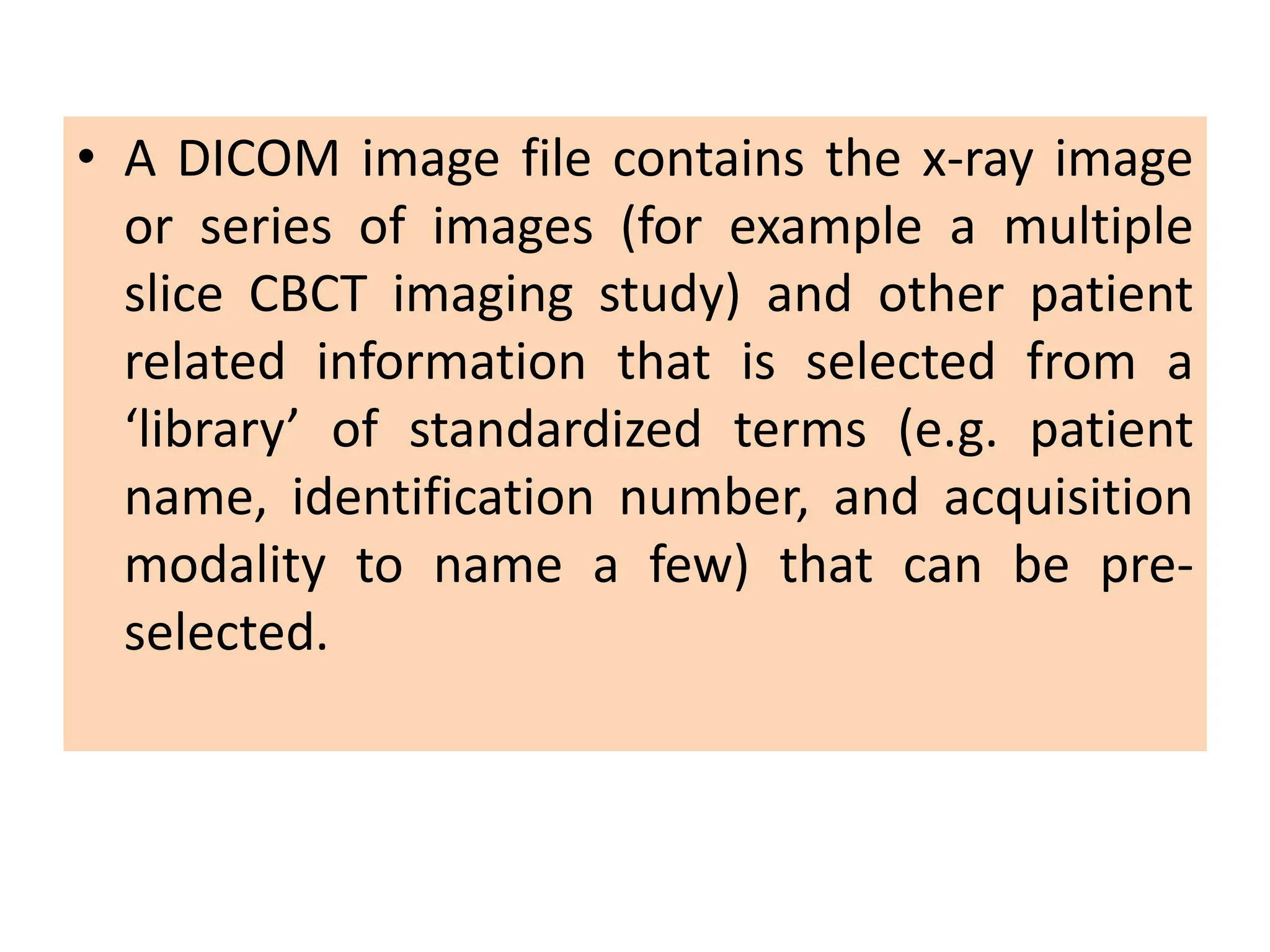
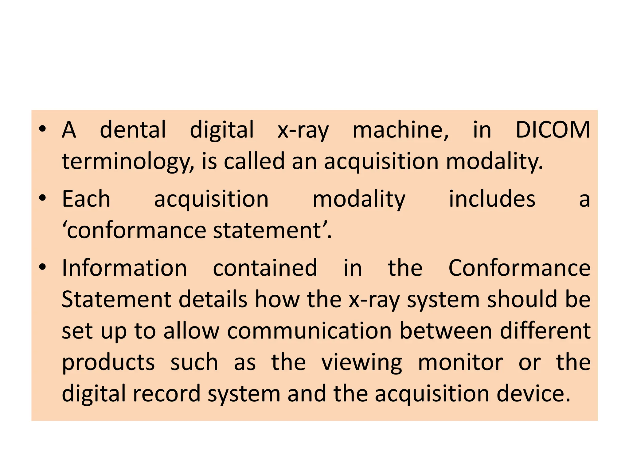
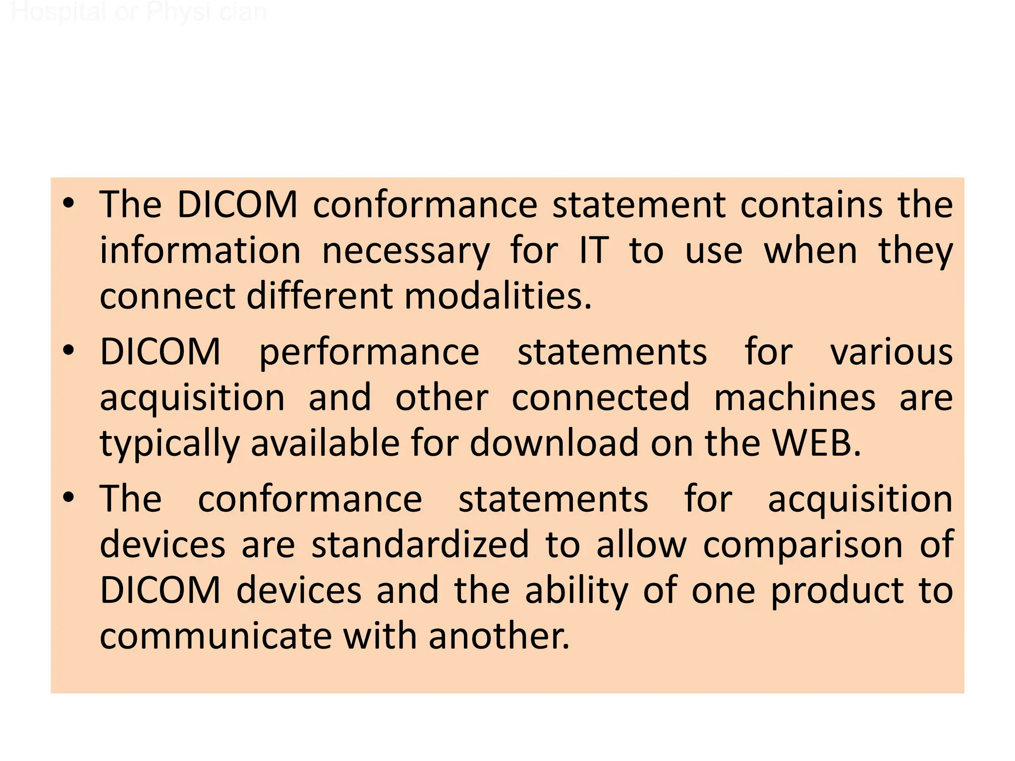
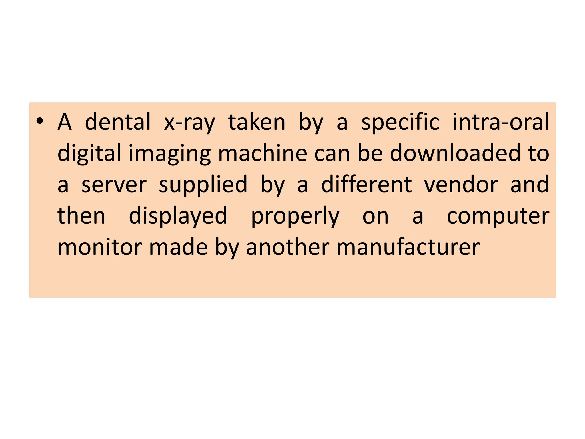
- DICOM is the international standard for transferring digital medical images and communication between devices. It allows images captured on one device to be viewed on another regardless of manufacturer.
- Digital images have advantages over film such as modification capabilities, electronic storage/transfer, and reduced radiation exposure.