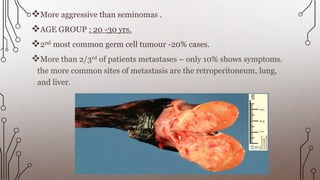
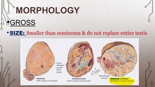
EMBRYONA
CARCINOM
ETIOLOGY
•Embryonal carcinoma is a NON-SEMINOMATOUS
germ cell tumour characterized by primitive epithelial
cells with marked pleomorphism and various histologic
patterns. It may present in pure form but often is part
of a mixed germ cell tumour.
The most accepted theory on the development of germ
cell tumours involves an initiating event that causes
foetal gonocytes to undergo abnormal cell division.
DEFINITION
More aggressive than seminomas .
AGE GROUP : 20 -30 yrs.
2nd most common germ cell tumour -20% cases.
More than 2/3rd of patients metastases – only 10% shows symptoms.
the more common sites of metastasis are the retroperitoneum, lung,
and liver.
MORPHOLOGY
•GROSS
•SIZE: Smaller than seminoma & do not replace entire testis
External surface:
smooth &
glistening
Cut surface:
1) has a
variegated
appearance
2)Poorly
demarcated
margins
3)Areas of
haemorrhage/necrosis
common
4) Tumour extends
into tunica albuginea ,
epididymis or
spermatic cord
MICROSCOPY
•Pattern – the tumour cells are arranged in ALVEOLAR / TUBULAR /
PAPILLARY patterns .
TUBULAR PAPPILLARY
They lack well formed glands with basally
situated nuclei and atypical cytoplasm.
More undifferentiated tumour shows
sheet/cluster of cells
Cells having epithelial appearance are large &
anaplastic.
Cell borders are usually indistinct.
Considerable variation in cell & nuclear size and
shape .
Nuclei are hyperchromatic with prominent
nucleoli .
Mitotic figures and tumour cells are frequent.
IMMUNOHISTOCHEMISTRY
Positive for : OCT ¾ & PLAP( placental alkaline phosphatase)
Positive for : cytokeratin & CD30
Negative for c-KIT(CD 117) & EMA
There is also increase in alpha fetoprotein & Lactic acid
dehydrogenase .
CD 30
OCT 3/4
embryonal carcinoma

embryonal carcinoma