Characterization strategies for primary cell culture term paper
•Download as DOCX, PDF•
4 likes•713 views

Characterization strategies for primary cell culture

Recommended
More Related Content
What's hot
What's hot (20)
Similar to Characterization strategies for primary cell culture term paper
Similar to Characterization strategies for primary cell culture term paper (20)
More from sworna kumari chithiraivelu
More from sworna kumari chithiraivelu (20)
Recently uploaded
Recently uploaded (20)
Characterization strategies for primary cell culture term paper
- 1. Characterization strategies for primary cell culture By Sworna kumari.c m.phil biotechnology Characterization strategies for primary cell culture Characterization of a cell line is vital for determining its functionality and in proving its authenticity as pure cell line. Special attention must be paid to the possibility that the cell line has become cross- contaminated with an existing continuous cell line or misidentified because of mislabeling or confusion in
- 2. handling DNA profiling. This has now become the major standard procedure for cell line identification, and a standard procedure with universal application. The various important factors for cell line characterization are: 1. It leads to authentication or confirmation that the cell line is not cross-contaminated or misidentified 2. It is confirmation of the species of origin 3. It is used for correlation with the tissue of origin, which comprises the following characteristics: a. Identification of the lineage to which the cell belongs b. Position of the cells within that lineage (i.e., the stem, precursor, or differentiated status) 4. For determination whether the cell line is transformed or not: a. Whether the cell line is finite or continuous? b. Whether the cell line expresses properties associated with malignancy? 5. It indicates whether the cell line is prone to genetic instability and phenotypic variation 6. Identification of specific cell lines within a group from the same origin, selected cell strains, or hybrid cell lines, all of which require demonstration of features unique to that cell line or cell strain Table 1: Decisive factors for characterization of cell lines and corresponding methods
- 3. Parameters of Characterization The nature of the technique used for characterization depends on the type of work being carried out. Some of the parameters are: In case molecular technology, DNA profiling or analysis of gene expression are most useful. A cytology laboratory may prefer to use chromosome analysis coupled with FISH (fluorescence in situ hybridization) and chromosome painting. Chromosomal analysis also known as karyotyping, is one of the best traditional methods for distinguishing among species. Chromosome banding patterns can be used to distinguish individual chromosomes. Chromosome painting, explicitly using combinations of specific molecular probes that hybridize to individual chromosomes, adds further resolution and specificity to this technique. These probes identify individual chromosome pairs and are species specific. Chromosome painting is a good method for distinguishing between human and mouse chromosomes in potential cross- contaminations. A laboratory with immunological capability may prefer to use MHC (Major Histo compatibility complex) analysis (e.g., HLA typing) coupled with lineage specific markers. Combined with a functional assay related to our own interests, these procedures should provide sufficient data to authenticate a cell line as well as confirm that it is suited to the concerned. Lineage or Tissue markers:
- 4. The progression of cells down a particular differentiation pathway towards a specific differentiated cell type and can be considered as a lineage, and as cells progress down this path they acquire lineage markers specific to the lineage and distinct from markers expressed by the stem cells. These markers often reflect the embryological origin of the cells from a particular germ layer. Lineage markers are helpful in establishing the relationship of a particular cell line to its tissue of origin. There are some lineage markers which are described as follows: Cell surface antigen: These markers are particularly useful in sorting hematopoietic cells and have also been effective in discriminating epithelium from mesenchymally derived stroma with antibodies such as anti- and anti- HMFG 1 and, distinguishing among epithelial lineages, and identifying neuroectodermally derived cells (e.g., with anti-A2B5). Intermediate filament proteins : These are among the most widely used lineage or tissue markers. Glial fibrillary acidic protein (GFAP) for astrocytes and desmin for muscle are the most specific, whereas cytokeratin marks epithelial cells and mesothelium. Differentiated products and functions : Haemoglobin for erythroid cells, myosin or tropomyosin for muscle, melanin for melanocytes, and serum albumin for hepatocytes are examples of specific cell type markers, but like all differentiation markers, they depend on the complete expression of the differentiated phenotype. Transport of inorganic ions, and the resultant transfer of water, is characteristic of absorptive and secretary epithelia. Polarized transport can also be demonstrated in epithelial and endothelial cells using Boyden chambers or filter well inserts. Other tissue-specific functions that can be expressed in vitro include muscle contraction and depolarization of nerve cell membrane. Enzymes: Three parameters are available in enzymatic characterization:
- 5. The constitutive level (in the absence of inducers or repressors) The induced or adaptive level (the response to inducers and repressors) Isoenzyme polymorphisms Table 1: Enzymatic markers used for cell line Regulation: The level of expression of many differentiated products is under the regulatory control
- 6. Unique markers Unique markers include specific chromosomal aberrations (e.g., deletions, translocations, polysomy), major histocompatibility (MHC) group antigens (e.g., HLA in humans), which are highly polymorphic, and DNA fingerprinting or SLTR DNA profiling. Enzymic deficiencies, such as thymidine kinase deficiency (TK−) and drug resistance such as vinblastine resistance (usually coupled to the expression of the P-glycoprotein by one of the mdr genes that code for the efflux protein) are not truly unique, but they may be used to distinguish among cell lines from the same tissues but different donors. Transformation: The transformation status forms a major element in cell line characterization and is dealt with separately. Cell Morphology: Observation of morphology is the simplest and most direct technique used to identify cells. Most of these are related to the plasticity of cellular morphology in response to different culture conditions. For example, epithelial cells growing in the center of a confluent sheet are usually regular, polygonal, and with a clearly defined birefringent edge, whereas the same cells growing at the edge of a patch may be more irregular and distended and, if transformed, may break away from the patch and become fibroblast-like in shape. Microscopy: The inverted microscope is one of the most important tools in the tissue culture laboratory, but it is often used incorrectly. As the thickness of the closed culture vessel makes observation difficult from above, because of the long working distance, the culture vessel is placed on the stage, illuminated of environmental influences, such as nutrients, hormones, the matrix, and adjacent cell. Hence the measurement of specific lineage markers may require preincubation of the cells in, for example, a hormone such as hydrocortisone, specific growth factors, or growth of the cells on extracellular matrix of the correct type. Lineage fidelity: L ineage markers are more properly regarded as tissue or cell type markers, as they are often more characteristic of the function of the cell than its embryonic origin.
- 7. from above, and observed from below. As the thickness of the wall of the culture vessel still limits the working distance, the maximum objective magnification is usually limited to 40X. The use of phase- contrast optics, where an annular light path is masked by a corresponding dark ring in the objective and only diffracted light is visible, enables unstained cells to be viewed with higher contrast than is available by normal illumination. Because this means that the intensity of the light is increased, an infrared filter should be incorporated for prolonged observation of cells.It is useful to keep a set of photographs at different cell densities for each cell line, prepared shortly after acquisition and at intervals thereafter, as a record in case a morphological change is subsequently suspected. Photographs of cell lines in regular use should be displayed above the inverted microscope. Photographic records can be supplemented with photographs of stained preparations and digital output from DNA profiling and stored with the cell line record in a database or stored separately and linked to the cell line database. Staining: A polychromatic blood stain, such as Giemsa, provides a convenient method of preparing a stained culture. Giemsa stain is usually combined with May–Grunwald stain when staining blood, but not when staining cultured cells. Alone, it stains the nucleus pink or magenta, the nucleoli dark blue, and the cytoplasm pale gray-blue. It stains cells fixed in alcohol or formaldehyde but will not work correctly unless the preparation is completely anhydrous. Chromosome Content: Chromosome content or karyotype is one of the most characteristic and best-defined criteria for identifying cell lines and relating them to the species and sex from which they were derived. Chromosome analysis can also distinguish between normal and transformed cells because the chromosome number is more stable in normal cells (except in mice, where the chromosome complement of normal cells can change quite rapidly after explantation into culture). Chromosome Banding: This group of techniques was devised to enable individual chromosome pairs to be identified when there is little morphological difference between them. For Giemsa banding, the chromosomal proteins are partially digested by crude trypsin, producing a banded appearance on subsequent staining. Trypsinization is not required for quinacrine banding. The banding pattern is characteristic for each chromosome pair. Other methods for banding are: a. Using trypsin and EDTA rather than trypsin alone b. Q-banding, which stains the cells in 5% (w/v) quinacrine dihydrochloride in 45% acetic acid, followed by rinsing Giemsa banding the slide, and mounting it in deionized water at pH 4.5 c. C-banding, which emphasizes the centromeric regions Techniques have been developed for discriminating between human and mouse chromosomes, principally
- 8. to aid the karyotypic analysis of human-mouse hybrids. These methods include fluorescent staining with Hoechst 33258, which causes mouse centromeres to fluoresce more brightly than human centromeres. Chromosome painting: Chromosome paints are available commercially from a number of sources. The hybridization and detection protocols vary with each commercial source, but a general scheme is available. Karyotypic analysis is carried out classically by chromosome banding, using dyes that differentially stain the chromosomes. Thus each chromosome is identified by its banding pattern. However, traditional banding techniques cannot characterize many complex chromosomal aberrations. New karyotyping methods based on chromosome painting techniques—namely spectral karyotyping (SKY) and multicolour fluorescence in situ hybridization (M-FISH)—have been developed. These techniques allow the simultaneous visualization of all 23 human chromosomes in different colours. Chromosome Analysis The following are methods by which the chromosome complement may be analyzed: 1. Chromosome count : Count the chromosome number per spread for between 50 and 100 spreads. (The chromosomes need not be banded.) 2. Karyotype : Digitally photograph about 10 or 20 good spreads of banded chromosomes. Image analysis can be used to sort chromosome images automatically to generate karyotypes. 3. Chromosome counting and karyotyping allow species identification of the cells and, when banding is used, distinguish individual cell line variations and marker chromosomes. However, karyotyping is time- consuming, and chromosome counting with a quick check on gross chromosome morphology may be sufficient to confirm or exclude a suspected cross-contamination. DNA Analysis: DNA content can be measured by propidium iodide fluorescence with a CCD camera or flow cytometry, although the generation of the necessary single-cell suspension will, of course, destroy the topography of the specimen. DNA can be estimated in homogenates with Hoechst 33258 and other DNA fluorochromes such as DAPI, propidium iodide, or Pico Green (Molecular Probes). Analysis of DNA content is particularly useful in the characterization of transformed cells that are often aneuploid and heteroploid.
- 9. DNA Hybridization: Hybridization of specific molecular probes to unique DNA sequences (Southern blotting) can provide information about species specific regions, amplified regions of the DNA, or altered base sequences that are characteristic to that cell line. Thus strain-specific gene amplifications, such as amplification of the dihydrofolate reductase (DHFR) gene, may be detected in cell lines selected for resistance to methotrexate; amplification of the MDR gene in vinblastine-resistant cells overexpression of a specific oncogene, or oncogenes in transformed cell lines or deletion, or loss, of heterozygosity in suppressor genes. Although DNA aberrations can be detected in restriction digests of extracts of whole DNA, this is limited by the amount of DNA required. It is more common to use the polymerase chain reaction (PCR) with a primer specific to the sequence of interest, enabling detection in relatively small numbers of cells. Alternatively, specific probes can be used to detect specific DNA sequences by in situ hybridization having the advantage of displaying topographical differences and heterogeneity within a cell population. DNA fingerprinting: DNA fingerprints appear to be quite stable in culture, and cell lines from the same origin, but maintained separately in different laboratories for many years, still retain the same or very similar DNA fingerprints. DNA fingerprinting is a very powerful tool in determining the origin of a cell line, if the original cell line, or DNA from it or from the donor individual, has been retained. This emphasizes the need to retain a blood, tissue, or DNA sample when tissue is isolated for primary culture. Furthermore, if a cross-contamination or misidentification is suspected, this can be investigated by fingerprinting the cells and all potential contaminant. Antigenic Markers: Immunostaining and ELISA assays are among the most useful techniques available for cell line characterization facilitated by the abundance of antibodies and kits which is commercially available. Antibody is essential to be certain of its specificity by using appropriate control material. This is true for monoclonal antibodies and polyclonal antisera alike; a monoclonal antibody is highly specific for a particular epitope.
- 10. Immunostaining: Antibody localization is determined by fluorescence, wherein the antibody is conjugated to a fluorochrome, such as fluorescein or rhodamine, or by the deposition of a precipitated product from the activity of horseradish peroxidase or alkaline phosphatase conjugated to the antibody. Various methods have been used to enhance the sensitivity of detection of these methods, particularly the peroxidase linked methods. In the peroxidase–anti-peroxidase (PAP) technique, a further amplifying tier is added by reaction with peroxidase conjugated to anti-peroxidase antibody from the same species as the primary antibody. Even greater sensitivity has been obtained by using a biotin-conjugated second antibody with streptavidin conjugated to peroxidase or alkaline phosphatase or gold-conjugated second antibody with subsequent silver intensification. Differentiation: Many of the characteristics described under antigenic markers or enzyme activities may also be regarded as markers of differentiation, and as such they can help to correlate cell lines with their tissue of origin as well as define their phenotypic status. Although sometimes constitutively expressed (e.g., melanin in B16 melanoma or Factor VIII in endothelial cells), expression of differentiated lineage markers may need to be induced before detection is possible.
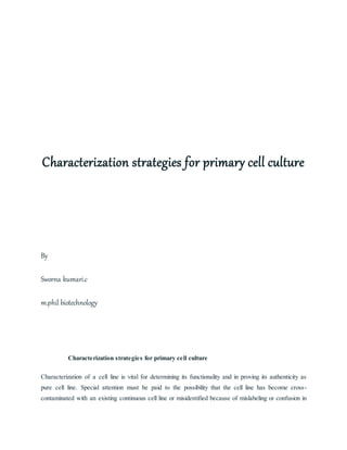
- 11. Figure 1: Karyotype Preparation Steps in the preparation of a karyotype from digital microphotographs of metaphase spread. Chinese hamster cells recloned from the Y-5 strain.