
Recommended
More Related Content
What's hot
What's hot (20)
Similar to SCFE Guide: Diagnosis and Management
Similar to SCFE Guide: Diagnosis and Management (20)
Recently uploaded
Recently uploaded (20)
SCFE Guide: Diagnosis and Management
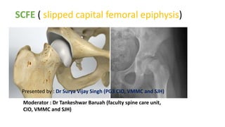
- 1. SCFE ( slipped capital femoral epiphysis) Presented by : Dr Surya Vijay Singh (PG3 CIO, VMMC and SJH) Moderator : Dr Tankeshwar Baruah (faculty spine care unit, CIO, VMMC and SJH)
- 2. SCFE • Introduction and definition • Epidemiology and risk factors • classification • Pathogenesis • Signs and symptoms • Diagnosis • Investigations • Management modalities • Complications
- 3. • INTRODUCTION • The capital femoral epiphysis is somewhat unique. It is one of the few epiphyses in the body that is inside the joint capsule. (The joint capsule is the tissue that Surround the joints). surrounds the joint.)
- 4. Definition of SCFE (slipped capital femoral epiphysis) • Slipped upper femoral epiphysis" term refer to slippage of the overlying epiphysis of proximal femur posteriorly and inferiorly due to weakness of the growth plate in relation to metaphysis. • Most often, it develops during periods of accelerated growth, shortly after the onset Of puberty. • The femoral epiphyses maintains its relation with acetabulum ,it’s the femoral neck and shaft upward and anterior movement on epiphyses thus epiphyses displaces relatively posterior
- 5. • EPIDEMIOLOGY AND RISK FACTORS • Incidence is 2-3 per 100000 population • Most common in adolescent period with rapid growth plate (boys aged 10-16 y, girls aged 12-14 y). • Very early onset[<10yrs] and late onset[>16] should be evaluated for endocrine disorders • Males have 2.4 times the risk as females. • Obesity is a risk factor because it places more shear forces around the proximal growth plate in the hip at risk. • Bilateral slippage is common of which 2nd slip is about 1218mths later to 1st (left hip is more common than right).
- 6. • ETIOLOGY-multifactorial • Local trauma , obesity • Endocrine disorders (e.g primary or secondary hypothyroidism, adiposogenital dystrophy(hypogonadal male), • Deficiency or increase of androgens. • Acute trauma • Growth hormone deficiency • ATYPICAL SCFE associated with renal failure,radiation therapy • Slipping of the upper femoral epiphysis occurs predominantly in obese children with underdeveloped sexual characteristics and less commonly, in tall, thin children.
- 7. MECHANICAL FACTORS Important features of the predisposed hip that may be the primary cause of slipped epiphysis are: 1.Thinning of the perichondrial ring complex with maturation 2.Relative or absolute retroversion of the femoral neck-making it more suceptable to AP shear forces 3.A change in the inclination of the adolescent proximal femoral physis relative to the femoral neck shaft . •Associated conditions with mechanical etiology- 1.Infantile and adolescent blount diasease 2.Patients with peroneal spastic flatfoot and Legg-Calve-Prthes disease
- 8. Classification of SCFE } Based on onset of symptoms [temporal classification] -acute ( <3 weeks) -Chronic (> 3 weeks) -acute on chronic (acute exacerbation of long standing symptoms) FUNCTIONAL CLASSIFICATION [ Loder classification] -stable (able to bear wt with or without crutches, min risk of osteonecrosis <10%) -Unstable (unable to ambulate – not even with crutches, high risk of osteonecrosis ~47%) MORPHOLOGICAL CLASSIFICATION [Southwick angle classification] -mild (<30°) -moderate (30-50°) -severe (>50°)
- 9. SYMPTOMS : <2WKS >2WKS grdual X-RAY : displaced epiphyses remodelling no remodelling healing noted ACUTE ON CHRONIC SLIPS-Symptoms lasting longer than 1mth and recent sudden exacerbation pain after trivial trauma
- 10. • FUNCTIONAL CLASSIFICATION • It is important to determine ability of the patient to bear weight. • Stable" SCFEs allow the patient to (walk) with or without crutches (walking aids). • "Unstable" SCFEs do not allow the patient to ambulate at all regardles of duration of symptoms; these cases carry a higher rate of complication, particularly of AVN.
- 11. • AP VIEW-145* -Best shows posterior slippage and LATERAL VIEW-170* subtle slipping also FROG LEG LATERAL POSITION- -Normally 10*posteriorly -Increases in slippage
- 12. • MORPHOLOGICAL CLASSIFICATION- • Grading Severity of SCFE according to AP and Lateral Xray • PRE SLIP-irregularity,widening,and indistinctness of physes Grade-1 Grade-II Grade-III
- 13. • the displacement is either superior and posterior (so-called valgus slip)or, even more rarely, anterior. • In valgus slips there is a restriction of adduction as well as of flexion. • In anterior slips there is a limitation of extension and external rotation—exactly the opposite of what is found in typical slips. • X-RAY of valgus slip show- • superior or lateral displacement of the capital epiphysis on the femoral neck on the AP projection -posterior displacement on the lateral projection. • Anterior slips may appear little different from typical slips on the AP projection, but the anterior displacement of the capital epiphysis is identified on the lateral projection.
- 14. Pathology • Grossly , with gradual slipping of the capital epiphysis in the typical posterior position • Periostium is stripped from the anterior and inferior surface of the femoral neck • So the area between the original femoral neck and the posterior periostium fills with callus which ossifies and become progresively more dense • The anterior and superior portion of the neck forms a hump or ridge that can impinge on the rim of acetabulum • Normally ,this ridge will remodel with anterior portion of the neck contouring into smoother surface • In case of acute slipping the periostium is torn anteriorly and haemarthrosis will be present.
- 15. Microscopically change in SCFE Microscopically : • Characteristc changes in PROLIFERATIVE and HYPERTROPHIC ZONES of epiphyses, • chondrocytes -number decrease and - irregularly arranged, • collagen fibres and Matrix are increased.
- 16. SYMPTOMS • Pain : in the groin and around the knee. • Antalgic Limp (intermittent). • Shortening of the affected limb (1-2 cm). • The limb is in external rotation.[frog leg position] • Flexion, abduction, medial rotation are limited • External rotation, adduction are increased. • The presence of hip flexion contracture points towards the possibility of chondrolysis. • Axis deviation – pathognomonic – when hip is flexed, the limb goes into external rotation
- 17. DIAGNOSIS • The diagnosis is a combination of clinical suspicion plus radiological investigation. • 20-50% of SCFE are missed or misdiagnosed on their first presentation to a medical facility. • This is because the common symptom is knee pain. This is referred pain from the hip. The knee is investigated and found to be normal • In acute cases it is essential to differentiate between SCFE and type • 1 epiphseal#as most of time both come with history injury/trauma • SCFE pt has prodromal pain in groin,thigh or knee.insidious onset whereas in type 1 epiphyseal # pt is normal acute pain associated with high energy trauma
- 18. SCFE PERTHE’S DISEASE Usually occurs in 10-14yrs age late onset in 14-16yrs Usually in 4-7yrs age late onset in 7-10yrs age Thin and tall adolescents or short and obese individuals Occurs in normal child Presents as pain with slippage and limping noted at later stage Initially the child limps and then at later stages complaints of pain Limb never has fixed flexion deformity It may be in hyperextension state Fixed flexion deformity is usually noted
- 19. • X-RAYS-AP VIEW • -thretowan’s sign[kleins line] • -steel metaphysel blanch sign • -sham’s sign • -capner’s sign • -widening of growth plate • -decrease of epiphyseal height • X-RAY -frog-leg [lowenstein]lateral view • CT-SCAN • MRI SCAN
- 20. A.P. VIEW- Posterior ,inferior, And medial translation of epiphyses FROG lateral view : To measure lateral epiphyseal shaft angle
- 21. • In normal hip a line drawn tangential to superior femoral neck[klein’s line] intersects small portion of lateral capital epiphyseal. • In posterior displacement of epiphyses the line doesn’t intersect.
- 22. • In AP VIEW-crescent-shaped area of increased density overlying thethe metaphysis adjacent to the physis • This increased density is due to the overlapping of the femoral neck and the posteriorly displaced capital epiphysis
- 23. • In the normal hip the inferiomedial femoral neck overlaps the posterior wall of the acetabulum producing triangular radiographic density. • With displacement of capital epiphysis this dense triangle is lost because this portion of the femoral neck is located lateral to the acetabulum.
- 24. CAPENERS SIGN- • In pelvic AP view in the normal hip, the posterior acetabular margin cuts across the medial corner of the upper femoral metaphysis With slipping, the entire metaphysis is lateral to the posterior acetabular margin.
- 25. • Very early slips may appear to be normal in AP VIEW but may be clearly noted in lateral view • CHRONIC CASE OF SCFE X-RAY- • Reactive bone formation along superolateral aspect of neck • Bone remodelling and broadening of neck resulting in PISTOL GRIP like appearance[hordons hump]
- 26. • USG-It has been useful in the detection of early slips -joint effusion and a ―step‖ between the femoral neck and the epiphysis created by slipping. • Absolute displacement of 6 mm, >2 mm is diagnostic of a slipped epiphysis. • CT-useful in documenting presence of decreased upper femoral neck anteversion or true retroversion. • it’s more accurate measure head–neck angle. } CT is useful in the management of slips. • First, CT of the hip can be very helpful in demonstrating whether penetration of the hip joint by fixation devices has occurred (Fig. 18-9). • CT is also used to confirm closure of the proximal femoral physis and also when reconstructive osteotomy is being considered. • MRI-useful to assess AVN.
- 27. COMPLICATIONS • Avascular necrosis • Chondrolysis • Osteoarthritis. • Coxa vara (is a deformity of the hip, whereby the angle between the ball and the shaft of the femur is reduced to less than 120 degrees). • Slipping of the opposite hip ≈ 20% to 80% of cases
- 28. NATURAL HISTORY •30-40% second slip asymptomatic (slow). •Premature OA (pistol grip deformity 40% primary OA) •Onset of OA directly related to severity of slip.
- 29. • IDEAL TREATMENT • •Prevent further slippage • •Stimulate early physeal closure • •Reduction of epiphyseal displacement • •Avoid complications like osteonecrosis , chondrolysis and osteoarthritis • •Any child with SCFE and open epiphyses needs treatment ,without stabilisation it progresses. • •In a patient with closed physes, the only surgical treatment in the absence of severe degenerative
- 30. Management of SCFE • Conservative management-rest and traction • Closed manipulative reduction • Operative management -In situ pinning -ORIF -BONE PEG Epiphysiodesis -Osteotomy - reconstruction by-Arthroplasty -Arthrodesis -Cheilectomy
- 31. Conservative treatment of SCFE • Rest for atleast 12wks and traction can be an alternative to surgical treatment • Indicated in – temporary measure before operative ℞, • - slip due to hypothyroidism • Rest in spica cast - ⇧ incidence of complications like chondrolysis
- 32. Percutaneous in situ fixation • Cannulated Screw placed percutaneously into center of epiphyses and perpendicular to physis. • Min 5 screw thread should be contained within the physis in order to provide adequate stability and prevent slip. • Screw s/b at least 5 mm from subchondral bone in all views. • The use of 1 vs 2 screw is contraversial. • Screw must be start on anterior surface of the neck in order to cross perpendicular to physis on both AP and Lateral view.
- 33. Prophylactic fixation of asymptomatic hip Indications • Children with HIGH RISK of contralateral slip, • Young at primary diagnosis ( <10years), • Have Endocrine disorder, or • Obese with delayed presentation. After in situ pinning: –early wt bearing in stable slips, -after 6-8wks in unstable slips
- 35. Open reduction for SCFE indications - Highly displaced, - Unstable scfe, - Not reduce in Closed maneuver - Sever slip Types 1) Modified Dunn procedure 2) Bone PEG Epiphysiodesis 3) Proximal femoral osteotomy
- 36. Modified Dunn procedure for SCFE -Used to reduce the epiphyses, performed anteriorly via Surgical Dislocation of Hip. -Opportunity to assess and confirm blood flow to femoral head thereby reducing risk of both AVN and Femoral acetabular impingement.
- 37. Bone PEG Epiphysiodesis for SCFE • Anterior approach to hip and H-shaped capsular incision, • Use of hollow mill to create tunnel across physis, • Sandwiched iliac bone grafts are driven across physis. -A portion of the residual physis is removed and a dowel or “peg” of autologous bone graft (ipsilateral iliac crest) is inserted into the epiphysis. -In unstable slips, supplementary internal fixation, postoperative traction, or spica cast immobilization for 3 to 8 weeks until early stabilization has occurred.
- 38. Bone PEG Epiphysiodesis for SCFE Disadvantages 1)Graft insufficiency 2)Increase in severity of slip 3)Failure of physeal fusion 4)longer operating time, increased blood loss, longer hospitalization, and longer rehabilitation.
- 39. Osteotomy in SCFE • There are two basic types of osteotomy: • 1)Closing wedge osteotomy through the femoral neck - correct the deformity. • 2)Compensatory osteotomy through the trochanteric region - produce a deformity in the opposite direction
- 41. Osteotomy in SCFE Four femoral neck osteotomies are described: • (1)the technique of Fish - just distal to the the physis, • (2) the technique of Dunn - just distal to the slip, • (3) the base of the neck technique of Kramer et al., and • (4) the technique of Abraham et al.- at the trochanteric region.
- 42. 1)Cuneiform osteotomy of femoral neck ( FISH) •Make the wedge anterior and superior to correct epiphyseal position subcapitally •The more severe the slip the more is wedge superiorly •Reduce the epiphysis by flexion, abduction, and internal rotation of the limb •After wedging, diameter of femoral head is greater than femoral neck. •Indication-severe chronic or acute on chronic slips
- 43. 2)Dunn osteotomy • Trapezoidal osteotomy of the femoral neck • Referred as “an open replacement of the displaced femoral head” should not be done if the physis is closed. • Reduce the capital femoral epiphysis on the femoral neck by resecting a portion of the superior femoral neck. • Advantage - the deformity itself is corrected • Results. High risk of complications, AVN and chondrolysis.
- 44. 3)Base-of-Neck Osteotomy (Kramer and Barmada Procedures) • Indicated to correct residual deformity after closure of the physis. • corrects the varus and retroversion components of moderate or severe chronic SCFE. • less risk to interruption of the blood supply to the femoral head than the Dunn procedure • Osteotomy held with threaded Steinmann pins, which extended into the capital epiphysis if the physis is still open.
- 45. 4)Intertrochanteric Osteotomy (Imhauser/ Southwick Procedure) • Preferable method to correct deformity associated with SCFE • Southwick osteotomy – chronic or healed slips with head–shaft deformities between 30 and 70 degrees. - Biplane osteotomy - Performed at the level of the lesser trochanter. • Imhauser's procedure - Intertrochanteric • COMPLICATIONS: I)Chondrolysis 2)Post operative narrowing of joint space
- 47. Complication 1) Chondrolysis Dissolution of articular cartilage with joint stiffness and pain Causes: • Persistent pin penitration • After trochantric osteotomy, open reduction,femoral neck osteotomy •Synovial malnutrition, ischaemia, excessive pressure •Autoimmune •Females>males
- 48. Chondrolysis... -Diagnosis: Joint pace of less than 3mm wide ( normal 4 to 6 mm), Decrease range of motion at hip joint. -TREATMENT : •Bed rest •Traction •Salicylates •Nsaids drugs •Intraarticular cortisone injections •Sugical manipulation in form of : Subtotal circumferential capsulectomy •Continuous passive motion and physical therapy
- 49. Complications... 2) Osteonecrosis of femoral head ( 4-6%) -Rare in untreated SCFE -Results from interruption of the retrograde blood supply by: •Original injury tamponade of the blood supply to the proximal femoral epiphysis as a result of acute hemorrhage within the capsule •Increase with severity of slip •increase in acute, unstable slips •increases with forcefull repititive manipulation, •pin placement in superior quadrant •Osteotomy of femoral neck
- 50. DIAGNOSIS: Early postoperative bone scan has excellent sensitivity and predictive value for detection of osteonecrosis after surgical treatment of SCFE TREATMENT • Remove metal work • Maintain ROM • Realignment • Shelf acetabuloplasty • Arthrodesis/THR
- 51. Complications... 3) Contralateral hip SCFE Most common complication after UNILATERAL surgical fixation of slip (20-80%). Risk factors for contralateral slip : • Male, • Obesity, • Young age of initial slip, • Endocrine disorders.
- 52. Complications.. 4) chronic pain 5) Infection 6) Degenerative arthritis 7) Residual proximal femoral deformity and LLD 8) Slip progression 9) Labral tear and degeneration.