
Recommended
More Related Content
Similar to Phsiyological structure and life process Suresh Chaudhary.docx
Similar to Phsiyological structure and life process Suresh Chaudhary.docx (20)
Recently uploaded
Recently uploaded (20)
Phsiyological structure and life process Suresh Chaudhary.docx
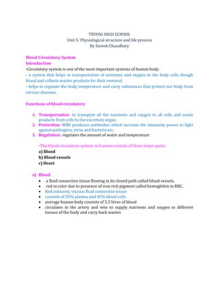
- 1. TRIYOG HIGH SCHOOL Unit 5: Physiological structure and life process By Suresh Chaudhary Blood Circulatory System Introduction -Circulatory system is one of the most important systems of human body. - a system that helps in transportation of nutrients and oxygen to the body cells though blood and collects waster products for their removal. - helps to regulate the body temperature and carry substances that protect our body from various diseases. Functions of blood circulatory 1. Transportation- to transport all the nutrients and oxygen to all cells and waste products from cells to the excretory organ. 2. Protection: WBC produces antibodies which increase the immunity power to fight against pathogens, virus and bacteria etc. 3. Regulation: regulates the amount of water and temperature -The blood circulatory system in human consists of three major parts: a) Blood b) Blood vessels c) Heart a) Blood a fluid connective tissue flowing in its closed path called blood vessels. red in color due to presence of iron rich pigment called hemoglobin in RBC. Red coloured, viscous fluid connective tissue consists of 55% plasma and 45% blood cells average human body consists of 5.5 litres of blood circulates in the artery and vein to supply nutrients and oxygen to different tissues of the body and carry back wastes
- 2. a. Figure of blood Composition of blood Blood is composed of 55% blood plasma and 45% blood cells Blood Plasma -transparent yellowish liquid -contains about 90% water, 2% different types of proteins and 2% clotting factors - blood cells are suspended in the plasma. Functions Transports digested food to different parts of the body Transports various waste materials to the excretory organs Maintains the amount of water in the body, regulates the body temperature, helps in blood clotting Blood Cells/ Blood corpuscles Blood cells are found suspended in the plasma
- 3. Occupy 45% of total volume of blood a) Red blood Cells also called erythrocytes Circular, biconcave and disc shaped cells, have no nuclei One cubic mm of blood contains about 4.5 million to 6 million RBCs Formed in the bone marrow and destroyed in the liver or spleen Life span is about 120 days They contain haemoglobin which is an iron pigmented substance due to which blood is red in colour. function of RBC is to transport oxygen and carbon dioxide between lungs and all parts of the body. Deficiency of RBC in the blood leads to the disease called anaemia are also called as oxygen-carriers. RBC is more than average it causes polythecimia. Symptoms of anaemia a. Loss in weight and appetite b. Slow and retarded growth c. Pale and dull colour of Skin etc d. Weakness and tiredness even in short walk. b) White blood cells
- 4. They are also called leukocytes Irregular in shape, contain nucleus but lack haemoglobin One cubic mm of blood contains about 4000 to 11000 WBCs Formed in the bone marrow lymph node s and destroyed in the spleen/ liver Life span is about 12 to 14 days They are the cells of immune system involved in defending the body against both pathogens and foreign materials Excess amount of WBCs in blood leads to the disease called Leukaemia ( blood cancer) Less WBCs in blood leads to leukopenia [ baldness] caused due to fall in neutrophil cells. Leucocytes are of 2 types: i) Granulocytes- having granules in their cytoplasm and lobed nucleus [neutrophiles, eosinophils and basophils] ii) Agranulocytes- without granules in their cytoplasm and unilobed nucleus [ monocytes and lymphocytes] c) Platelets They are also called thrombocytes Smallest blood cells, oval in shape and no nucleus Formed in the bone marrow and destroyed in the spleen One cubic mm of blood contains about 1.5 lakhs to 4.5 lakhs platelets Life span 5 to 10 days, formed in the bone marrow destroyed in the spleen They contain a pigment called fibrinogen which helps in blood clotting and thus prevent the excess loss of blood Less platelets causes thrombopenia – excessive bleeding can occur (Haemophilia)
- 5. Excess platelets causes thrombocytosis- obstruct blood vessels Functions of blood a) transport oxygen from lungs and all parts of the body b) carries waste products from different tissues to the excretory organs c) carries hormones produced by endocrine glands to the target cells d) helps to destroy various microorganism and pathogens e) helps to regulate body temperature f) helps in blood clotting and minimize the excess loss of blood Differences between RBC and WBC RBC WBC 1.They are oval, biconcave and without nucleus 1.They are irregular and nucleated 2.They are small in size 2.They are large in size 3.They are red in colour due to the presence of haemoglobin 3.They are colourless 4.They help in respiration 4.They help in the defence of the body Differences between Blood and plasma Blood Plasma 1.It contains haemoglobin 1.It does not contain haemoglobin 2.It is formed of blood cells 2.It is formed of water and solid materials 3.It is red in colour 3.It is light yellow in colour 4.Its function is to transport materials inside the body 4.It acts as a mediator between blood and body cells Youtube https://youtu.be/wqndItCcWMw blood cells Website blood cellhttps://en.m.wikipedia.org/wiki/Blood_cell TEST YOURSELF 1) What is blood? Write its composition and mention any three functions of it. 2) Differentiate between Erythrocytes and leukocytes on the basis of their structure and function. 3) Write down the functions of plasma. 4) What is leukaemia?
- 6. 5) A person suffering from anaemia feels weak even after a short walk, why? 6) Write down the function of RBC and WBC BLOOD VESSELS Blood vessels are the narrow tubular vessels through which blood flows They circulate the blood from the heart to body tissues and form tissues to the heart They are of three types: 1) Arteries The blood vessels which carry blood away from the heart are called arteries They are thick walled and elastic in nature to prevent from rupturing as pure blood flows at high pressure They don’t have valves and are deep seated The blood flows at high pressure in arteries All arteries carry pure blood except pulmonary artery which carries impure blood from heart to the lungs Arteries are divided into arterioles Valves are absent in arteries except at their origin in the heart. Arteries have narrow lumen. 2) Veins The blood vessels which bring blood into the heart from different parts of the body are called veins They are thin walled and non-elastic and are superficial. They have valves to prevent the back flow of blood The blood flows with low pressure since it carries the impure blood except pulmonary vein. All veins carry impure blood except pulmonary vein which carries pure blood from lungs to the heart. The larger structure of veins is called Venacava and smaller branches of veins are called venules. Veins have wider lumen so that the blood flows easily with low speed under low pressure through them. 3) Capillaries They are formed as a result of branching of arteries They are a very fine network of blood vessels which connect arteries to the veins
- 7. Capillaries provide a definite path for the flow of the blood. Their wall is made up of a single layer of endothelium. #website: https://www.thoughtco.com/blood-vessels-373483 #video: https://youtu.be/whtNDBIhczQ https://youtu.be/iqRTd1NY-pU Questions a. Why arteries deep seated in the body and veins are superficial? b. Why are veins provided with valves but arteries don't have valve? c. Write any two differences between arteries and veins?
- 8. Heart Heart is a hollow muscular organ which consists of cardiac muscles It is situated in the middle of chest cavity just above the diaphragm and between two lungs or situated obliquely in the middle mediastinum. It is cone shaped and as big as the owner’s closed fist. It is not the median plane and located about 2/3rd to the left and 1/3rd to the right. Heart wall composed of three layers i.e. pericardium [outer], myocardium [middle] and endocardium. Functions of pericardium-protect and anchor the heart and prevent overfilling of the heart with blood. It is covered with a double layered membranous sac called the pericardium A narrow cavity between two layers is called pericardial cavity which is filled with a watery fluid called pericardial fluid. The function of pericardial fluid is to allow frictionless movement of the heart and protect the heart from mechanical injury and shocks. Its weight is 280-340 gm male adult [10-12 ounce] and 240-280 gm in adult female [8-10 ounces]. External Structure of heart
- 9. Internal Structure of heart Internally, the heart is divided into two equal parts longitudinally by a muscular layer called septum They are the right part and the left part. These two parts are further divided into two parts each. These two parts are divided by right tricuspid valve and left bicuspid valve Human heart is four chambered. The upper chambers are called auricles and the lower chambers are called ventricles.
- 10. Functions of four chambers of heart a) Right auricle: It receives deoxygenated blood from different parts of the body and pumps it into the right ventricle
- 11. b) Right ventricle: It receives deoxygenated blood from right auricle and pumps it into the lungs for purification c) Left auricle: It receives oxygenated blood from the lungs and pumps it into the left ventricle. d) Left ventricle: It receives oxygenated blood from the left auricle and pumps it into different parts of the body. Functions of four blood vessels of the heart a) Venacavae [ Superior and Inferior venecava]: Transport deoxygenated blood from different parts of the body into the right auricle of the heart b) Pulmonary Arteries: Carry deoxygenated blood from the right ventricle of heart into the lungs for purification c) Pulmonary veins: Carry oxygenated blood from the lungs to the left auricle of the heart. d) Aorta: Carry pure blood from the left ventricle of the heart to different parts of the body. Function of four valves of the heart a) Tricuspid/right autrio-ventricular valve: It blocks the back flow of blood form the right ventricle to the right auricles b) Bicuspid/left autrio-ventricular/mitral valve: It blocks the back flow of blood from the left ventricle to left auricle c) Pulmonary/Pulmonic valve [Semi-lunar]: It blocks the back flow of blood from pulmonary arteries to the right ventricle d) Aortic valve[Semi-lunar]: It blocks the back flow of blood from aorta to the left ventricle Differences between Auricles and ventricles Auricles Ventricles 1.They are thin-walled upper chambers 1.They are thin walled lower chamber 2. They are smaller in size. 2. They are bigger in size. 3.They receive blood from different parts of the body and push it into ventricles 3.They receive blood from auricles and pump it to different parts of the body
- 12. The wall of left ventricle is thicker that the wall of right ventricle. Why? Or There is more muscular tissue in the wall of left ventricle than that in the right ventricle. Why? [SLC 2071 MP, 2065 B, 2062 A, Government Textbook] Ans: Left ventricle pumps out more amount of oxygenated (pure) blood to different parts body in longer distance with the aorta in more pressure and speed. For this greater force is required which provided by thick and strong muscle but the right ventricles receives the impure blood from right amides and supplies to lungs by pulmonary artery in low speed and low pressure, for this greater force is not is not required. Therefore, the wall of left ventricle is thicker (muscular) than that of wall of right ventricle. The wall of right ventricle is thicker than that of right auricle. Why? Ans: The right ventricle pumps the deoxygenated blood through pulmonary artery to lungs for purification with more pressure and force whereas right auricle receives the deoxygenated blood from whole parts body through venacava (superior and inferior) with low pressure and force. Therefore, to resist the more pressure the wall of right ventricle is thicker than that of right auricle Working Method of heart The main function the heart is to pump blood in different parts of the body by the rhythmic contraction and relaxation of auricles and ventricles. The contraction phase of the auricles is called systole and the relaxation phase is called diastole. When auricles contract, ventricles relax. It makes impure blood from the right auricle pass into the right ventricle by opening the tricuspid valve. Similarly, pure blood from the left auricle passes into the left ventricle by opening the bicuspid valve. After this, when auricles relax and ventricles contract, the impure blood from different parts of the body is brought into the right auricle through venacava and pure blood is brought into the left auricle through pulmonary vein. During this process, impure blood of the right auricle passes into the lungs for purification through pulmonary artery
- 13. Finally, the pure blood left in the left ventricle is pumped into different parts of the body through aorta by opening aortic valve. Approximately, 5 litres of blood is pumped out by the heart in one minute. In short working mechanism of heart Impure blood in right auricle ----- >Right ventricle ( by opening of Tricuspid valve) ------> Pulmonary artery (by the opening of pulmonary valve) -------> Lungs ( Oxygenation) -------> Pulmonary vein ------> Left auricle ------> Left ventricle ( by opening of bicuspid valve) ------ -> Aorta ( by opening of Aortic valve) -------> tissues of body (exchange of gases and waste products) --------> Venacava -------> Right auricle. Youtube https://youtu.be/UMTDmP81mG4 human heart https://youtu.be/pyI4Kz01ZR4 https://youtu.be/HYr2NiOvjZE Website https://www.webmd.com/heart-disease/high-cholesterol-healthy-heart https://bio.libretexts.org/Bookshelves/Introductory_and_General_Biology/Book%3 A_General_Biology_(Boundless)/40%3A_The _Circulatory_System/40.3%3A_Mammalian_Heart_and_Blood_Vessels/40.3A%3A_St ructures_of_the_Heart structure of hear Questions 1) What is septum? Name four chambers of human heart. 2) Where are tricuspid valve and bicuspid valve located? Mention their function. 3) Draw a internal structure of human heart. 4) The wall of left ventricle is thicker than the wall of right ventricle, why? 5) What is systole and diastole? 6) Write down the function of pulmonary artery and pulmonary vein. 7) What is pericardial fluid? What is its function? Heart beat Heart is the major pumping organ of the body.
- 14. This rhythm of contraction and relaxation of atrium and ventricles is called heart beat. The normal heart beat rate for a healthy person is 72 to 80 beats per minute [60- 72] per minute. Heart beat also varies from person to person depending on age, sex mental condition, physical condition etc. Blood Circulation The human heart has four chambers with double circulation which means blood passes twice through heart to supply once to the body. The circulation in human takes place through two routes mainly. They are systemic circulation and pulmonary circulation. Types of circulation Circulation in human beings is called double circulation as the blood enters into the heart for two times in one complete course of circulation. Mainly there are two parts of circulation: 1. Pulmonary circulation:
- 15. The exchange of blood between heart and lungs is called pulmonary circulation. When ventricles contract, the impure blood front the right ventricle is pumped to the lungs through pulmonary artery for purification After exchange of oxygen and carbon dioxide, the blood is purified and it comes back into the left auricle of the heart. It is comparatively short route of circulation. During pulmonary circulation blood flows through these paths: Impure blood in right auricle Right ventricle (by opening of Tricuspid valve) Pulmonary artery (by the opening of pulmonary valve) Lungs (Oxygenation) Pulmonary veinLeft auricle 2. Systemic circulation: The exchange of blood between heart and various parts of the body except lungs is called systemic circulation. When the ventricles contract, the pure blood form the left ventricles is pumped into different parts of the body through aorta. After providing necessary components to the cells, the blood returns to the right auricle though superior and inferior venacavae. The auricles are relaxed in this situation This route is longer than pulmonary circulation. In systemic circulation blood flows through: Left auricle Left ventricle (by opening of bicuspid valve) Aorta (by opening of Aortic valve) Superior and inferior arteries tissues of body (exchange of gases and waste products) (through capillaries) Veins Venacava Right auricle # Website: https://www.proprofs.com/discuss/q/503351/match-the-types-of- circulation-with-their- description # Video: https://youtu.be/K57qjYYjgIY https://youtu.be/qWti317qb_w Questions a. Write two differences between systemic and pulmonary circulation. b. Draw a well labelled figure to show systemic blood circulation. c. Show in flow chart both systemic and pulmonary circulation.
- 16. Blood groups A blood group is a classification of blood based on the presence or absence of certain antigens on the surface of red blood cells. Antigen appears early in the fetal life. Antigens are the protein molecules found on the surface of the RBC. They are responsible for: transporting molecules in and out of the cell, maintain structure of RBC and find the unwanted cells that can cause illness. Two main types of antigens to classify blood types- ABO antigens and Rh antigens, ABO System ABO blood grouping was discovered by German biochemist Karl Landsteiner in 1900AD. According to him: 2 types proteins: antigen (agglutinogen) and antibody(agglutinin) 2 types antigens A and B and to types of antibodies A and B in human body. ABO blood group system was assigned to chromosome- ABO blood groups system has 4 phenotype [ main blood groups] and 6 genotypes. ABO grouping: i. Cell typing/ front typing
- 17. ii. Serum typing/back typing/reverse typing Most common blood group in the world is group O. Most common blood group n Nepal group A. Universal donor→ O [-ve] Universal recipients→ Ab[+ve] Blood groups and their compatibility Blood Group A: People with blood group A have the A antigen on the surface of their red blood cells, and produce antibodies against the B antigen. This means that they can receive blood from someone with blood group A or O (who doesn't have the B antigen), but not from someone with blood group B or AB (who has the B antigen). Blood Group B: People with blood group B have the B antigen on the surface of their red blood cells, and produce antibodies against the A antigen. This means that they can receive blood from someone with blood group B or O (who doesn't have the A antigen), but not from someone with blood group A or AB (who has the A antigen). Blood Group AB: People with blood group AB have both the A and B antigens on the surface of their red blood cells, and don't produce antibodies against either antigen. This means that they can receive blood from someone with any blood group (A, B, AB, or O), but can only donate blood to someone with blood group AB. Blood Group O: People with blood group O don't have either the A or B antigens on the surface of their red blood cells, but produce antibodies against both antigens. This means that they can only receive blood from someone with blood group O, but can donate blood to anyone (A, B, AB, or O). The blood group O can be given to person with blood group O, A, B or AB. So, person having blood group O are universal donors. The persons with blood group AB can receive blood from A, B, AB or O blood groups as their bloods lacks antibodies in their plasma. Therefore, person with blood group AB is called universal recipients.
- 18. The antigens present in the donor’s blood can reacts with antibodies present in the recipient’s blood and cause clumping or agglutination of RBC. Hence, knowledge of blood groups is essential for the safe transfusions o that the antigens of the donor’s blood match with the antibodies f the recipient’s blood. Blood pressure The pressure exerted by blood on the walls of blood vessel is called blood pressure. OR The pressure exerted by the flow of blood on the walls of arteries is called blood pressure. The normal blood pressure of a healthy person is 120/80 mm of Hg. 120 represent systolic blood pressure (blood pressure at the time of contraction of heart) and 80 represents diastolic blood pressure (blood pressure at the time of relaxation of heart). Blood pressure is measured by sphygmomanometer. Blood pressure varies according to age, sex , mental condition, physical condition etc. Blood pressure depends upon Force created by the pumping of the heart Volume of circulating blood Size of the blood vessels. Types of blood pressure a. Systolic pressure It is the pressure of blood on the artery when the ventricles [left ventricles] are contracted. It is also called as the upper or higher limit of arterial blood pressure. Since it is the maximum pressure of flow of blood on the walls of arteries. In a healthy person, its value is 95-140 mm of Hg or 90-130 mm of Hg and the average is taken as 120 mm of Hg. b. Diastolic pressure
- 19. It is the pressure of blood on the walls of the arteries when the ventricles are relaxed. It is the minimum pressure of flow of blood on the walls of arteries. So, it is also called as lower limit of arterial blood pressure. In a healthy person, its value is 70-90 mm of Hg or 60-90mm of Hg and the average is 80mm of Hg. High blood pressure Anyone whose blood pressure is 140/90 mmHg or more for a sustained period is said to have high blood pressure, or hypertension. Blood pressure is usually divided into five categories: Hypotension (low blood pressure) Systolic mmHg 90 or less, or Diastolic mmHg 60 or less Normal Systolic mmHg 90-119, and Diastolic mmHg 60-79 Prehypertension Systolic mmHg 120-139, or Diastolic mmHg 80-89 Stage 1 Hypertension Systolic mmHg 140-159, or Diastolic mmHg 90-99 Stage 2 Hypertension Systolic mmHg over 160, or Diastolic mmHg over 100 Causes of High Blood Pressure 1. Smoking and alcohol 2. Overweight or obesity 3. Lack of physical activities 4. Stress 5. Chronic kidney diseases and thyroid disorder 6. Sleep apnea caused snoring 7. Diabetes 8. Ethnic background 9. Temperature 10. Family History Prevention of high blood pressure
- 20. 1. Regular Exercise 2. Reducing alcohol and smoking 3. Eating healthily 4. Lowering salt intake 5. Losing weight 6. Lowering caffeine consumption 7. Relaxation Techniques 8. Sleep Heart beat The contraction and relaxation of heart muscles pumps the blood. One complete contraction phase [systolic] and relaxation phase [diastolic] of the heart muscles is called a heartbeat. The number of heart beat in one minute in called heart beat rate. The normal heart beat rate in an adult is 72-80 times per minute But the rate varies according to position and condition of the body. The rate of heart beat is generally higher in children then in adults. Diabetes [diabetes mellitus] It is a disease caused due to increase of sugar level or due to high concentration of sugar in blood above the normal level, which is occurred due to deficiency of insulin hormones pancreas. Insulin is hormones that convert excess glucose stored in form of glycogen. In the case of diabetes, pancreas either does not make sufficient insulin at all or does not make enough insulin. Due to the deficiency of insulin, blood glucose cannot be converted into glycogen and hence absorbed in the blood and passes through urine. This condition is called diabetes mellitus type. (Insipidus is genetic type). Normal blood glucose level (tested while fasting) for non-diabetics is between 3.9 and 7.1 mmol/L (70 to 130 mg/dL) Hyperglycemia-more sugar level-body suppresses appetite over the short term. Long-term hyperglycaemia causes many health problems including heart disease, cancer, eye, kidney, and nerve damage. Hypoglycemia-less sugar level in blood- lethargy, impaired mental functioning; irritability; shaking, twitching, weakness in arm and leg muscles; pale complexion; sweating; loss of consciousness.
- 21. Symptoms of diabetes 1. Extreme thirst(polydipsia) 2. Frequent urination (polyuria) 3. Blurry vision 4. Extreme hunger(polyphagia) 5. Increased tiredness 6. Unusual weight loss 7. Slow healing of wounds 8. Irritability Prevention of Diabetes 1. Changing life styles 2. Reducing body weight 3. Reduce stress, avoiding alcohol, smoking tobacco etc. 4. Regular exercise and physical activity 5. Healthy food focused on green vegetables and fruits. Uric Acid Uric acid is a chemical waste product produced during the partial breakdown of purines [liver break down the purines]. Or It is a product of the metabolic breakdown of purines nucleotides [proteins] and it is a normal components of urine. Purines are naturally occurring substances found in food such as liver, mushroom, dried beans, peas, beer, etc. It is a component of carbon, nitrogen, oxygen and hydrogen with the molecular formula C5H4N4O3. Its function is to removes the toxic substances and protects the inner walls of the blood vessels. Kidney is the main source of uric acid. Most of the uric acid produced in our body is dissolved in the blood and come to the kidney. From there, it is passed out in the form of urine. But if there is high production of uric acid in the body, the kidney will not be able to remove all these acids out of our body. So, the high level of uric acid will accumulate in our body. This condition is known as hyper-uricemia High concentration of uric acid in the blood will convert into urate crystals and
- 22. accumulate around joints and soft tissues. It can lead to painful symptoms, permanent damage of joints and kidneys. The condition when the needle shaped urate crystals start to deposit to the joints and cause severe pain is called Gout. Hyperuricemia- kidney stone as well. Causes of gout/ gouty arthritis Gout is the most common form of inflammatory arthritis. It is caused by an elevation of serum uric acid levels that cause crystals to accumulate in the joints, which bring on a gout flare. 1. Having a family history of gout 2. Being overweight 3. Having kidney problems 4. Lead exposure 5. Drinking too much alcohol 6. Taking certain medications like diuretics or niacin. Prevention of Uric acid 1. Apple cider vinegar 2. Excess water 3. Lemon juices 4. Cherries 5. Olive oils 6. Low purine diet should be taken 7. Legumes, animal protein, yeast, mushroom, asparagus, beer etc should avoid. Heart Attack /myocardial infarction It is a serious and dangerous medical emergency in which supply of blood to the heart is suddenly blocked. It occurs when a muscle of heart does not get enough blood. Causes of heart attack Major Cause: Blockage in one of the arteries [coronary artery] near to heart. Occurs due to coronary heart-plaque [made of cholesterol and other substances] collects in artery- narrowing them.
- 23. Minor Cause: Misuse of drugs [cocaine]-blood vessels narrow. Low oxygen level Symptoms /Sign of heart attack Sharper chest pain in the middle/left side. Shortness of breath. Discomfort/ pain in both arms. Pain neck, jaw/ back. Fatigue, sweating, nausea and vomiting. Risk factors of heart attack Smoking, alcohol consumption, lack of physical exercise. Obesity, old age, diabetes, genetics, high cholesterol and blood pressure. Diagnosis and tests of heart attack ECG [ electrocardiography]- measure hearts electrical activity Imaging test. Treatment of heart attack a. Medication: 1. Aspirin- makes blood thin and helps to flow blood in a narrowed artery. 2. Thrombolytics/ fibrinolytics: break the clotted blood that are blocking blood flow to the heart. 3. Heparin: given by IV/ injection-makes blood less sticky. 4. Nitroglycerin: pills under tongue/ pills to swallow/injection- widen the blood vessels and improves the flow of blood in heart. 5. Morphine- to relive chest pain 6. Beta blockers and ACE inhibitors: Beta blockers slow the heart beat and decrease blood pressure. ACE inhibitors lower blood pressure and reduce stress in the heart. 7. Statins: drugs help lower unhealthy cholesterol.
- 24. b. Treatment of heart attack/surgical and other procedure 1. Cardiac catheterization: It is used for procedures to open narrowed or blocked arteries. 2. Balloon angioplasty: It is done during cardiac catheterization. In this method, a balloon tipped catheter [thin, hollow tube] is inserted into the blocked artery in the heart. The balloon is inflated gently to press plaque outward against the walls of artery. It helps to open the blocked artery and improves the blood flow. 3. Stent Placement: In this method, a small tube is inserted through a catheter into a blocked artery to “prop” it open. The stent is commonly made f metal and is permanent. Some stents have medicine which helps keep the artery from blocked again. 4. Open heart surgery/ Bypass surgery: Open heart surgery is also called a coronary artery bypass surgery. It is done in the days after a heart attack to restore the supply of the blood to the heart. The surgeon reroutes the flow of blood around the
- 25. blocked artery using a blood vessel from leg or chest. Doctors may bypass multiples arteries. Preventive measures for heart attack Avoiding smoking Consuming healthy diet Low intake of sodium, sugar and saturated fats. Avoiding alcohol and consumption Increase physical activity Decrease stress and obesity Questions a. What blood pressure? b. Which instruments is used for measuring blood pressure human body? c. Write two methods to prevent uric acid. d. What do you mean by blood pressure 120/80mm of Hg?. e. Define gout. Key Take away Blood is a fluid connective tissue circulating throughout the body composed of 55% of blood plasma and 45% of blood cell. The major functions of blood are transportation, regulation and protection. Plasma is a transparent, yellow fluid that consists of 90% of water, 10% of plasma protein and other dissolved substances like salt, glucose, amino acids etc. The conical and hallow muscular organ that by means of regular contraction, pumps blood through the circulatory system is called the heart. Arteries are thick-walled blood vessels, which are deep seated and carry the pure blood away from the heart besides the pulmonary artery. Veins are thin-walled blood vessels, which are superficial and carry the impure blood towards the heart besides pulmonary vein.
- 26. Capillaries are narrow, thin-walled network of blood vessels, where the exchange of substances takes place. Pulmonary artery is that artery which carries deoxygenated blood from the right ventricle of the heart to the lungs for oxygenated. Pulmonary vein carries oxygenated blood from lungs to the left auricle. Tricuspid valve consisting of three palps, situated between the right auricle and the right ventricle that prevents the back flow of blood from right ventricle to right auricle when ventricles are contracted. Bicuspid valve (mitral valve) consisting of two flaps situated between the left auricle and left ventricle that blocks the back flow of blood from the left ventricle to left auricle when ventricles are contracted. The pressure exerted by the flow of blood on the walls of the articles is called blood pressure. Systemic blood circulation is the circulation of blood between the heart and different parts of the body other than the lungs. Pulmonary circulation is the circulation of blood between the heart and lungs. The combined form of one contraction and one relaxation of the heart muscles are called heartbeat. High blood pressure, diabetes and increased uric acid level are some diseases of blood and blood circulatory system. Useful information's Blood does not clot inside the body due to the presence of heparin produced in the liver. RBCs (erythrocytes) are red in colour due to the presence of the most important pigment called haemoglobin. A healthy individual has 12-16 gm of haemoglobin in every 100 ml of blood. RBCs are formed in bone marrow, destroyed in the liver and spleen, and life span is about 127 days. WBCs are colourless due to the absence of haemoglobin. WBCs (leukocytes) are formed in bone marrow and lymph nodes, destroyed in liver and spleen and the life span is about 15 days. Platelets (thromobocytes) are formed in bone marrow, die in the spleen and life span is about 2 – 3 days. Pericardium is a thin transparent double layered membranous sac which covered the heart. Pericardial fluid is a watery flurid filled in pericardial cavity which allows frictionless movement of the heart. Septum is a muscular layer which divides the heart into two parts longitudinally.
- 27. Systolic pressure is the pressure of blood on the artery of the heart when ventricles are contracted. Diastolic pressure is the pressure of blood on the artery when the ventricles are relaxed. The normal blood pressure in human body is 120/80 mm of Hg. The normal heart beats is 72 times per minute. Sphygmomanometer is an instrument used for measuring the blood pressure. Decreasement of RBCs in blood leads anaemia, increase of WBCs more than average causes leukaemia and WBCs less than average cause leukopenia. Purines are a specific type of molecules made up of carbon and nitrogen present in food like seafood, organ meat etc which break in our body to form uric acids. Gout is a form of acute arthritis that causes severe pain and swelling in the joints. Hyperuricemia is the high level of uric acid in blood. Differences between Artery and vein. Artery Vein 1. It carries blood from the heart to different parts of the body besides the pulmonary artery. 1. It carries blood from different parts to the heart besides the pulmonary vein. 2. It doesn’t have valves. 2. It has valves to prevent the back flow of blood. Bleeding from artery is more dangerous than that from veins. Why? Ans: Artery carries the pure (oxygenated blood) in a very high speed and very high pressure. So, there is no possibility of clotting of blood and in short time more blood will lost from the body that may lead to death of person but the veins carry the impure blood in very low pressure so there is no possibility of losing more blood from the body in short time. Hence, the rupturing (bleeding) from the artery is more dangerous that from veins. Any two differences between pulmonary artery and pulmonary veins are:- Pulmonary Artery Pulmonary Vein 1. It carries deoxygenated (impure) blood from (right ventricle) heart to the lungs 1. It carries oxygenated (pure) blood from lungs to left auricle of heart.
- 28. for purification. 2. It is guarded by semi-lunar (pulmonic) valve. 2. It is not guarded by any valve. Right auricle is larger (bigger) than left auricle. Why? The right auricle receives the large amount of deoxygenated blood brought by the superior and inferior venacava from the different parts of the body whereas left auricle receives the pure blood from the lungs with the help of pulmonary veins. So, to store the large amount of collected blood the right auricle is larger than left auricle. The entire artery carries pure blood but pulmonary artery carries impure blood. Why? The entire artery besides the pulmonary artery carries pure blood from heart to different parts of the body but pulmonary artery carries the impure blood from right ventricle to lungs for purification. Therefore, the entire arteries carry pure blood but pulmonary artery carries ‘impure blood’. Happy Learning
- 29. Overview questions a. Write two functions of white blood cells. b. What is the normal blood pressure of human body. c. Write one difference between RBC and WBC on the basis of their function. d. Write one function of left ventricle and right ventricle of human heart. e. Write down a function of blood related to transportation? f. What do you mean by oxygenated blood and deoxygenated blood? g. What is leukopenia and leukaemia? h. There is more muscular tissue in the wall of left ventricle than that in the right ventricle. Why? i. A person suffering from anaemia feels tired in short walk. Why? j. Write any two differences between pulmonary artery and pulmonary veins. k. According to the given diagram. Write the function of A and B. Write an effect due to lack of fibrinogen in blood. l. Right auricle is larger (bigger) than left auricle. Why?