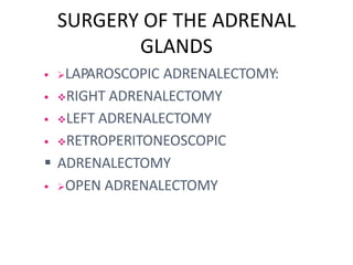
ADRENALECTOMY
Dr. SUBBAIAH. M.P.T
ADRENALECTOMY
• DISORDERSS OF ADRENAL GLAND
ANATOMY
 Weight = 4g
 2 adrenal gland,
right and left
 2 component ; inner
adrenal medulla and
outer adrenal cortex
 Situated near upper
poles of kidneys in
retro peritoneum,
within Gerota’s
capsule
ANATOMY CON’T
 Right adrenal
gland
– between
right liver
lobe and
diaphragm
 Left adrenal gland
– close to upper
pole of left kidney
and renal pedicle,
covered by
pancreatic tail and
spleen
ANATOMY CON’T
 Arterial blood suppl
–superior suprarenal
artery (from inferior
phrenic artery)
-middle suprarenal
artery (from abdomin
aorta)
- inferior suprarenal
artery (from renal
artery)
DEFINITION
 Adrenalectomy is the surgical removal of
one or both (bilateraladrenalectomy) adrenal
glands.
• It is usually advised for patients with tumors
of the adrenal glands.
• The procedure can be performed using an
open incision (laparotomy)
or laparoscopic technique.
INDICATIONS
• Conn syndrome.
• Cushing syndrome.
• Pheochromocytoma.
• Large myelolipoma.
• Metastatic tumors.
• Adrenocortical carcinoma.
• Neuroblastoma (pediatric population)
1. Conn syndrome
• Primary aldosteronism, also
known as primary
hyperaldosteronism or Co
nn's syndrome, is excess
production of the hormone
aldosterone by the adrenal
glands resulting in low renin
levels.
• Often it produces
few symptoms. Most
people have high blood
pressure which may cause
poor vision or headaches.
2. CUSHING’S SYNDROME
• Cushing's syndrome is
caused by either
excessive cortisol-like
medication such as
prednisone or a tumor
that either produces or
results in the
production of excessive
cortisol by the adrenal
glands.
3.PHEOCHROMOCYTOMA
• Pheochromocytoma is
a rare tumor of adrenal
gland tissue.
• It results in the release
of too much
epinephrine and
norepinephrine,
hormones that control
heart rate, metabolism,
and blood pressure.
4. LARGE MYELOLIPOMA
• Myelolipoma (myelo-,
from the ancient greek
marrow;
lipo, meaning of, or
pertaining to, fat; -
oma meaning tumor or
mass) is a benign
tumor-like lesion
composed of mature
adipose (fat) tissue and
haematopoietic (blood-
forming) elements in
4. METASTASTIC TUMORS
• Metastasis is the
spread of cancer cells to
new areas of the body
(often by way of the
lymph system or
bloodstream).
• A metastatic cancer,
or metastatic tumor, is
one which has spread
from the primary site of
origin (where it started)
into different area(s) of
5. ADRENO CORTICAL
CARCINOMA
• Adrenocortical
carcinoma is a rare
disease in which
malignant (cancer) cells
form in the outer layer
of theadrenal gland.
• There are
two adrenal glands.
Theadrenal glands are
small and shaped like a
triangle.
One adrenal gland sits
6. NEUROBLASTOMA
• Neuroblastoma (NB) is
a type of cancer that
forms in certain types of
nerve tissue. It most
frequently starts from
one of the adrenal
glands, but can also
develop in the neck,
chest, abdomen, or
spine. ..
PRIMARY
HYPERALDOSTERONISM (PHA)
 Hypertension , Hypokalemia,
Hypersecretion
• of aldosterone
 Hypertensive patient with hypokalaemic
PHA~
• 2%
 Hypertensive
patie potassium :
12%
SURGERY OF THE ADRENAL
GLANDS
 LAPAROSCOPIC ADRENALECTOMY:
 RIGHT ADRENALECTOMY
 LEFT ADRENALECTOMY
 RETROPERITONEOSCOPIC
 ADRENALECTOMY
 OPEN ADRENALECTOMY
TYPES OF ADRENALECTOMY
The two types of adrenalectomy
1. Open adrenalectomy
2. Laparoscopic adrenalectomy
OPEN ADRENALECTOMY
 An open adrenalectomy is often required
when either the adrenal glands or the
tumors are abnormally large.
In this procedure a single incision is made
either in the abdominal wall just under
the ribcage, or the back or sides.
OPEN ADRENALECTOMY
 Malignant adrenal tumour suspected
 Rt side: hepatic flexure of the colon is
mobilised & the rt liver lobe is cranially
retracted to achieve an optimal
exposure of the IVC & the adrenal gland
 Lf side: AG can be exposed after
mobilisation of the splenic flexure of the
colon,through the transverse mesocolon/
the gastrocolic ligament
 Resection of regional lymph node is
recommended and should include
resection of the tissue between the renal
LAPROSCOPIC ADRENALECTOMY
Laparoscopic adrenalectomy is the procedure
of choice for benign (non-cancerous) adrenal
tumors.
 Laparoscopic surgery has proved to be a
major advancement for the management of
adrenal tumors.
RIGHT ADRENALECTOMY
 Position: right side up,with table brake
 Dissection start at the level of the periadrenal
fat
 Peritoneum is divided 2cm below the edge of
liver
• from medial(IVC) to the lateral abdominal wall
 Flap of peritoneum can be used to retract the
liver up & off the adrenal
 Identify the gland and mobilise gently,
securing the vein with a clip/using one of
the available energy device
RIGHT ADRENALECTOMY
LAPROSCOPIC PROCEDURE
LEFT ADRENALECTOMY
 Position: right side
 Mobilisation of the spleen will displace
the pancreatic tail medially
 Incison of Gerota’s fascia is followed by
• identification of the adrenal vein
 The resection is completed by mobilising the
• adrenal gland at the level of the periadrenal
fat
 Remove the gland in a bag
LEFT ADRENALECTOMY LAPROSCOPIC
PROCEDURE
RETROPERITONEOSCOPIC
ADRENALECTOMY
 1ST port: distal end of the 12th rib(prone
• position)
 Digital dissection into the retroperitoneum,
• Gerota’s fascia is displaced ventrally
 RAV is covered by the retrocaval posterior
aspect of the adrenal gland
 LAV is located at the medial inferior pole of the
• adrenal gland
 High inflation pressures allow bloodless
• dissection effectively tamponading the veins
COMPLICATIONS
• Insufficient cortisol production.
• Decreases in blood pressure.
• Bleeding.
• Infections in the wound, urinary tract, or
lungs.
• Blood clots in the legs.
• Injury to nearby organs or structures.
• Adverse reaction to anesthesia.

Adrenalectomy new

  • 1.
  • 2.
  • 3.
    ANATOMY  Weight =4g  2 adrenal gland, right and left  2 component ; inner adrenal medulla and outer adrenal cortex  Situated near upper poles of kidneys in retro peritoneum, within Gerota’s capsule
  • 4.
    ANATOMY CON’T  Rightadrenal gland – between right liver lobe and diaphragm  Left adrenal gland – close to upper pole of left kidney and renal pedicle, covered by pancreatic tail and spleen
  • 5.
    ANATOMY CON’T  Arterialblood suppl –superior suprarenal artery (from inferior phrenic artery) -middle suprarenal artery (from abdomin aorta) - inferior suprarenal artery (from renal artery)
  • 6.
    DEFINITION  Adrenalectomy isthe surgical removal of one or both (bilateraladrenalectomy) adrenal glands. • It is usually advised for patients with tumors of the adrenal glands. • The procedure can be performed using an open incision (laparotomy) or laparoscopic technique.
  • 7.
    INDICATIONS • Conn syndrome. •Cushing syndrome. • Pheochromocytoma. • Large myelolipoma. • Metastatic tumors. • Adrenocortical carcinoma. • Neuroblastoma (pediatric population)
  • 8.
    1. Conn syndrome •Primary aldosteronism, also known as primary hyperaldosteronism or Co nn's syndrome, is excess production of the hormone aldosterone by the adrenal glands resulting in low renin levels. • Often it produces few symptoms. Most people have high blood pressure which may cause poor vision or headaches.
  • 9.
    2. CUSHING’S SYNDROME •Cushing's syndrome is caused by either excessive cortisol-like medication such as prednisone or a tumor that either produces or results in the production of excessive cortisol by the adrenal glands.
  • 10.
    3.PHEOCHROMOCYTOMA • Pheochromocytoma is arare tumor of adrenal gland tissue. • It results in the release of too much epinephrine and norepinephrine, hormones that control heart rate, metabolism, and blood pressure.
  • 11.
    4. LARGE MYELOLIPOMA •Myelolipoma (myelo-, from the ancient greek marrow; lipo, meaning of, or pertaining to, fat; - oma meaning tumor or mass) is a benign tumor-like lesion composed of mature adipose (fat) tissue and haematopoietic (blood- forming) elements in
  • 12.
    4. METASTASTIC TUMORS •Metastasis is the spread of cancer cells to new areas of the body (often by way of the lymph system or bloodstream). • A metastatic cancer, or metastatic tumor, is one which has spread from the primary site of origin (where it started) into different area(s) of
  • 13.
    5. ADRENO CORTICAL CARCINOMA •Adrenocortical carcinoma is a rare disease in which malignant (cancer) cells form in the outer layer of theadrenal gland. • There are two adrenal glands. Theadrenal glands are small and shaped like a triangle. One adrenal gland sits
  • 14.
    6. NEUROBLASTOMA • Neuroblastoma(NB) is a type of cancer that forms in certain types of nerve tissue. It most frequently starts from one of the adrenal glands, but can also develop in the neck, chest, abdomen, or spine. ..
  • 15.
    PRIMARY HYPERALDOSTERONISM (PHA)  Hypertension, Hypokalemia, Hypersecretion • of aldosterone  Hypertensive patient with hypokalaemic PHA~ • 2%  Hypertensive patie potassium : 12%
  • 16.
    SURGERY OF THEADRENAL GLANDS  LAPAROSCOPIC ADRENALECTOMY:  RIGHT ADRENALECTOMY  LEFT ADRENALECTOMY  RETROPERITONEOSCOPIC  ADRENALECTOMY  OPEN ADRENALECTOMY
  • 17.
    TYPES OF ADRENALECTOMY Thetwo types of adrenalectomy 1. Open adrenalectomy 2. Laparoscopic adrenalectomy
  • 18.
    OPEN ADRENALECTOMY  Anopen adrenalectomy is often required when either the adrenal glands or the tumors are abnormally large. In this procedure a single incision is made either in the abdominal wall just under the ribcage, or the back or sides.
  • 19.
    OPEN ADRENALECTOMY  Malignantadrenal tumour suspected  Rt side: hepatic flexure of the colon is mobilised & the rt liver lobe is cranially retracted to achieve an optimal exposure of the IVC & the adrenal gland  Lf side: AG can be exposed after mobilisation of the splenic flexure of the colon,through the transverse mesocolon/ the gastrocolic ligament  Resection of regional lymph node is recommended and should include resection of the tissue between the renal
  • 20.
    LAPROSCOPIC ADRENALECTOMY Laparoscopic adrenalectomyis the procedure of choice for benign (non-cancerous) adrenal tumors.  Laparoscopic surgery has proved to be a major advancement for the management of adrenal tumors.
  • 21.
    RIGHT ADRENALECTOMY  Position:right side up,with table brake  Dissection start at the level of the periadrenal fat  Peritoneum is divided 2cm below the edge of liver • from medial(IVC) to the lateral abdominal wall  Flap of peritoneum can be used to retract the liver up & off the adrenal  Identify the gland and mobilise gently, securing the vein with a clip/using one of the available energy device
  • 22.
  • 23.
    LEFT ADRENALECTOMY  Position:right side  Mobilisation of the spleen will displace the pancreatic tail medially  Incison of Gerota’s fascia is followed by • identification of the adrenal vein  The resection is completed by mobilising the • adrenal gland at the level of the periadrenal fat  Remove the gland in a bag
  • 24.
  • 25.
    RETROPERITONEOSCOPIC ADRENALECTOMY  1ST port:distal end of the 12th rib(prone • position)  Digital dissection into the retroperitoneum, • Gerota’s fascia is displaced ventrally  RAV is covered by the retrocaval posterior aspect of the adrenal gland  LAV is located at the medial inferior pole of the • adrenal gland  High inflation pressures allow bloodless • dissection effectively tamponading the veins
  • 27.
    COMPLICATIONS • Insufficient cortisolproduction. • Decreases in blood pressure. • Bleeding. • Infections in the wound, urinary tract, or lungs. • Blood clots in the legs. • Injury to nearby organs or structures. • Adverse reaction to anesthesia.