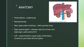
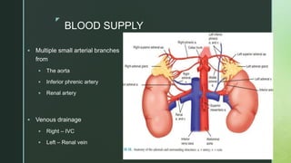
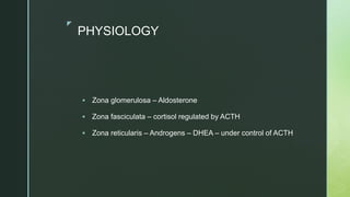
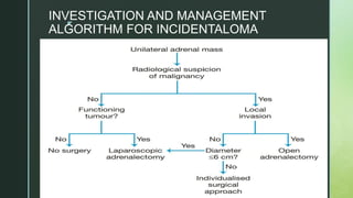
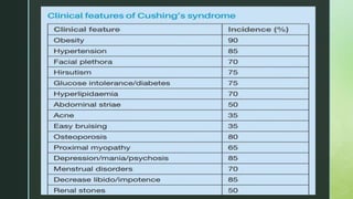
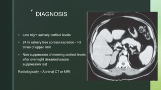
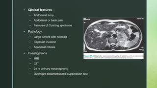
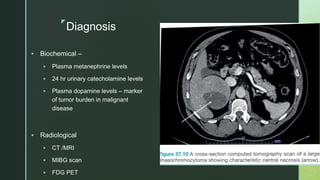
z
ADRENAL TUMORS
z
ANATOMY
 Paired glands , weighing 4g
 Retroperitoneal
 Near upper poles of kidneys , within gerotas facial
 Right adrenal gland – between right love of liver and
diaphragm, partly behind IVC
 Left – superomedial to upper poles of left kidney ,
covered by pancreatic tail and spleen
z
BLOOD SUPPLY
 Multiple small arterial branches
from
 The aorta
 Inferior phrenic artery
 Renal artery
 Venous drainage
 Right – IVC
 Left – Renal vein
z
PHYSIOLOGY
 Zona glomerulosa – Aldosterone
 Zona fasciculata – cortisol regulated by ACTH
 Zona reticularis – Androgens – DHEA – under control of ACTH
z
INCIDENTALOMA
 An asymptomatic adrenal mass detected on imaging not performed for suspected
adrenal disease
 Etiology
 Benign and malignant tumors of cortex , medulla , extra adrenal origin
 Functional or non functional – determined through
 clinical assessment
 Dexamethasone suppression test
 measurement of urinary metanephrins
 Plasma Aldosterone renin ratio
 2% incidence
z
 Benign or Malignant
 Clinically cushings and virilising tumors – Malignant
 Non contrast CT – Benign tumors <10HU
 PET /MRI
z
INVESTIGATION AND MANAGEMENT
ALGORITHM FOR INCIDENTALOMA
z
CORTICAL TUMORS
 ALDOSTERONE PRODUCING ADENOMA( Primary hyperaldosteronism )
 Arises from Zona glomerulosa
 Max diameter 10 to 20mm
 Macroscopically – Well circumscribed , golden yellow in color ( canary tumor )
 Mutations in potassium coded channels , triggering calcium influx into
glomerulosa cells
 C/F
 HTN
 Hypokalemia
 Can be asymptomatic
z
 Diagnosis
 Non suppressed plasma Aldosterone and suppressed plasma renin
activity
 Radiologically – CT and PET
 Treatment
 Prior to surgery – HTN and hypokalemia to be corrected
 Surgery – Adrenalectomy
z
ADRENAL ADENOMA
 Most common cause of adrenal Cushings syndrome
 Well circumscribed, modular in appearance
 Results in hypercortisolim leading to suppressed ACTH ,
causing atrophy of fasciculata , reticularis not glomerulosa
z
z
z
DIAGNOSIS
 Late night salivary cortisol levels
 24 hr urinary free cortisol excretion - >3
times of upper limit
 Non suppression of morning cortisol levels
after overnight dexamethasone
suppression test
Radiologically – Adrenal CT or MRI
z DEXAMETHASONE SUPPRESSION TEST
 In this test, 1 mg of a synthetic glucocorticoid (dexamethasone)
is given at 11 p.m. and plasma cortisol levels are measured at 8
a.m. the following morning.
 Physiologically normal adults suppress cortisol levels to <3
μg/dL, whereas most patients with Cushing’s syndrome do not.
 In patients with a negative test but a high clinical suspicion, the
classic low-dose dexamethasone (0.5 mg every 6 hours for eight
doses, or 2 mg over 48 hours) suppression test or urinary
cortisol measurement should be performed.
z
TREATMENT
 Unilateral adenoma – Adrenalectomy
 Bilateral Adrenalectomy – in severe and symmetrically enlarged
adrenal
 Asymmetric disease – excision of larger gland
z
ADRENOCORTICAL CARCINOMA
 Rare aggressive malignancy from adrenal cortex
 Presents at late stage with poor prognosis
 Sporadic – majority , or as a part of tumor
syndromes ( MEN 1 )
 Incidence – 1 in 1 lakh
 Female preponderance
 Non functional > functional
z
 Clinical features
 Abdominal lump ,
 Abdominal or back pain
 Features of Cushing syndrome
 Pathology
 Large tumors with necrosis
 Capsular invasion
 Abnormal mitosis
 Investigations
 MRI
 CT
 24 hr uninary metanephrins
 Overnight dexamethasone suppression test
z
Treatment
 Indeterminate or probably malignant tumor < 6cm : laparoscopic
Adrenalectomy
 Indeterminate or probably Malignant>6cm : Open radical
resection with en bloc dissection of involved adjacent organs
 Metastatic disease
 Limited disease – open resection of primary tumor with adjuvant
treatment of mets
 Widespread metastatic disease – palliative treatment
z
ADRENAL MEDULLA TUMORS
 PHEOCHROMOCYCTOMA AND PARAGANGLIOMA (PPGL )
 Tumors arising from the neuroectoderm tissue of adrenal
medulla are pheochromocytomas( PCC )
 Those arising from extraadrenal parasympathetic ( carotid body
) and sympathetic ganglia( organ of zuckerkandl) are
paragangliomas ( PGL )
 70% sporadic , remaining –familial
 Rule of 10 – 10% Malignant, 10% familial , 10%bilateral ,10%
Extraadrenal,10%Children .
z
 Pathology
 Highly vascularised with greyish pink
on cut surface
 Areas of necrosis and hemorrhage
 PCC produce calcitonin, ACTH,
vasoactive intestinal polypeptide
(VIP) and parathyroid hormone-
related protein (PTHrP)
z
Diagnosis
 Biochemical –
 Plasma metanephrine levels
 24 hr urinary catecholamine levels
 Plasma dopamine levels – marker
of tumor burden in malignant
disease
 Radiological
 CT /MRI
 MIBG scan
 FDG PET
z
Treatment
 Medical Management
 Pre-op control of blood pressure with phenoxybenzamine ( Alpha
adrenergic receptor blocker ) 20mg initially followed by 10mg until
daily dose of 100 – 160 mg is reached
 Beta adrenergic blockers if tachycardia or arrhythmias present
 Post operatively – hypoglycemia and hypovolemic should be
checked
 Surgical Treatment
 Adrenalectomy
z
Neuroblastoma
 Most common and deadliest extracranial malignancy in children
 Derived from primitive nerve cells of sympathetic nervous
system of neural crest cells
 Clinical enigma
 Peak incidence – 5yrs of age
 Clinical features – lump across midline of abdomen , Diarrhea,
dehydratio due to VIP
 Metastatic lesions – blue berry muffin lesions , racoon eyes
z
 Investigations – MRI > CECT
 Treatment – Surgical Resection of primary tumor
Chemotherapy – Etoposide , Cisplatin
z
GANGLIONEUROMA
 Ganglioneuromas (GNs) are benign diferentiated tumours of neural
crest-derived cells in the autonomic nerves.
 Sporadic , assosciated with MEN 2b
 Pathology – admixture of ganglion and schwann cells
 Sites – Mediastinum , retroperitoneal, Adrenal gland
 Clinical features – benign , present as non secreting slow growing
tumors
 Usually asymptomatic, <30years , when large in size cause
pressure symptoms
z
 Diagnosis – CT /MRI – well defined with capsule and
calcification
 Treatment – Surgical excision – laparoscopic method preferred
 Excellent prognosis
z
THANK YOU

adrenal tumors ppt.pptx presentation....

  • 1.
  • 2.
    z ANATOMY  Paired glands, weighing 4g  Retroperitoneal  Near upper poles of kidneys , within gerotas facial  Right adrenal gland – between right love of liver and diaphragm, partly behind IVC  Left – superomedial to upper poles of left kidney , covered by pancreatic tail and spleen
  • 3.
    z BLOOD SUPPLY  Multiplesmall arterial branches from  The aorta  Inferior phrenic artery  Renal artery  Venous drainage  Right – IVC  Left – Renal vein
  • 4.
    z PHYSIOLOGY  Zona glomerulosa– Aldosterone  Zona fasciculata – cortisol regulated by ACTH  Zona reticularis – Androgens – DHEA – under control of ACTH
  • 5.
    z INCIDENTALOMA  An asymptomaticadrenal mass detected on imaging not performed for suspected adrenal disease  Etiology  Benign and malignant tumors of cortex , medulla , extra adrenal origin  Functional or non functional – determined through  clinical assessment  Dexamethasone suppression test  measurement of urinary metanephrins  Plasma Aldosterone renin ratio  2% incidence
  • 6.
    z  Benign orMalignant  Clinically cushings and virilising tumors – Malignant  Non contrast CT – Benign tumors <10HU  PET /MRI
  • 7.
  • 8.
    z CORTICAL TUMORS  ALDOSTERONEPRODUCING ADENOMA( Primary hyperaldosteronism )  Arises from Zona glomerulosa  Max diameter 10 to 20mm  Macroscopically – Well circumscribed , golden yellow in color ( canary tumor )  Mutations in potassium coded channels , triggering calcium influx into glomerulosa cells  C/F  HTN  Hypokalemia  Can be asymptomatic
  • 9.
    z  Diagnosis  Nonsuppressed plasma Aldosterone and suppressed plasma renin activity  Radiologically – CT and PET  Treatment  Prior to surgery – HTN and hypokalemia to be corrected  Surgery – Adrenalectomy
  • 10.
    z ADRENAL ADENOMA  Mostcommon cause of adrenal Cushings syndrome  Well circumscribed, modular in appearance  Results in hypercortisolim leading to suppressed ACTH , causing atrophy of fasciculata , reticularis not glomerulosa
  • 11.
  • 12.
  • 13.
    z DIAGNOSIS  Late nightsalivary cortisol levels  24 hr urinary free cortisol excretion - >3 times of upper limit  Non suppression of morning cortisol levels after overnight dexamethasone suppression test Radiologically – Adrenal CT or MRI
  • 14.
    z DEXAMETHASONE SUPPRESSIONTEST  In this test, 1 mg of a synthetic glucocorticoid (dexamethasone) is given at 11 p.m. and plasma cortisol levels are measured at 8 a.m. the following morning.  Physiologically normal adults suppress cortisol levels to <3 μg/dL, whereas most patients with Cushing’s syndrome do not.  In patients with a negative test but a high clinical suspicion, the classic low-dose dexamethasone (0.5 mg every 6 hours for eight doses, or 2 mg over 48 hours) suppression test or urinary cortisol measurement should be performed.
  • 15.
    z TREATMENT  Unilateral adenoma– Adrenalectomy  Bilateral Adrenalectomy – in severe and symmetrically enlarged adrenal  Asymmetric disease – excision of larger gland
  • 16.
    z ADRENOCORTICAL CARCINOMA  Rareaggressive malignancy from adrenal cortex  Presents at late stage with poor prognosis  Sporadic – majority , or as a part of tumor syndromes ( MEN 1 )  Incidence – 1 in 1 lakh  Female preponderance  Non functional > functional
  • 17.
    z  Clinical features Abdominal lump ,  Abdominal or back pain  Features of Cushing syndrome  Pathology  Large tumors with necrosis  Capsular invasion  Abnormal mitosis  Investigations  MRI  CT  24 hr uninary metanephrins  Overnight dexamethasone suppression test
  • 18.
    z Treatment  Indeterminate orprobably malignant tumor < 6cm : laparoscopic Adrenalectomy  Indeterminate or probably Malignant>6cm : Open radical resection with en bloc dissection of involved adjacent organs  Metastatic disease  Limited disease – open resection of primary tumor with adjuvant treatment of mets  Widespread metastatic disease – palliative treatment
  • 19.
    z ADRENAL MEDULLA TUMORS PHEOCHROMOCYCTOMA AND PARAGANGLIOMA (PPGL )  Tumors arising from the neuroectoderm tissue of adrenal medulla are pheochromocytomas( PCC )  Those arising from extraadrenal parasympathetic ( carotid body ) and sympathetic ganglia( organ of zuckerkandl) are paragangliomas ( PGL )  70% sporadic , remaining –familial  Rule of 10 – 10% Malignant, 10% familial , 10%bilateral ,10% Extraadrenal,10%Children .
  • 20.
    z  Pathology  Highlyvascularised with greyish pink on cut surface  Areas of necrosis and hemorrhage  PCC produce calcitonin, ACTH, vasoactive intestinal polypeptide (VIP) and parathyroid hormone- related protein (PTHrP)
  • 21.
    z Diagnosis  Biochemical – Plasma metanephrine levels  24 hr urinary catecholamine levels  Plasma dopamine levels – marker of tumor burden in malignant disease  Radiological  CT /MRI  MIBG scan  FDG PET
  • 22.
    z Treatment  Medical Management Pre-op control of blood pressure with phenoxybenzamine ( Alpha adrenergic receptor blocker ) 20mg initially followed by 10mg until daily dose of 100 – 160 mg is reached  Beta adrenergic blockers if tachycardia or arrhythmias present  Post operatively – hypoglycemia and hypovolemic should be checked  Surgical Treatment  Adrenalectomy
  • 23.
    z Neuroblastoma  Most commonand deadliest extracranial malignancy in children  Derived from primitive nerve cells of sympathetic nervous system of neural crest cells  Clinical enigma  Peak incidence – 5yrs of age  Clinical features – lump across midline of abdomen , Diarrhea, dehydratio due to VIP  Metastatic lesions – blue berry muffin lesions , racoon eyes
  • 24.
    z  Investigations –MRI > CECT  Treatment – Surgical Resection of primary tumor Chemotherapy – Etoposide , Cisplatin
  • 25.
    z GANGLIONEUROMA  Ganglioneuromas (GNs)are benign diferentiated tumours of neural crest-derived cells in the autonomic nerves.  Sporadic , assosciated with MEN 2b  Pathology – admixture of ganglion and schwann cells  Sites – Mediastinum , retroperitoneal, Adrenal gland  Clinical features – benign , present as non secreting slow growing tumors  Usually asymptomatic, <30years , when large in size cause pressure symptoms
  • 26.
    z  Diagnosis –CT /MRI – well defined with capsule and calcification  Treatment – Surgical excision – laparoscopic method preferred  Excellent prognosis
  • 27.