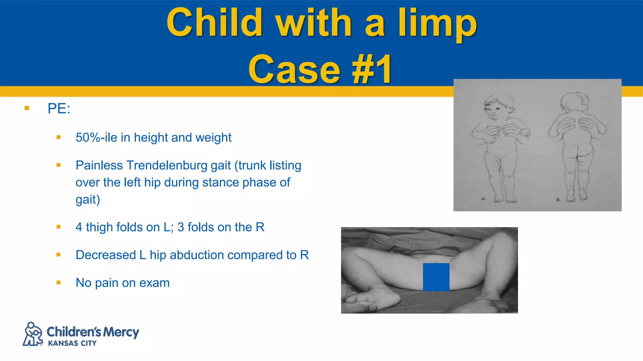
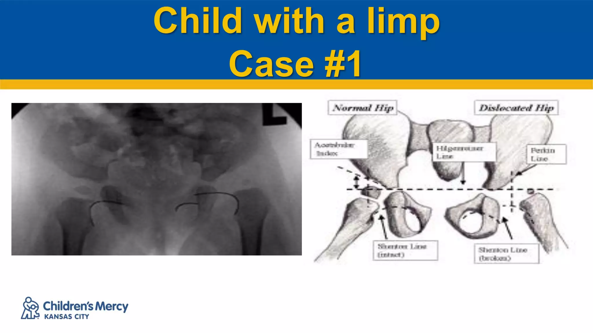
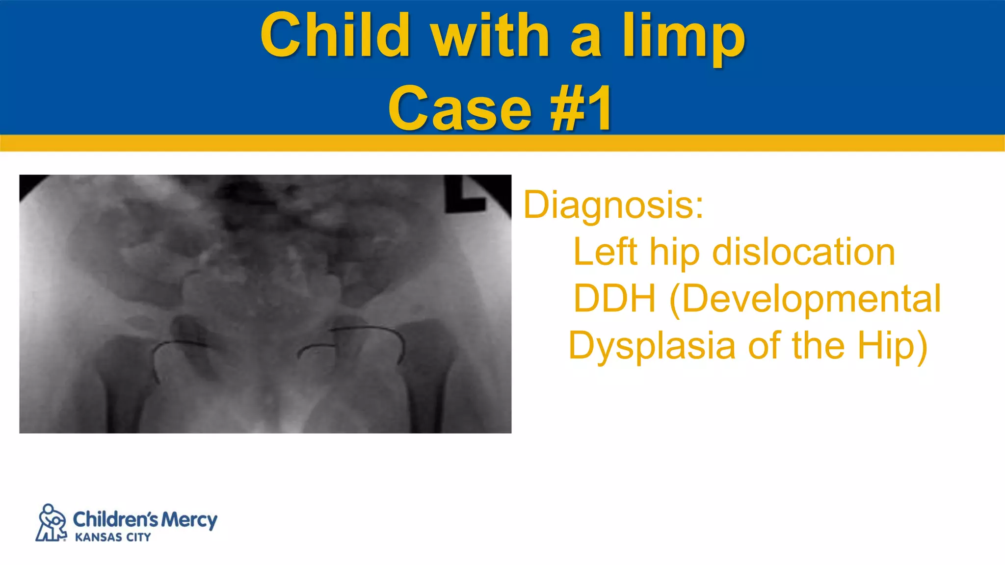
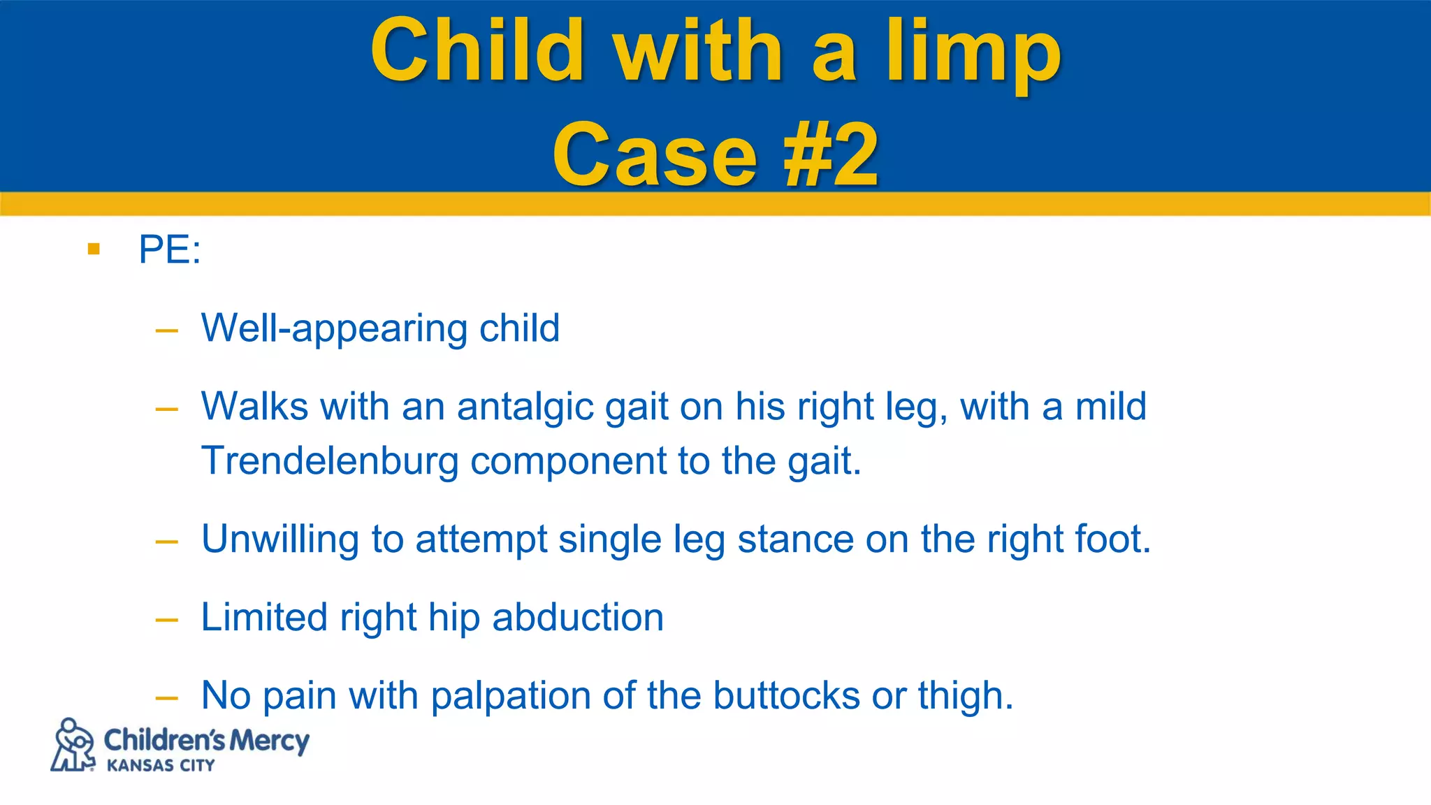
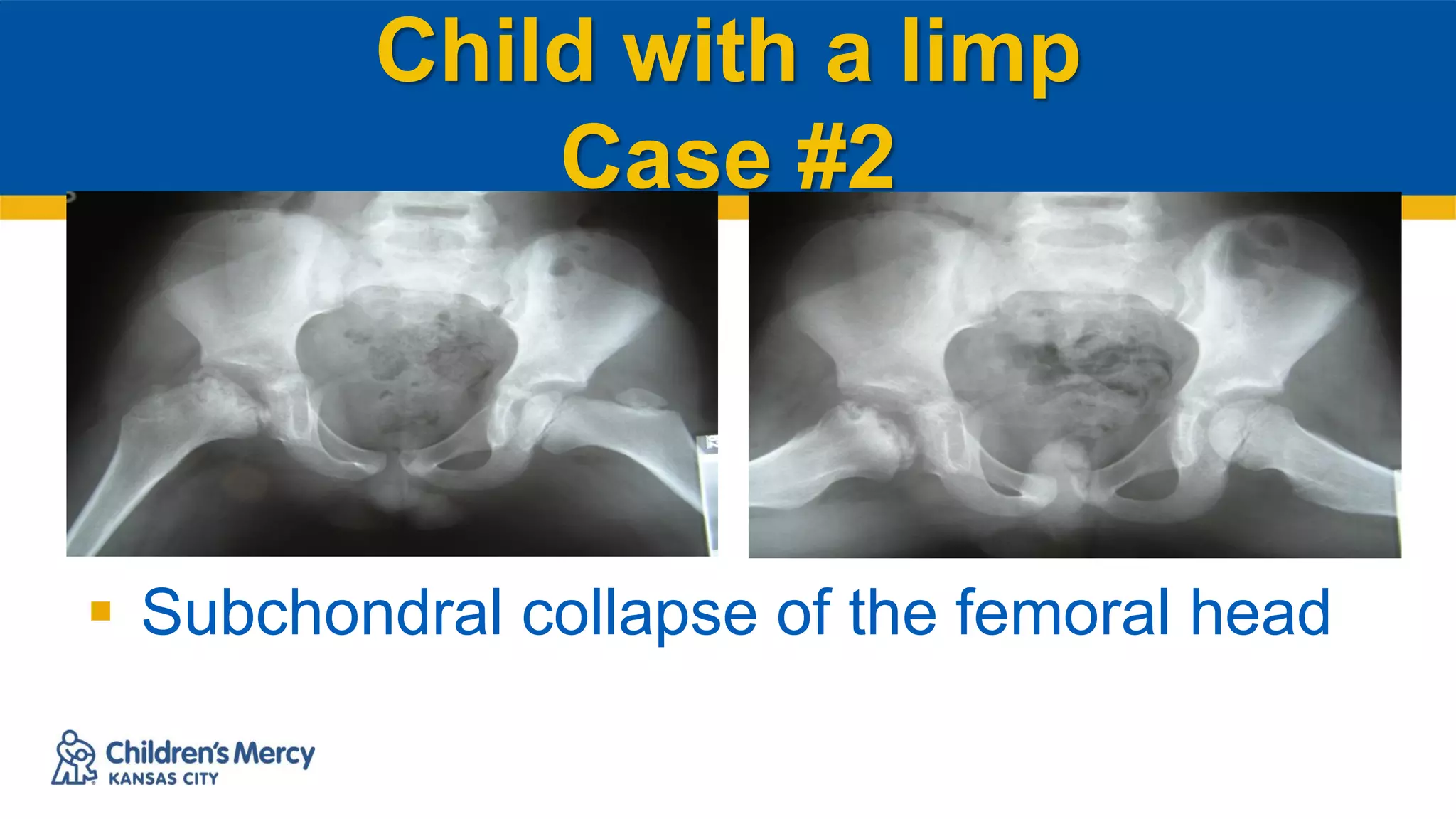
© The Children's Mercy Hospital, 2016
The Limping Child:
Differential Diagnosis
Kathryn A Keeler, MD
Assistant Professor
University of Missouri-Kansas City School of Medicine, Department of Orthopaedic Surgery and
Department of Pediatrics
Children’s Mercy – Kansas City, Division of Orthopaedics and Section of Sports Medicine
Disclosures
 I have no relevant financial relationships with the
manufacturer(s) of any commercial product(s) and/or
provider(s) of commercial services discussed in this CME
activity.
 I do not intend to discuss an unapproved/investigative use
of a commercial product/device in my presentation
2
Child with a limp
 What is a limp?
 1 a: to walk lamely; especially: to walk
favoring one leg. b: to go unsteadily
 2: to proceed slowly with difficulty
Child with a limp
 Describe the gait pattern:
– Antalgic gait…painful
– Trendelenberg gait…torso shifts
Child with a limp
 Localize:
– Spine
– Hip
– Knee
– Foot
Child with a limp
 Consider patient factors:
– Age
– PMH
– Fam Hx
Child with a limp
 V
 I
 N
 D
 I
 C
 A
 T
 E
Child with a limp
 Vascular (Legg-Calve-Perthes)
 Infection (septic arthritis, psoas abscess, diskitis)
 Neoplasm (tumor, lymphoma, leukemia)
 Developmental or Neuromuscular disorders
 Inflammatory (transient synovitis, RA, SLE)
 Congenital (DDH)
 Autoimmune
 Trauma
 Endocrine / Metabolic
Child with a limp
Case #1
 18 month old female
 CC: limp
 No pain
 No trauma
Child with a limp
Case #1
Birth/Developmental Hx:
 Uncomplicated pregnancy
 Breech delivery
 9.5# at birth
– Sitting at 6 months
– Walking at 13 months
Child with a limp
Case #1
 PE:
 50%-ile in height and weight
 Painless Trendelenburg gait (trunk listing
over the left hip during stance phase of
gait)
 4 thigh folds on L; 3 folds on the R
 Decreased L hip abduction compared to R
 No pain on exam
Child with a limp
Case #1
Child with a limp
Case #1
Diagnosis:
Left hip dislocation
DDH (Developmental
Dysplasia of the Hip)
Child with a limp
Case #1
 Treatment: Surgical reduction of the hip
– Permits acetabular development due to femoral head deeply in
acetabulum
Child with a limp
Case #1
 Factors associated with DDH:
– Breech positioning (intrauterine molding)
– High birth weight
– Family history
– Female
– L hip
Child with a limp
Case #2
 4 y/o male
 CC: Limp
 Painful limp for 3 months
 Increased pain on weight-bearing for the past 2 weeks.
Child with a limp
Case #2
 PE:
– Well-appearing child
– Walks with an antalgic gait on his right leg, with a mild
Trendelenburg component to the gait.
– Unwilling to attempt single leg stance on the right foot.
– Limited right hip abduction
– No pain with palpation of the buttocks or thigh.
Child with a limp
Case #2
 Subchondral collapse of the femoral head
Child with a limp
Case #2
– Diagnosis: Legg-Calve-Perthes disease
– Idiopathic avascular/ischemic necrosis of the
femoral head
Child with a limp
Case #2
 Treatment: Optimize sphericity (round vs. flattened) of
femoral head during healing process
– Limit weight bearing
– Regain hip range of motion
 Physiotherapy
 Surgery
 Surgical containment of femoral head in acetabulum
Child with a limp
Case #3
 4 y/o female
 CC: Limp
 3 day h/o of a left increasing groin pain and limp
 2 weeks prior: uncomplicated URI that resolved 10 days
prior to the onset of limp.
Child with a limp
Case #3
 PE:
– T = 38.5 C
– Does not appear sick/toxic.
– Left hip is held in abduction, external rotation and slight flexion.
– Resists pROM of the left hip
Child with a limp
Case #3
Slight widening of the left hip joint.
Child with a limp
Case #3
 Labs
– CBC: Normal WBC, no shift
– ESR: 19
– CRP: 0.6
Child with a limp
Case #3
 Hip aspiration / arthrocentesis
– Ultrasonographic guidance
– Clear yellowish fluid
– + Lymphocytes
– Gram’s stain: no organisms
Child with a limp
Case #3
 Diagnosis: Transient synovitis
– Must r/o septic/bacterial arthritis of the hip and osteomyelitis of the proximal
femur.
– Toxic synovitis is a diagnosis of exclusion.
 Treatment:
– Restriction of weight-bearing (short-term)
– Gentle range of motion
– NSAIDs and antipyretics.
Child with a limp
Case #4
 7 y/o male
 CC: Limp
 Increasing left groin / buttock pain and limp for 5 days.
 Now, unable to bear weight
 Uncomplicated URI of one week’s duration that resolved ten days
prior to the onset of his limp.
Child with a limp
Case #4
– T: 39.5 C
– Appears sick/toxic.
– Left hip is held in abduction, external rotation and slight flexion.
– Painful arc of hip motion (all planes), with the greatest discomfort
felt on extension and internal rotation.
– Substantial active patient resistance to passive motion
Child with a limp
Case #4
 Labs
– CBC neutrophilia, with left shift to immature cells in
the peripheral smear.
– ESR: 40
– CRP: 3
Child with a limp
Case #4
 Hip ultrasound with aspiration if a hip
effusion
– Arthrocentesis:
 Cloudy yellow (purulent) material.
 Neutrophils >100,000/ml are present.
Child with a limp
Case #4
 Diagnosis: Septic or bacterial arthritis of
the hip.
 Treatment: Emergent hip arthrotomy for
irrigation and drainage (I&D).
 This is a surgical emergency!!
Child with a limp
Case #4
Child with a limp
Case #5
 12 y/o male with 3 week h/o increasingly painful limp
 Right groin/knee pain increasing over the preceding 3
days.
 Pain localized to thigh and knee joint.
Child with a limp
Case #5
– Obese
– Afebrile.
– Unable to elevate right leg off exam table
– Passive motion of the hip joint causes severe knee, groin and buttock
pain.
– Leg held in adducted and externally rotated position
Child with a limp
Case #5
Child with a limp
Case #5
 Diagnosis: SCFE (Slipped capital femoral epiphysis)
 Treatment: In-situ screw fixation
 Complications of untreated SCFEs:
– LLD: limb length discrepancy
– Femoral retrotorsion (excessive external rotation)
– Early onset osteoarthritis,
– Ischemic necrosis of proximal femoral epiphysis
Child with a limp
Case #6
 14 y/o female soccer player with a 4 month h/o a mild limp.
 Multiple episodes of painful locking of the knee (an inability to
straighten out the knee because she feels that it is ‘stuck’)
 Knee swelling
 h/o a twisting injury 18 months ago while playing basketball
Child with a limp
Case #6
– Athletic female
– Normal hip and knee AROM/PROM
– Mild knee effusion
– Tender to palpation over medial femoral condyle adjacent to the
patella.
– No ligamentous laxity
– No tenderness at medial or lateral joint lines.
Child with a limp
Case #6
Child with a limp
Case #6
 Dx: Osteochondritis dissecans (OCD)
– Subchondral bone resorption
– Initially cartilage is intact
– Later stages cartilage may fracture and detach forming a loose body
– A loose body is freely mobile and can cause locking and knee effusions.
– In general terms, the treatment for OCD loose bodies is arthroscopic
excision or repair.
Child with a limp
Case #7
 16 y/o female volleyball player
 4 month h/o mild limp associated with diffuse and poorly
localized knee pain.
 No history of clicking, locking, or instability of the knee.
 Persistent mild swelling of the knee is present.
 No history of trauma.
Child with a limp
Case #7
– Athletic appearing female
– Normal hip and knee AROM/PROM
– Mild knee effusion is present.
– Tender to palpation over the distal femur 6 cm superior to the
patella.
– No ligamentous laxity
– No tenderness at medial or lateral joint lines.
Child with a limp
Case #7
 Permeative
osteolytic/osteoblastic
lesion
 Indistinct borders
 Soft tissues invasion
 Periosteal elevation
Child with a limp
Case #7
 Work-up
– Plain radiographs (ENTIRE bone)
– Technetium bone scan
– Chest CT
– MRI
– Biopsy (open vs. needle)
 Dx: Osteosarcoma vs. Ewing’s sarcoma
Child with a limp
 ALWAYS get an x-ray. Every patient with a musculoskeletal
complaint deserves a normal x-ray, after all.
 KNEE PAIN in an adolescent patient is SCFE until proven otherwise.
 ALWAYS get a plain AP and lateral x-ray before vague ill-defined
knee pain is ascribed to ‘idiopathic anterior knee pain of
adolescence.’

Approach to a limping child.pdf

  • 1.
    © The Children'sMercy Hospital, 2016 The Limping Child: Differential Diagnosis Kathryn A Keeler, MD Assistant Professor University of Missouri-Kansas City School of Medicine, Department of Orthopaedic Surgery and Department of Pediatrics Children’s Mercy – Kansas City, Division of Orthopaedics and Section of Sports Medicine
  • 2.
    Disclosures  I haveno relevant financial relationships with the manufacturer(s) of any commercial product(s) and/or provider(s) of commercial services discussed in this CME activity.  I do not intend to discuss an unapproved/investigative use of a commercial product/device in my presentation 2
  • 3.
    Child with alimp  What is a limp?  1 a: to walk lamely; especially: to walk favoring one leg. b: to go unsteadily  2: to proceed slowly with difficulty
  • 4.
    Child with alimp  Describe the gait pattern: – Antalgic gait…painful – Trendelenberg gait…torso shifts
  • 5.
    Child with alimp  Localize: – Spine – Hip – Knee – Foot
  • 6.
    Child with alimp  Consider patient factors: – Age – PMH – Fam Hx
  • 7.
    Child with alimp  V  I  N  D  I  C  A  T  E
  • 8.
    Child with alimp  Vascular (Legg-Calve-Perthes)  Infection (septic arthritis, psoas abscess, diskitis)  Neoplasm (tumor, lymphoma, leukemia)  Developmental or Neuromuscular disorders  Inflammatory (transient synovitis, RA, SLE)  Congenital (DDH)  Autoimmune  Trauma  Endocrine / Metabolic
  • 9.
    Child with alimp Case #1  18 month old female  CC: limp  No pain  No trauma
  • 10.
    Child with alimp Case #1 Birth/Developmental Hx:  Uncomplicated pregnancy  Breech delivery  9.5# at birth – Sitting at 6 months – Walking at 13 months
  • 11.
    Child with alimp Case #1  PE:  50%-ile in height and weight  Painless Trendelenburg gait (trunk listing over the left hip during stance phase of gait)  4 thigh folds on L; 3 folds on the R  Decreased L hip abduction compared to R  No pain on exam
  • 12.
    Child with alimp Case #1
  • 13.
    Child with alimp Case #1 Diagnosis: Left hip dislocation DDH (Developmental Dysplasia of the Hip)
  • 14.
    Child with alimp Case #1  Treatment: Surgical reduction of the hip – Permits acetabular development due to femoral head deeply in acetabulum
  • 15.
    Child with alimp Case #1  Factors associated with DDH: – Breech positioning (intrauterine molding) – High birth weight – Family history – Female – L hip
  • 16.
    Child with alimp Case #2  4 y/o male  CC: Limp  Painful limp for 3 months  Increased pain on weight-bearing for the past 2 weeks.
  • 17.
    Child with alimp Case #2  PE: – Well-appearing child – Walks with an antalgic gait on his right leg, with a mild Trendelenburg component to the gait. – Unwilling to attempt single leg stance on the right foot. – Limited right hip abduction – No pain with palpation of the buttocks or thigh.
  • 18.
    Child with alimp Case #2  Subchondral collapse of the femoral head
  • 19.
    Child with alimp Case #2 – Diagnosis: Legg-Calve-Perthes disease – Idiopathic avascular/ischemic necrosis of the femoral head
  • 20.
    Child with alimp Case #2  Treatment: Optimize sphericity (round vs. flattened) of femoral head during healing process – Limit weight bearing – Regain hip range of motion  Physiotherapy  Surgery  Surgical containment of femoral head in acetabulum
  • 21.
    Child with alimp Case #3  4 y/o female  CC: Limp  3 day h/o of a left increasing groin pain and limp  2 weeks prior: uncomplicated URI that resolved 10 days prior to the onset of limp.
  • 22.
    Child with alimp Case #3  PE: – T = 38.5 C – Does not appear sick/toxic. – Left hip is held in abduction, external rotation and slight flexion. – Resists pROM of the left hip
  • 23.
    Child with alimp Case #3 Slight widening of the left hip joint.
  • 24.
    Child with alimp Case #3  Labs – CBC: Normal WBC, no shift – ESR: 19 – CRP: 0.6
  • 25.
    Child with alimp Case #3  Hip aspiration / arthrocentesis – Ultrasonographic guidance – Clear yellowish fluid – + Lymphocytes – Gram’s stain: no organisms
  • 26.
    Child with alimp Case #3  Diagnosis: Transient synovitis – Must r/o septic/bacterial arthritis of the hip and osteomyelitis of the proximal femur. – Toxic synovitis is a diagnosis of exclusion.  Treatment: – Restriction of weight-bearing (short-term) – Gentle range of motion – NSAIDs and antipyretics.
  • 27.
    Child with alimp Case #4  7 y/o male  CC: Limp  Increasing left groin / buttock pain and limp for 5 days.  Now, unable to bear weight  Uncomplicated URI of one week’s duration that resolved ten days prior to the onset of his limp.
  • 28.
    Child with alimp Case #4 – T: 39.5 C – Appears sick/toxic. – Left hip is held in abduction, external rotation and slight flexion. – Painful arc of hip motion (all planes), with the greatest discomfort felt on extension and internal rotation. – Substantial active patient resistance to passive motion
  • 29.
    Child with alimp Case #4  Labs – CBC neutrophilia, with left shift to immature cells in the peripheral smear. – ESR: 40 – CRP: 3
  • 30.
    Child with alimp Case #4  Hip ultrasound with aspiration if a hip effusion – Arthrocentesis:  Cloudy yellow (purulent) material.  Neutrophils >100,000/ml are present.
  • 31.
    Child with alimp Case #4  Diagnosis: Septic or bacterial arthritis of the hip.  Treatment: Emergent hip arthrotomy for irrigation and drainage (I&D).  This is a surgical emergency!!
  • 32.
    Child with alimp Case #4
  • 33.
    Child with alimp Case #5  12 y/o male with 3 week h/o increasingly painful limp  Right groin/knee pain increasing over the preceding 3 days.  Pain localized to thigh and knee joint.
  • 34.
    Child with alimp Case #5 – Obese – Afebrile. – Unable to elevate right leg off exam table – Passive motion of the hip joint causes severe knee, groin and buttock pain. – Leg held in adducted and externally rotated position
  • 35.
    Child with alimp Case #5
  • 36.
    Child with alimp Case #5  Diagnosis: SCFE (Slipped capital femoral epiphysis)  Treatment: In-situ screw fixation  Complications of untreated SCFEs: – LLD: limb length discrepancy – Femoral retrotorsion (excessive external rotation) – Early onset osteoarthritis, – Ischemic necrosis of proximal femoral epiphysis
  • 37.
    Child with alimp Case #6  14 y/o female soccer player with a 4 month h/o a mild limp.  Multiple episodes of painful locking of the knee (an inability to straighten out the knee because she feels that it is ‘stuck’)  Knee swelling  h/o a twisting injury 18 months ago while playing basketball
  • 38.
    Child with alimp Case #6 – Athletic female – Normal hip and knee AROM/PROM – Mild knee effusion – Tender to palpation over medial femoral condyle adjacent to the patella. – No ligamentous laxity – No tenderness at medial or lateral joint lines.
  • 39.
    Child with alimp Case #6
  • 40.
    Child with alimp Case #6  Dx: Osteochondritis dissecans (OCD) – Subchondral bone resorption – Initially cartilage is intact – Later stages cartilage may fracture and detach forming a loose body – A loose body is freely mobile and can cause locking and knee effusions. – In general terms, the treatment for OCD loose bodies is arthroscopic excision or repair.
  • 41.
    Child with alimp Case #7  16 y/o female volleyball player  4 month h/o mild limp associated with diffuse and poorly localized knee pain.  No history of clicking, locking, or instability of the knee.  Persistent mild swelling of the knee is present.  No history of trauma.
  • 42.
    Child with alimp Case #7 – Athletic appearing female – Normal hip and knee AROM/PROM – Mild knee effusion is present. – Tender to palpation over the distal femur 6 cm superior to the patella. – No ligamentous laxity – No tenderness at medial or lateral joint lines.
  • 43.
    Child with alimp Case #7  Permeative osteolytic/osteoblastic lesion  Indistinct borders  Soft tissues invasion  Periosteal elevation
  • 44.
    Child with alimp Case #7  Work-up – Plain radiographs (ENTIRE bone) – Technetium bone scan – Chest CT – MRI – Biopsy (open vs. needle)  Dx: Osteosarcoma vs. Ewing’s sarcoma
  • 45.
    Child with alimp  ALWAYS get an x-ray. Every patient with a musculoskeletal complaint deserves a normal x-ray, after all.  KNEE PAIN in an adolescent patient is SCFE until proven otherwise.  ALWAYS get a plain AP and lateral x-ray before vague ill-defined knee pain is ascribed to ‘idiopathic anterior knee pain of adolescence.’