Lumbar and sacral Biomechanics
•
108 likes•17,677 views

The document discusses the anatomy and biomechanics of the lumbar spine and sacrum. It describes the typical lumbar vertebrae, intervertebral discs, spinal ligaments, muscles, and motions of the lumbar spine. It also summarizes common spinal disorders like back strain, herniated discs, spondylolysis, and scoliosis. The sacroiliac joint and sacral anatomy are also briefly covered.

Recommended
Recommended
More Related Content
What's hot
What's hot (20)
Viewers also liked
Similar to Lumbar and sacral Biomechanics
Similar to Lumbar and sacral Biomechanics (20)
More from Sreeraj S R
More from Sreeraj S R (20)
Recently uploaded
Recently uploaded (20)
Lumbar and sacral Biomechanics
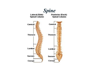
- 1. Spine
- 3. Sreeraj S R Lumbar Anatomy 5 vertebrae L1-L5 5 intervertebral discs 5 pair of exiting nerve roots Lumbar lordosis L1-S1 ranges from 30°–80° The apex of lumbar lordosis L3-L4 1 2 3 4 5
- 4. Sreeraj S R Lumbar Spine Anatomy Typical lumbar vertebra (L2) Body Vertebral foramen/canal Intervertebral foramen Pedicle Transverse process Lamina Spinous process Facet joints Pars interarticularis inferior Superior Anterior (oblique) A Lateral P Posterior (oblique) Superior Inferiorsuperior
- 5. Sreeraj S R Intervertebral Disc Soft fibro-cartilaginous cushions Between two vertebra Allows some motion Serve as shock absorbers Total – 23 discs ¼ th of the spinal column's length Avascular Nutrients diffuse through end plates Collagen
- 6. Sreeraj S R Intervertebral Disc NUCLEUS PULPOSUS Has more water and PGs PG are macro-molecules Attract and retain water Hydrophilic gel–like matter Resists compression Amount of water Activity related Varies throughout the day
- 7. Sreeraj S R Intervertebral Disc NUCLEUS PULPOSUS Eccentrically positioned posteriorly Young & healthy, 90% water, bound to proteoglycans Aging> desiccation> increase viscosity> fissuring Young nucleus> even distribution of load Old nucleus> undue concentration on vertebral body edges Small displacement w/ ROM, ball- bearing like Compressive stress predominates
- 8. Sreeraj S R Intervertebral Disc NUCLEUS PULPOSUS Pascal’s law: Fluid mass within closed container> local increase in pressure> transmit around entire side wall (annulus) Nucleus pulpous imbibes water Develops internal pressure Pressure exerted in all directions Lateral forces against annulus Superiorly and inferiorly directed forces against end plates Increases stiffness of end plate and annulus fibrosus
- 9. Sreeraj S R Intervertebral Disc ANNULUS FIBROSUS Strong radial tire–like structure Series of lamellae Concentric sheets of collagen fibers Connected to end plates Orientated at various angles Under compression Become horizontal Encloses nucleus pulposus
- 10. Sreeraj S R Intradiscal Pressure INTRADISCAL PRESSURE Compressive loads in vivo: 500N standing, 700N sitting Increased to 3000 to 6000N during lifting of moderate weights, decreases with load closer to body Estimate of P = 1.5X compressive load divided by the cross sectional area Disk pressure is usually uniform Pressure lowest in supine position Disk usually does not fail, but end plates fracture
- 11. Sreeraj S R Spinal Ligaments Anterior Longitudinal Posterior Longitudinal Ligamentum Flavum Interspinous Ligaments Supraspinous Ligaments Intertransverse Ligaments
- 12. Sreeraj S R Lumbar Spine
- 13. Sreeraj S R Lumbar Spine Thoraco lumbar fascia Stabilizing corset Transmit load longitudinally to the spinous process Ilio lumbar ligament Stabilize 5th lumbar vertebrae from ant. Displacement
- 14. Types of motion
- 15. Sreeraj S R Stress-Strain Curve
- 16. Sreeraj S R The Motion Segment Functional Spinal Unit 2 adjacent vertebrae & intervening soft tissue Anterior Vertebral body Disk ALL, PLL Support, absorb impact, restrict vertical translation Posterior Neural arch & its processes Facet joint
- 17. Sreeraj S R STABILITY The vertebral column subject to Axial compression Bending Torsion Shear
- 18. Sreeraj S R STABILITY Primary load-transmitting element, 80-90% Bone Mineral Content, Size Osteoporosis> loss of horizontal trabeculae Increasing size from C to L spine Compressive load> pressure higher in center of end plates than periphery In vivo, filled with blood> greater strength, hydraulic shock absorber
- 19. Sreeraj S R STABILITY POSTERIOR ELEMENTS pedicles, lamina, facet joints, spinous & transverse processes Bony processes> lengthen moment arms of muscles Forces on processes> transmitted to Lamina Forces on posterior elements> transmitted to vertebral bodies from Pedicles Pars Interarticularis Large bending forces; excessive extension Thicker than rest of lamina Common site of stress/fatigue fractures> weakens motion segment> spondylolithesis
- 20. Sreeraj S R STABILITY Facet Joints Major role in controlling motion Resist torsion & shear, role in compression Lumbar FSU – facets 40% torque resistance, 40% disk, 20% ligaments Load sharing varies with flexion & extension Seated position> decreased lumbar lordosis> increased intradiscal pressure & decreased load-bearing of the facets Orientation of facets C spine - 45º transverse, parallel frontal T spine - 60º transverse, 20º frontal L spine - 90º transverse, 45º frontal Capsules lax> allow gliding
- 21. Sreeraj S R MOBILITY Flexion-Extension large, due to sizable disks & lack of facet restraint posterior half of disk, moves w/ flex- ext Lateral bending Axial rotation
- 22. Sreeraj S R MOBILITY Lumbo pelvic rhythm Coordinated simultaneous activity of lumbar flexion and tilting of pelvis LPR can increase the range of forward flexion, anterior pelvic tilt and flexion of lumbar spine
- 23. Sreeraj S R Lumbo sacral angle Ferguson’s angle Is formed by the fifth lumbar vertebra and first sacral segment The first sacral segment , which inclined anteriorly and inferiorly forms an angle with the horizontal 35-40⁰ considered normal
- 24. Sreeraj S R Sacral Anatomy The sacrum is a series of 3, 4, or 5 fused coccygeal vertebrae The coccyx articulates with the inferior aspect of the sacrum1 2 3 4 C
- 25. Sreeraj S R SACROILIAC JOINT A joint that connects the spinal column with the pelvis. The V- shaped sacrum near the base of the spine fits like a wedge between the wide wings of the ilium (hipbone).
- 26. Sreeraj S R SACROILIAC JOINT
- 27. Sreeraj S R MOBILITY AND STABILITY Poorly understood Permits a small amount of motion Stiff, coarse interdigitating articular surfaces Complete ankylosis in up to 76% over age of 50 Nutation, as described by Kapandji, is the anterior inferior motion of the sacral base. counter- nutation as the movement of the sacral base posteriorly and superiorly. This nutation and counter- nutation motion of the sacrum is a pivoting type of motion, so that when the base moves forward, the sacral apex (inferior part of the sacrum) moves posteriorly.
- 29. Sreeraj S R Iliocostalis Lumborum O Common tendon origin in sacrum, iliac crest, lumber vertebrae I Lower borders ribs 6-12 N Dorsal rami of spinal nerves F Bilateral Spinal extension Maintenance of erect posture Stabilization of spine during flexion Unilateral Lateral flexion Ipsilateral rotation
- 30. Sreeraj S R Longissimus Thoracis O Common tendon origin in sacrum, iliac crest, lumber vertebrae I T1-12 transverse processes N Dorsal rami of spinal nerves F Same as above
- 31. Sreeraj S R Spinalis Thoracis O Common tendon origin in sacrum, iliac crest, lumber vertebrae I T3-8 spinous processes N Dorsal rami of spinal nerves F Same as above
- 32. Sreeraj S R Multifidus O Transverse processes C4-L5 Sacrum PSIS I Spinous process of vert above origin N Spinal nerve roots F Extend and lateral flexion of vertebral column
- 33. Sreeraj S R Quadratus Lumborum O Iliolumbar Ligament Iliac crest I Lower border 12th rib L1-L4 transverse processes N ventral branches of T12 and L1 to L4. F Pelvis elevation Trunk extension Trunk lateral flexion Pulls down rib 12 to fix origin of diaphragm
- 34. Sreeraj S R Rotatores O Transverse processes from axis to sacrum I Laminae of vert above N Direct branches over spinal nerve roots F Spine extension Rotation to opposite side
- 35. Sreeraj S R Disorders Of The Back/Spine Back Strain/Sprain Ankylosing Spondylitis Cauda Equina Herniated Nucleus Pulposus (HNP) Spinal Stenosis Kyphosis/Scoliosis Low Back Pain (LBP): Spondylolysis, Spondylolisthesis
- 36. Sreeraj S R Back Strain/Sprain LBP is the most frequent cause of lost work time and disability in adults <45 years Most symptoms of limited duration 85% of patients improve and returning to work within 1 month
- 37. Sreeraj S R Ankylosing Spondylitis Progressive spinal flexion deformities (may progress to a chin-on-chest deformity)
- 38. Sreeraj S R Cauda Equina symdrome
- 39. Sreeraj S R Herniated Nucleus Pulposus (HNP) of the Lumbar Spine Displacement of the central area of the disc (nucleus) resulting in impingement on a nerve root
- 40. Sreeraj S R Kyphosis Defined: abnormally increased convexity in the curvature of the thoracic spine as viewed from side Scheuermann’s Disease Hyperkyphosis that does not reverse on attempts at hyperextension
- 41. Sreeraj S R Scoliosis Lateral curvature of the spine of greater than 10 degrees, usually thoracic or lumbar, associated with rotation of the vertebrae and sometimes excessive kyphosis or lordosis Idiopathic scoliosis Lateral deviation and rotation of the spine without an identifiable cause
- 42. Sreeraj S R Low Back Pain Spondylolysis Unilateral Pars defect is the result of a fatigue fracture from repetitive hyperextension
- 43. Sreeraj S R Low Back Pain Spondylolisthesis Bilateral Pars Interarticularis defect Forward slippage of one vertebra on another Usually L5-S1