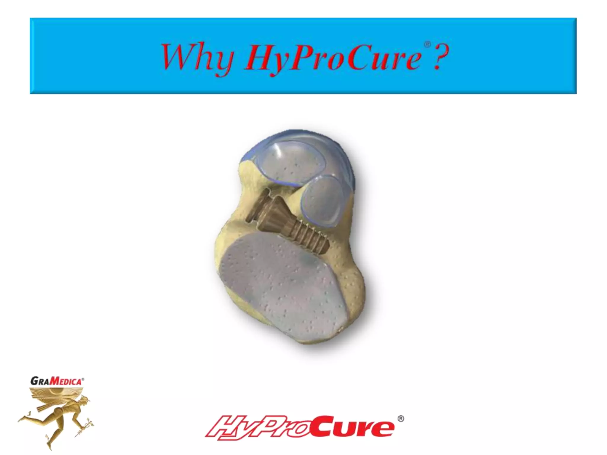
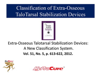
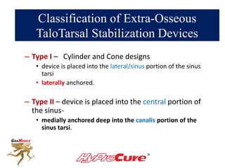
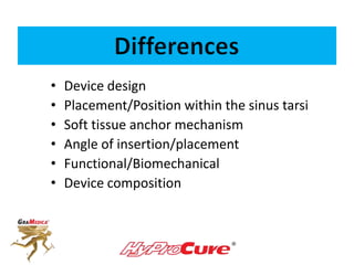
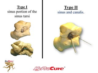
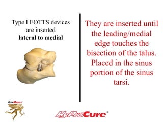
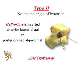
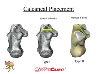
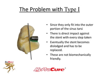
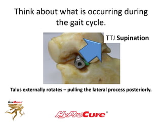
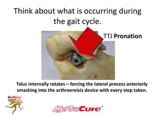
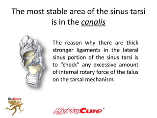
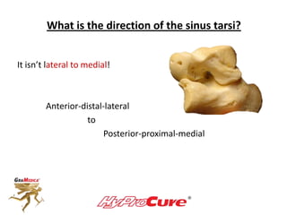
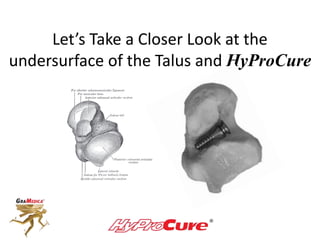
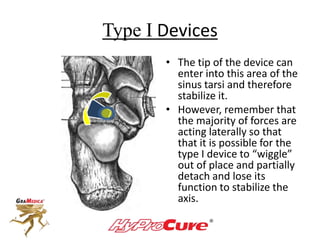
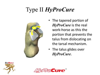
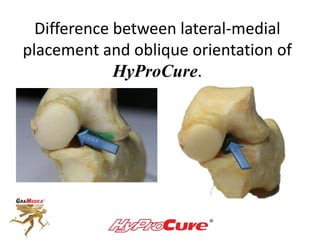
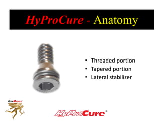
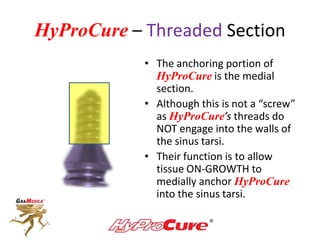
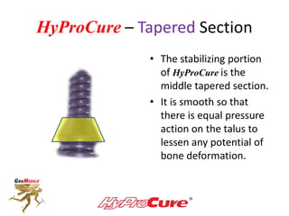
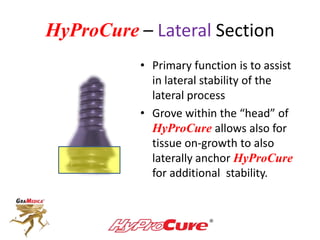
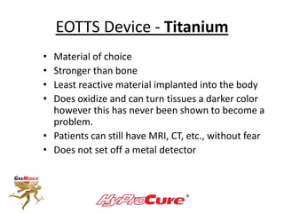
Sinus tarsi devices are not all the same!
What makes HyProCure®
so special?
Classification of Extra-Osseous
TaloTarsal Stabilization Devices
Extra-Osseous Talotarsal Stabilization Devices:
A New Classification System.
Vol. 51, No. 5, p. 613-622, 2012.
Classification of Extra-Osseous
TaloTarsal Stabilization Devices
– Type I – Cylinder and Cone designs
• device is placed into the lateral/sinus portion of the sinus
tarsi
• laterally anchored.
– Type II – device is placed into the central portion of
the sinus-
• medially anchored deep into the canalis portion of the
sinus tarsi.
• Device design
• Placement/Position within the sinus tarsi
• Soft tissue anchor mechanism
• Angle of insertion/placement
• Functional/Biomechanical
• Device composition
Type I
sinus portion of the
sinus tarsi
Type II
sinus and canalis.
Type I EOTTS devices
are inserted
lateral to medial
They are inserted until
the leading/medial
edge touches the
bisection of the talus.
Placed in the sinus
portion of the sinus
tarsi.
Type II
Notice the angle of insertion.
HyProCure is inserted
anterior-lateral-distal
to
posterior-medial-proximal
Calcaneal Placement
Type I Type II
Lateral to Medial Oblique & deep
The Problem with Type I
• Since they only fit into the outer
portion of the sinus tarsi
• There is direct impact against
the stent with every step taken
• Eventually the stent becomes
dislodged and has to be
replaced.
• These are not biomechanically
friendly.
Think about what is occurring during
the gait cycle.
TTJ Supination
Talus externally rotates – pulling the lateral process posteriorly.
Think about what is occurring during
the gait cycle.
TTJ Pronation
Talus internally rotates – forcing the lateral process anteriorly
smashing into the arthroereisis device with every step taken.
The most stable area of the sinus tarsi
is in the canalis
The reason why there are thick
stronger ligaments in the lateral
sinus portion of the sinus tarsi is
to “check” any excessive amount
of internal rotary force of the talus
on the tarsal mechanism.
Let’s Think About This!
If the canalis portion of the sinus tarsi is the
most stable…
…doesn’t it make
sense that this is the most important area
to anchor our talotarsal stabilization device?
What is the direction of the sinus tarsi?
It isn’t lateral to medial!
Anterior-distal-lateral
to
Posterior-proximal-medial
Let’s Take a Closer Look at the
undersurface of the Talus and HyProCure
What/where is the most important
area to stabilize the talus on the
tarsal mechanism?
This is about the most important question that can be
asked as this is what we are trying to achieve.
This is the exact area where the talus MUST be prevented from
slipping off its position on the posterior aspect of the calcaneus.
So, how do the various stent do
this?
Type I Devices
• The tip of the device can
enter into this area of the
sinus tarsi and therefore
stabilize it.
• However, remember that
the majority of forces are
acting laterally so that
that it is possible for the
type I device to “wiggle”
out of place and partially
detach and lose its
function to stabilize the
axis.
Type II HyProCure
• The tapered portion of
HyProCure is the real
work-horse as this the
portion that prevents the
talus from dislocating on
the tarsal mechanism.
• The talus glides over
HyProCure.
Difference between lateral-medial
placement and oblique orientation of
HyProCure.
HyProCure - Anatomy
• Threaded portion
• Tapered portion
• Lateral stabilizer
HyProCure – Threaded Section
• The anchoring portion of
HyProCure is the medial
section.
• Although this is not a “screw”
as HyProCure’s threads do
NOT engage into the walls of
the sinus tarsi.
• Their function is to allow
tissue ON-GROWTH to
medially anchor HyProCure
into the sinus tarsi.
HyProCure – Tapered Section
• The stabilizing portion
of HyProCure is the
middle tapered section.
• It is smooth so that
there is equal pressure
action on the talus to
lessen any potential of
bone deformation.
HyProCure – Lateral Section
• Primary function is to assist
in lateral stability of the
lateral process
• Grove within the “head” of
HyProCure allows also for
tissue on-growth to also
laterally anchor HyProCure
for additional stability.
Tissue Adherence
In-growth On-growth
• Tissue incorporates into the
device.
• Not only does the tissue
completely surround the
device it grows internally.
• Sounds good except if the
device has to be removed
these tissue are removed as
well.
• Tissues grow onto the
device but not deep into the
device.
• Advantage is that if the
device has to be removed
the tissues are not.
HyProCure’s unique design allows for
multiple points of peripheral soft tissue
on-growth by not in-growth.
Does it make a difference what
material makes up the device?
You bet it does!
TaloTarsal Stabilization
• Bone
• Silicone
• High Molecular Weight Polyethene
• Combination Titanium and Polyethene
• Titanium
• Poly-lactate Acid (absorbable)
Device Material
• Bone – gets absorbed, not reliable, not strong enough
• Silicone – not strong enough, fragments, silicone shards,
not reliable.
• Polyethene – not strong enough, fragments, not reliable.
• Polylactate – absorbs, temporary, loss of correction.
EOTTS Device - Titanium
• Material of choice
• Stronger than bone
• Least reactive material implanted into the body
• Does oxidize and can turn tissues a darker color
however this has never been shown to become a
problem.
• Patients can still have MRI, CT, etc., without fear
• Does not set off a metal detector
Titanium and Tissue Adherence
• Titanium has extremely small micropores that
allow partial tissue attachment.
• If a HyProCure device has to be remove, twist
it 360 degrees which frees-up the tissue
attachments.
Conclusion:
Why HyProCure?
HyProCure has the
best anatomic design
& biomechanical function.
“Changing Lives, One Step at a Time”

Why HyProCure

  • 2.
    Sinus tarsi devicesare not all the same! What makes HyProCure® so special?
  • 3.
    Classification of Extra-Osseous TaloTarsalStabilization Devices Extra-Osseous Talotarsal Stabilization Devices: A New Classification System. Vol. 51, No. 5, p. 613-622, 2012.
  • 4.
    Classification of Extra-Osseous TaloTarsalStabilization Devices – Type I – Cylinder and Cone designs • device is placed into the lateral/sinus portion of the sinus tarsi • laterally anchored. – Type II – device is placed into the central portion of the sinus- • medially anchored deep into the canalis portion of the sinus tarsi.
  • 5.
    • Device design •Placement/Position within the sinus tarsi • Soft tissue anchor mechanism • Angle of insertion/placement • Functional/Biomechanical • Device composition
  • 6.
    Type I sinus portionof the sinus tarsi Type II sinus and canalis.
  • 7.
    Type I EOTTSdevices are inserted lateral to medial They are inserted until the leading/medial edge touches the bisection of the talus. Placed in the sinus portion of the sinus tarsi.
  • 8.
    Type II Notice theangle of insertion. HyProCure is inserted anterior-lateral-distal to posterior-medial-proximal
  • 9.
    Calcaneal Placement Type IType II Lateral to Medial Oblique & deep
  • 10.
    The Problem withType I • Since they only fit into the outer portion of the sinus tarsi • There is direct impact against the stent with every step taken • Eventually the stent becomes dislodged and has to be replaced. • These are not biomechanically friendly.
  • 11.
    Think about whatis occurring during the gait cycle. TTJ Supination Talus externally rotates – pulling the lateral process posteriorly.
  • 12.
    Think about whatis occurring during the gait cycle. TTJ Pronation Talus internally rotates – forcing the lateral process anteriorly smashing into the arthroereisis device with every step taken.
  • 13.
    The most stablearea of the sinus tarsi is in the canalis The reason why there are thick stronger ligaments in the lateral sinus portion of the sinus tarsi is to “check” any excessive amount of internal rotary force of the talus on the tarsal mechanism.
  • 14.
    Let’s Think AboutThis! If the canalis portion of the sinus tarsi is the most stable… …doesn’t it make sense that this is the most important area to anchor our talotarsal stabilization device?
  • 15.
    What is thedirection of the sinus tarsi? It isn’t lateral to medial! Anterior-distal-lateral to Posterior-proximal-medial
  • 16.
    Let’s Take aCloser Look at the undersurface of the Talus and HyProCure
  • 17.
    What/where is themost important area to stabilize the talus on the tarsal mechanism? This is about the most important question that can be asked as this is what we are trying to achieve.
  • 18.
    This is theexact area where the talus MUST be prevented from slipping off its position on the posterior aspect of the calcaneus.
  • 19.
    So, how dothe various stent do this?
  • 20.
    Type I Devices •The tip of the device can enter into this area of the sinus tarsi and therefore stabilize it. • However, remember that the majority of forces are acting laterally so that that it is possible for the type I device to “wiggle” out of place and partially detach and lose its function to stabilize the axis.
  • 21.
    Type II HyProCure •The tapered portion of HyProCure is the real work-horse as this the portion that prevents the talus from dislocating on the tarsal mechanism. • The talus glides over HyProCure.
  • 22.
    Difference between lateral-medial placementand oblique orientation of HyProCure.
  • 23.
    HyProCure - Anatomy •Threaded portion • Tapered portion • Lateral stabilizer
  • 24.
    HyProCure – ThreadedSection • The anchoring portion of HyProCure is the medial section. • Although this is not a “screw” as HyProCure’s threads do NOT engage into the walls of the sinus tarsi. • Their function is to allow tissue ON-GROWTH to medially anchor HyProCure into the sinus tarsi.
  • 25.
    HyProCure – TaperedSection • The stabilizing portion of HyProCure is the middle tapered section. • It is smooth so that there is equal pressure action on the talus to lessen any potential of bone deformation.
  • 26.
    HyProCure – LateralSection • Primary function is to assist in lateral stability of the lateral process • Grove within the “head” of HyProCure allows also for tissue on-growth to also laterally anchor HyProCure for additional stability.
  • 27.
    Tissue Adherence In-growth On-growth •Tissue incorporates into the device. • Not only does the tissue completely surround the device it grows internally. • Sounds good except if the device has to be removed these tissue are removed as well. • Tissues grow onto the device but not deep into the device. • Advantage is that if the device has to be removed the tissues are not.
  • 28.
    HyProCure’s unique designallows for multiple points of peripheral soft tissue on-growth by not in-growth.
  • 29.
    Does it makea difference what material makes up the device? You bet it does!
  • 30.
    TaloTarsal Stabilization • Bone •Silicone • High Molecular Weight Polyethene • Combination Titanium and Polyethene • Titanium • Poly-lactate Acid (absorbable)
  • 31.
    Device Material • Bone– gets absorbed, not reliable, not strong enough • Silicone – not strong enough, fragments, silicone shards, not reliable. • Polyethene – not strong enough, fragments, not reliable. • Polylactate – absorbs, temporary, loss of correction.
  • 32.
    EOTTS Device -Titanium • Material of choice • Stronger than bone • Least reactive material implanted into the body • Does oxidize and can turn tissues a darker color however this has never been shown to become a problem. • Patients can still have MRI, CT, etc., without fear • Does not set off a metal detector
  • 33.
    Titanium and TissueAdherence • Titanium has extremely small micropores that allow partial tissue attachment. • If a HyProCure device has to be remove, twist it 360 degrees which frees-up the tissue attachments.
  • 34.
    Conclusion: Why HyProCure? HyProCure hasthe best anatomic design & biomechanical function.
  • 35.
    “Changing Lives, OneStep at a Time”