1. Anatomy & Physiology
By Kimberly Mancillas October 18, 2012 Volume 1, Issue 1
Gila and Neurons Information:
In this issue:
Gila and Neurons 1
Potentials and 2
Impulses
Organization of the 3
Nervous System
Voluntary and 4
Involuntary Muscles
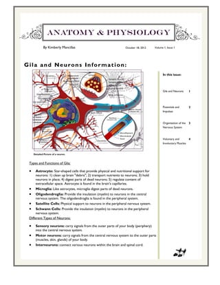
Detailed Picture of a neuron.
Types and Functions of Glia:
Astrocyte: Star-shaped cells that provide physical and nutritional support for
neurons: 1) clean up brain "debris"; 2) transport nutrients to neurons; 3) hold
neurons in place; 4) digest parts of dead neurons; 5) regulate content of
extracellular space. Astrocyte is found in the brain’s capillaries.
Microglia: Like astrocytes, microglia digest parts of dead neurons.
Oligodendroglia: Provide the insulation (myelin) to neurons in the central
nervous system. The oligodendroglia is found in the peripheral system.
Satellite Cells: Physical support to neurons in the peripheral nervous system.
Schwann Cells: Provide the insulation (myelin) to neurons in the peripheral
nervous system.
Different Types of Neurons:
Sensory neurons: carry signals from the outer parts of your body (periphery)
into the central nervous system.
Motor neurons: carry signals from the central nervous system to the outer parts
(muscles, skin, glands) of your body.
Interneurons: connect various neurons within the brain and spinal cord.
2. Anatomy & Physiology
By Myika Thompson October 18, 2012 Volume 1, Issue 1
I m p u l s e s a n d Po t e n t i a l s :
In this issue:
Nerve Impulses:
Nerve Impulses- Nerve Impulses are electrical currents that move by dendrites or
Gia and Neurons 1
axons due to ions moving towards the Neuron’s plasma membrane.
Different Potentials and their functions:
Membrane Potentials- made from a separation of positive and negative charges Impulses and 2
Potentials
(ions) across the membrane.
· are determined by three different factors
Organization of the 3
1) the concentration of ions on the inside and outside of the cell
Nervous System
2) the permeability of the cell membrane to those ions through specific ion channels
3) by the activity of electrogenic pumps that maintain the ion concentrations across
Voluntary and 4
the membrane
Involuntary Muscles
Resting Membrane Potentials-distinguishes the steady-state electrical condition
of all cells from the electrical transients that are the “action potentials” of
excitable cells neurons and muscle cells
Local Field Potential- also known as LFP is a class of electrophysiological signals
which is figured out by the electrical current from the dendritic synaptic activity within
a volume of tissue.
Action Potential- part of the neural membrane opens to allow positively charged
ions inside the cell and negatively charged ions out.
1) Sodium Channels open up and the positive sodium cells go into the cells.
Detailed Picture of action and local field potential.
3. Anatomy & Physiology
By October 18, 2012 Volume 1, Issue 1
Or ga niza tion of th e Ner vou s System:
In this issue:
Organization of the Nervous System:
The nervous system is split into two central and peripheral divisions.
Gila and Neurons 1
Within the skull vertebrae is the brain and spinal cord that forms the central
nervous system which functions as the body’s Command and control center.
The nerves networking creates the peripheral nervous system and carries signals
Potentials and 2
that allows the brain and spinal cord to communicate with the body’s tissue
Impulses
and organs.
In a nerve groups of nerve cells are bundled together by connective tissue to form
Organization of the 3
fascicles. Nervous System
The nerve cells are the ones that link to CNS to the peripheral organ.
Voluntary and 4
Involuntary Muscles
Detailed Picture of the nervous system and the
processes by which it functions.
4. Anatomy & Physiology
By October 18, 2012 Volume 1, Issue 1
Vo l u n t a r y a n d I n v o l u n t a r y M u s c l e s : In this issue:
Gila and Neurons 1
Voluntary Muscles:
Voluntary is when you are thinking about moving your muscle Potentials and 2
Impulses
Involuntary Muscles: Organization of the 3
Nervous System
Involuntary is when you hit the tendon and it makes it move without any
thought of moving the muscle Voluntary and 4
Involuntary Muscles
Data Recorded: Based on the data for voluntary movement it takes longer for the brain to
send the message to the muscle rather than involuntary movement which is physical contact.
Kick 1 Kick 2 Kick 3 Kick 4 Kick 5 Average
Time of muscle contractions 3.27 5.90 10.26 15.33 21.29 11.31
Time of Stimulus 3.09 5.88 9.97 15.00 21.40 11.07
0.18 0.02 0.29 0.33 -0.11 0.24
table1:
Table 2:
Reflex 1 Reflex 2 Reflex 3 Reflex 4 Reflex 5 Average
time of muscle contrac- 4.05 6.52 9.18 11.93 14.86 9.31
tions
time of stimulus 3.83 6.39 9.02 11.77 14.62 9.13
0.22 0.13 0.16 0.16 0.24 0.18
Table 3:
Reflex without reinforcement Reflex with reinforcement
Reflex response Max (mV) Min (mV)
1 1.33 0.73 0.6 1.13 0.82 0.31
2 1.34 0.79 0.55 1.49 0.85 0.64
3 1.15 0.85 0.3 1.24 0.83 0.41
4 1.63 0.75 0.88 1.15 0.87 0.28
5 1.31 0.79 0.52 1.30 0.84 0.46
Average values 1.58 0.78 0.8 1.26 0.84 0.42