Vitreomacular traction is when the vitreous doesn't successfully detach from the retina, which is part of the normal aging process. Some factors can increase your risk of developing this eye condition, such as age-related macular degeneration, which is common with aging and is when your macula starts to deteriorate.
Vitreous Detachment
The eye is a very complex functional and anatomic organ. The retina is a thin, delicate and transparent sheet of tissue that lines the inside of the back of the eye. Directly in front of the retina is also a cavity that contains a gel called vitreous. The structure responsible for the bulk and shape of our eye is Vitreous part. It is a jelly-like body that fills the posterior chamber of the eye, giving the eyeball its round shape and keeping the retina in place against the back of the eye.
It is made up of millions of tiny collagen fibrils along with ground substance mucopolysaccharides such as hyaluronic acid, which form a gel. The vitreous is mostly water, which makes up 98% of it. The collagen strands connect to the superficial layers of the retina especially around the macula, the retinal vessels or sites at the retinal periphery.
Posterior vitreous detachment (PVD), also known as hyaloid detachment, occurs when the retinal layer and vitreous body/posterior hyaloid membrane dissociate, with an intervening fluid collection forming in the subhyaloid space. It is thought to be a common consequence of aging, occurring in more than 70% of the population over the age of 60
Who is at risk of posterior vitreous detachment?
The risk factors for vitreous detachment include:
Older age.
Nearsightedness.
Past eye trauma.
Prior Cataract Surgery.
Vitreous detachment in one eye.
The vitreous is a transparent, gel-like substance that fills the eye, giving your eye shape. It is protected by a thin shell, which is known as the vitreous cortex. The cortex is sealed to the retina in those with healthy eyes.
18th International conference on OPHTHALMOLOGY AND VISION SCIENCE,April 24-25...OphthalmologyCongres
18th International Conference on
Ophthalmology and Vision Science
April 24-25, 2023 Amsterdam, Netherlands
Theme- Upgradation and modernization of ophthalmologists via new innovation and Research, which focuses on the most recent innovative improvements and research in the field of Ophthalmology
To know about definition , causes, types, signs and symptoms, diagnosis, treatment home remedies for floating these can give knowledge by this presentation
The retina is the light-sensitive layer of
tissue that lines the inside of the eye and sends visual messages through the
optic nerve to the brain. When the retina detaches, it is lifted or pulled from
its normal position. If not promptly treated, retinal detachment can
cause permanent vision loss.
In some cases there may be small areas of
the retina that are torn. These areas, called retinal tears or retinal breaks,
can lead to retinal detachment.
Explore natural remedies for syphilis treatment in Singapore. Discover alternative therapies, herbal remedies, and lifestyle changes that may complement conventional treatments. Learn about holistic approaches to managing syphilis symptoms and supporting overall health.
More Related Content
Similar to Vitreomacular Traction Treatment, Causes, and Symptoms.pdf
The retina is the light-sensitive layer of
tissue that lines the inside of the eye and sends visual messages through the
optic nerve to the brain. When the retina detaches, it is lifted or pulled from
its normal position. If not promptly treated, retinal detachment can
cause permanent vision loss.
In some cases there may be small areas of
the retina that are torn. These areas, called retinal tears or retinal breaks,
can lead to retinal detachment.
Similar to Vitreomacular Traction Treatment, Causes, and Symptoms.pdf (20)
Explore natural remedies for syphilis treatment in Singapore. Discover alternative therapies, herbal remedies, and lifestyle changes that may complement conventional treatments. Learn about holistic approaches to managing syphilis symptoms and supporting overall health.
Title: Sense of Smell
Presenter: Dr. Faiza, Assistant Professor of Physiology
Qualifications:
MBBS (Best Graduate, AIMC Lahore)
FCPS Physiology
ICMT, CHPE, DHPE (STMU)
MPH (GC University, Faisalabad)
MBA (Virtual University of Pakistan)
Learning Objectives:
Describe the primary categories of smells and the concept of odor blindness.
Explain the structure and location of the olfactory membrane and mucosa, including the types and roles of cells involved in olfaction.
Describe the pathway and mechanisms of olfactory signal transmission from the olfactory receptors to the brain.
Illustrate the biochemical cascade triggered by odorant binding to olfactory receptors, including the role of G-proteins and second messengers in generating an action potential.
Identify different types of olfactory disorders such as anosmia, hyposmia, hyperosmia, and dysosmia, including their potential causes.
Key Topics:
Olfactory Genes:
3% of the human genome accounts for olfactory genes.
400 genes for odorant receptors.
Olfactory Membrane:
Located in the superior part of the nasal cavity.
Medially: Folds downward along the superior septum.
Laterally: Folds over the superior turbinate and upper surface of the middle turbinate.
Total surface area: 5-10 square centimeters.
Olfactory Mucosa:
Olfactory Cells: Bipolar nerve cells derived from the CNS (100 million), with 4-25 olfactory cilia per cell.
Sustentacular Cells: Produce mucus and maintain ionic and molecular environment.
Basal Cells: Replace worn-out olfactory cells with an average lifespan of 1-2 months.
Bowman’s Gland: Secretes mucus.
Stimulation of Olfactory Cells:
Odorant dissolves in mucus and attaches to receptors on olfactory cilia.
Involves a cascade effect through G-proteins and second messengers, leading to depolarization and action potential generation in the olfactory nerve.
Quality of a Good Odorant:
Small (3-20 Carbon atoms), volatile, water-soluble, and lipid-soluble.
Facilitated by odorant-binding proteins in mucus.
Membrane Potential and Action Potential:
Resting membrane potential: -55mV.
Action potential frequency in the olfactory nerve increases with odorant strength.
Adaptation Towards the Sense of Smell:
Rapid adaptation within the first second, with further slow adaptation.
Psychological adaptation greater than receptor adaptation, involving feedback inhibition from the central nervous system.
Primary Sensations of Smell:
Camphoraceous, Musky, Floral, Pepperminty, Ethereal, Pungent, Putrid.
Odor Detection Threshold:
Examples: Hydrogen sulfide (0.0005 ppm), Methyl-mercaptan (0.002 ppm).
Some toxic substances are odorless at lethal concentrations.
Characteristics of Smell:
Odor blindness for single substances due to lack of appropriate receptor protein.
Behavioral and emotional influences of smell.
Transmission of Olfactory Signals:
From olfactory cells to glomeruli in the olfactory bulb, involving lateral inhibition.
Primitive, less old, and new olfactory systems with different path
TEST BANK for Operations Management, 14th Edition by William J. Stevenson, Ve...kevinkariuki227
TEST BANK for Operations Management, 14th Edition by William J. Stevenson, Verified Chapters 1 - 19, Complete Newest Version.pdf
Knee anatomy and clinical tests 2024.pdfvimalpl1234
This includes all relevant anatomy and clinical tests compiled from standard textbooks, Campbell,netter etc..It is comprehensive and best suited for orthopaedicians and orthopaedic residents.
NVBDCP.pptx Nation vector borne disease control programSapna Thakur
NVBDCP was launched in 2003-2004 . Vector-Borne Disease: Disease that results from an infection transmitted to humans and other animals by blood-feeding arthropods, such as mosquitoes, ticks, and fleas. Examples of vector-borne diseases include Dengue fever, West Nile Virus, Lyme disease, and malaria.
Ozempic: Preoperative Management of Patients on GLP-1 Receptor Agonists Saeid Safari
Preoperative Management of Patients on GLP-1 Receptor Agonists like Ozempic and Semiglutide
ASA GUIDELINE
NYSORA Guideline
2 Case Reports of Gastric Ultrasound
Flu Vaccine Alert in Bangalore Karnatakaaddon Scans
As flu season approaches, health officials in Bangalore, Karnataka, are urging residents to get their flu vaccinations. The seasonal flu, while common, can lead to severe health complications, particularly for vulnerable populations such as young children, the elderly, and those with underlying health conditions.
Dr. Vidisha Kumari, a leading epidemiologist in Bangalore, emphasizes the importance of getting vaccinated. "The flu vaccine is our best defense against the influenza virus. It not only protects individuals but also helps prevent the spread of the virus in our communities," he says.
This year, the flu season is expected to coincide with a potential increase in other respiratory illnesses. The Karnataka Health Department has launched an awareness campaign highlighting the significance of flu vaccinations. They have set up multiple vaccination centers across Bangalore, making it convenient for residents to receive their shots.
To encourage widespread vaccination, the government is also collaborating with local schools, workplaces, and community centers to facilitate vaccination drives. Special attention is being given to ensuring that the vaccine is accessible to all, including marginalized communities who may have limited access to healthcare.
Residents are reminded that the flu vaccine is safe and effective. Common side effects are mild and may include soreness at the injection site, mild fever, or muscle aches. These side effects are generally short-lived and far less severe than the flu itself.
Healthcare providers are also stressing the importance of continuing COVID-19 precautions. Wearing masks, practicing good hand hygiene, and maintaining social distancing are still crucial, especially in crowded places.
Protect yourself and your loved ones by getting vaccinated. Together, we can help keep Bangalore healthy and safe this flu season. For more information on vaccination centers and schedules, residents can visit the Karnataka Health Department’s official website or follow their social media pages.
Stay informed, stay safe, and get your flu shot today!
Surat @ℂall @Girls ꧁❤8527049040❤꧂@ℂall @Girls Service Vip Top Model Safe
Vitreomacular Traction Treatment, Causes, and Symptoms.pdf
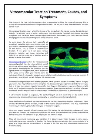
1. Vitreomacular Traction Treatment, Causes, and
Symptoms
The vitreous is the clear, jelly-like substance that is responsible for filling the center of your eye. This is
connected to the macula and retina using millions of fibers. The macular is what is responsible for detailed
central vision.
Vitreomacular traction occurs when the vitreous of the eye pulls on the macula, causing damage to your
macula. The vitreous starts to shrink, pulling away from the macula. Eventually the vitreous detaches
completely from the retina, which is known as posterior vitreous detachment (PVD). This is a normal part of
the aging process and not something to be overly concerned about.
In some cases, the vitreous isn't successful at
completely detaching with some staying stuck to
your macula. When this happens, it constantly pulls
on the macula. This is known as vitreomacular
traction. If you ignore it, it can cause serious
problems, including scar tissue, swimming, and
holes. It can pull the retina out of position, which is
known as retinal detachment.
Vitreomacular traction is when the vitreous doesn't
successfully detach from the retina, which is part of
the normal aging process. Some factors can increase
your risk of developing this eye condition, such as
age-related macular degeneration, which is common
with aging and is when your macula starts to
deteriorate. Diabetic retinopathy also puts you in a higher risk bracket to develop vitreomacular traction. If
you are very near-sighted, you will find your risk also increases.
Vitreomacular degeneration does lead to symptoms, which you may be able to identify, often it changes
your vision. You may notice your vision sharpness has decreased or you experience distortions when you
look at straight lines, objects may also look smaller than they are. Often patients experience flashes of light
in the eye. It is not uncommon for the symptoms to develop slowly over time and they can mimic other eye
conditions, which is why you need to have your eyes checked by an optometrist or ophthalmologist.
If vitreomacular traction is suspected, the ophthalmologist will look inside the eye to confirm their
diagnosis using ultrasound, fluorescein angiography, or optical coherence tomography (oct).
Once they have confirmed that you have vitreomacular traction, they will recommend a treatment. There
are four treatment options available, based on the severity of your condition. They may recommend
monitoring, medication, pneumatic vitreolysis, or vitrectomy surgery.
Vitrectomy surgery is usually offered in severe cases. This is when the ophthalmologist makes a small cut in
your eye and removes the vitreous carefully from the retina. They will repair any damage to the retina
before filling your eye with saline or gas, helping the eye retain its shape.
They will recommend monitoring your condition if it doesn't cause vision changes. In some cases,
medication has proven successful. It dissolves the fibers that keep the vitreous stuck to the macular. It is
given as a single injection into the center of the eye. If this fails, they may recommend vitreomacular
traction surgery.
2. Pneumatic vitreolysis is a procedure where the ophthalmologist injects a gas bubble into the eye. The
bubble breaks the bond between the vitreous and the macular. You look down several times an hour over
days to ensure the bubble stays in the right position. Sometimes the eye doctor may recommend
pneumatic vitreolysis with medication to separate the vitreous gel.
About us: mahi muqit is a leading
consultant ophthalmologist, cataract, and
vitreoretinal surgeon at two private clinics in
london, united kingdom. He provides patients
with superior service and support with a range
of surgical procedures to meet their eyesight
requirements. He has built up a solid reputation for his eye services in the london area as an expert eye
doctor and surgeon offering surgical retina, medical retina, and complex cataract surgery. He also offers
surgery to patients suffering from diabetic retinopathy. Mahi muqit is a member of the royal college of
ophthalmologists, a member of the british and eire association of vitreoretinal surgeons, and the uk and
ireland society of cataract and refractive surgeons. To find out more,
visit https://www.retinasurgeon.uk.com/.