
Recommended
More Related Content
Similar to CATARACTS.pptx
Similar to CATARACTS.pptx (20)
Recently uploaded
Recently uploaded (20)
CATARACTS.pptx
- 1. CATARACT : “EXPERIENCE IS A KEEN KNIFE THAT HURTS WHILE IT EXTRACTS THE CATARACT THAT BLINDS”
- 2. INDEX • What is Cataract ? • Types Of Cataracts • Difference Between Immature & Mature Cataract • Causes • Symptoms • Risk Factors • Diagnosis • Complications • Treatment • Surgery • Prevention • Cataracts In India • Why cataract is prevalent in India • Case study • Bibliography
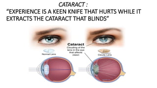
- 3. WHAT IS CATARACT ? A cataract is a cloudy area in the lens of your eyes. Cataracts are very common in older people. Over time, cataracts can make your vision blurry, hazy, or less colourful. Cataracts can lead to vision loss. Surgery can get rid of cataracts. Cataract surgery is safe and corrects vision problems caused by cataracts. One can get cataracts in one eye or both eyes — but they can’t spread from one eye to the other. Blurry or dim vision is a symptom of cataracts. A cataract is a dense, cloudy area that forms in the lens of the eye. A cataract begins when proteins in the eye forms clumps that prevent the lens from sending clear images to the retina. The retina works by converting the light that comes through the lens into signals. It sends the signals to the optic nerve which carries them to the brain. A cataract is a clouding of the normally clear lens of your eye
- 4. TYPES OF CATARACTS • Cataracts affecting the centre of the lens Or Nuclear Cataracts: A nuclear cataract may at first cause more near sightedness or even a temporary improvement in your reading vision. But with time, the lens gradually turns more densely yellow and further clouds your vision. As the cataract slowly progresses, the lens may even turn brown. Advanced yellowing or browning of the lens can lead to difficulty distinguishing between shades of colour. • Cataracts that affect the edges of the lens Or Cortical Cataracts: A cortical cataract begins as whitish, wedge-shaped opacities or streaks on the outer edge of the lens cortex. As it slowly progresses, the streaks extend to the centre and interfere with light passing through the centre of the lens. Develops in the edges of the lens and then make their way towards the center in a spoke-like manner. A cortical cataract occurs in the edges of the lens.
- 5. • Cataracts that affect the back of the lens Or Posterior Sub-capsular Cataracts: A posterior sub-capsular cataract starts as a small, opaque area that usually forms near the back of the lens, right in the path of light. A posterior sub-capsular cataract often interferes with your reading vision, reduces your vision in bright light, and causes glare or halos around lights at night. These types of cataracts tend to progress faster than other types do. • Cataracts you're born with Or Congenital Cataracts: Some people are born with cataracts or develop them during childhood. These cataracts may be genetic, or associated with an intrauterine infection or trauma. These cataracts may also be due to certain conditions, such as myotonic dystrophy, galactosemia, neurofibromatosis type 2 or rubella. Congenital cataracts don't always affect vision, but if they do, they're usually removed soon after detection. Cataracts present from birth are sometimes caused by a faulty gene being passed to a child from their parents. This fault means that the lens does not develop properly. It's estimated there's a family history of congenital cataracts.
- 6. DIFFERENCE BETWEEN IMMATUE & MATURE CATARACT IMMATURE CATARACT • Visual acuity is reduced to counting fingers • Lens is slightly opaque • Iris shadow is present • Fundus may be visible MATURE CATARACT • Visual acuity is reduced to hand movement or perception of light • Lens is totally opaque • No iris shadow is present • No fundus details
- 7. Causes Most cataracts happen because of normal changes in the eyes as you get older. When a person is young, the lens in the eye is clear. The proteins in the lens of your eye start to break down and clump together. This clump makes a cloudy area on your lens — known as a cataract. Over time, the cataract gets worse and makes more of your lens cloudy. Most cataracts develop when aging or injury changes the tissue that makes up the eye's lens. Proteins and fibers in the lens begin to break down, causing vision to become hazy or cloudy . Some inherited genetic disorders that cause other health problems can increase the risk of cataracts. Cataracts can also be caused by other eye conditions, past eye surgery or medical conditions such as diabetes. Long-term use of steroid medications, too, can cause cataracts to develop.
- 8. Symptoms • Clouded, blurred or dim vision • Increasing difficulty with vision at night • Sensitivity to light and glare • Need for brighter light for reading and other activities • Seeing "halos" around lights • Frequent changes in eyeglass or contact lens prescription • Double vision in a single eye • Seeing double or a ghosted image out of the eye with cataract • Seeing bright colours as faded or yellow instead • Lamps, sunlight, or headlights seem too bright
- 9. RISK FACTORS • Diabetes • Trauma • Smoking • Obesity • Increasing age • High blood pressure • Excessive exposure to sunlight • Drinking excessive amount of alcohol • Nutritional deficiency of vitamin - C • Previous eye injury or inflammation • Prolonged use of corticosteroid medications
- 10. DIAGONOSIS • VISUAL ACUITY TEST : A visual acuity test uses an eye chart to measure how well you can read a series of letters. It is used to determine the smallest letters you can read on a standardized chart or a card held 20 feet (6 meters) away. Special charts are used when testing at distances shorter than 20 feet. Visual acuity is a term that means clarity or sharpness of vision and that the objects you see are crisply outlined and not blurry. • SLIT – LAMP EXAMINATION : A slit lamp allows your eye doctor to see the structures at the front of your eye under magnification. The microscope is called a slit lamp because it uses an intense line of light, a slit, to illuminate your cornea, iris, lens, and the space between your iris and cornea. The slit allows your doctor to view these structures in small sections, which makes it easier to detect any tiny abnormalities. • Retinal exam: To prepare for a retinal exam, your eye doctor puts drops in your eyes to open your pupils wide (dilate). This makes it easier to examine the back of your eyes (retina). Using a slit lamp or a special device called an ophthalmoscope, your eye doctor can examine your lens for signs of a cataract. Your pupils are dilated beforehand. Your eye doctor will shine a bright light and look through a microscope to assess the optic nerve, retina, and blood vessels. A cataract scatters and blocks the light as it passes through the lens, preventing a sharply defined image from reaching your retina. As a result, the vision becomes blurred. Applanation Tonometry: This test measures fluid pressure in your eye. the pressure inside an ideal, dry, thin-walled sphere equals the force necessary to flatten its surface divided by the area of flattening. Tonometry is a diagnostic test that measures your intraocular pressure (IOP), or the pressure inside your eye. Tonometry can help your healthcare provider determine if you're at risk for glaucoma. People with glaucoma have high intraocular pressure because the fluid inside the eye drains too slowly.
- 11. COMPLICATIONS OF CATARACT SURGERY • Infection- Germs that get in your eye during surgery can lead to an infection. One might feel sensitive to light or have pain • Inflammation-A little swelling and redness after surgery is normal. • Retinal Detachment-The retina sits way back in your eye, sensing light and sending messages to the brain. • Lens Fragments-When your doctor removes your cloudy lens during cataract surgery, some pieces may fall into your eye and get left behind. • Fluid Build-up in the Retina- Sometimes after surgery, blood vessels in the retina leak. As fluid collects in your eye, it blurs your vision. • Dislocated Intraocular Lens- The IOL is the artificial lens your doctor puts in your eye during surgery. It can slip out of place, causing blurred or double vision. It can also lead to more serious issues like bleeding and swelling. • Secondary Cataract-The lens capsule surrounds the eye's lens. Cataract surgery removes the front part of the lens but leaves the back in place. That's where you may get a secondary cataract, also called posterior capsule opacification (PCO). • Swelling in the Cornea-The cornea is the clear, front part of the eye. It may get swollen and hazy after surgery, making it harder to see.
- 12. TREATMENT Cataracts can be removed only with surgery. Medical treatment: No medical treatment is effective. Surgical treatment: Intra-capsular Cataract Extraction (ICCE)- Intra-capsular cataract extraction (ICCE) involves the removal of the lens and the surrounding lens capsule in one piece. The lens is then replaced with an artificial plastic lens of appropriate power which remains permanently in the eye. ICCE is a surgical procedure used to correct cataracts. The lens and the thin capsule surrounding the lens are removed. Extra-capsular Cataract Extraction (ECCE)- Extra-capsular cataract extraction involves the removal of the lens while the elastic lens capsule is left partially intact to allow implantation of an intraocular lens (IOL). This procedure requires a much smaller incision. The main objective of modern cataract surgery is to remove this hazy lens and replace it with a tiny plastic prescription lens that will be permanently implanted in your eye.
- 13. Lens replacement: Aphakic contact lenses - Contacts lenses may be fitted on eyes in all age groups and are a highly effective device in the visual rehabilitation of pediatric aphakia. Aphakic contact lenses are specially designed for adults who have recently had cataract surgery without an intraocular lens implant. Made of a silicone hydrogel lens material, these contacts allow high levels of oxygen to reach the eye. Contact lenses- A person might have sustained drying of the surface of the eye (the cornea) during the operation and a "bandage" soft contact lens could make the eye more comfortable while the results of the drying heal. IOL Implants-An intraocular lens implant is an artificial replacement for the lens of your eye. It's part of the surgery to fix cataracts. An intraocular lens implant, or IOL, is made of a clear plastic, and it's about a third the size of a dime. It'll take about 8 to 12 weeks to fully heal. The lens bends light rays that enter the eye, helping you to see. Your lens should be clear. But if you have a cataract, your lens has become cloudy. Things look hazy or less colourful with a cataract. Cataract surgery removes this cloudy lens and replaces it with a clear IOL to improve your vision.
- 14. SURGERY : Cataract surgery is a procedure to remove the clouded lens of an eye and, in most cases, replace it with an artificial lens. • Phacoemulsification: A cataractous lens could be emulsified through a small incision of 2-3 mm, giving perfect visual outcomes. It involves the creation of a superior or temporal clear corneal incision of 2-3 mm, two side port incisions of at 2-3 clock hours on either side of the main wound. An ultrasonic probe is used to the trench, emulsify, and aspirate the cataractous lens from the main wound. The creation of small wounds revolutionized modern cataract surgery as it was self- sealing , anatomically better-wound strength, and less incidence of complications. • Femtosecond laser-assisted cataract surgery (FLACS): A computer-guided laser linked to an optical imaging system (e.g. OCT) performs the corneal incision, capsulotomy, and lens fragmentation steps, thus changing the requirements associated with traditional techniques by removing the need for blade incisions and reducing phacoemulsification time and energy. • Extracapsular cataract extraction (ECCE): A computer-guided laser linked to an optical imaging system performs the corneal incision, capsulotomy, and lens fragmentation steps, thus changing the requirements associated with traditional techniques by removing the need for blade incisions and reducing phacoemulsification time and energy. The potential gains in precision associated with FLACS may improve safety and clinical outcomes.
- 15. PREVENTION • Have regular eye examinations- Eye examinations can help detect cataracts and other eye problems at their earliest stages.Ask your doctor how often you should have an eye examination. • Quit smoking- Medications, counselling and other strategies are available to help. • Manage other health problems • Choose a healthy diet that includes plenty of fruits and vegetables. Adding a variety of colorful fruits and vegetables to your diet ensures that you're getting many vitamins and nutrients. Fruits and vegetables have many antioxidants, which help maintain the health of your eyes. Fruits and vegetables have many proven health benefits and are a safe way to increase the amount of minerals and vitamins in your diet. • Wear sunglasses. Ultraviolet light from the sun may contribute to the development of cataracts. Wear sunglasses that block ultraviolet B (UVB) rays. • Reduce alcohol use-Excessive alcohol use can increase the risk of cataracts.
- 16. CATARACT IN INDIA Cataract is one of the leading causes of visual impairment in diabetic patients, and India is known as the world’s diabetic capital . One of the primary causes of the crisis is a lack of manpower in terms of the number of ophthalmologists. The factors that are responsible for cataract formation in young people is due to exposure to ultraviolet (UV) rays, eye injury, genetic disorders, steroid use, radiation therapy, smoking, and diabetes . Cataract has been documented to be the most significant cause of bilateral blindness in India . In India cataract has been reported to be responsible for 50- 80% of the bilaterally blind in the country .The prevalence of any cataracts was similar in north (73.8%) and south India (71.8%).
- 17. Major focuses of interest in cataract research are potential underlying defects in the protein formation and structure of the lens as well as risk factors and control of the disease . Cataract is one of the leading causes of blindness worldwide . The prevalence of cataracts increases with age, ranging from 3.9% among 55– 64 years to 92.6% among those 80 years and older. Age is the most common cause of cataracts. Lens proteins denature and degrade over time, and this process is accelerated by diseases such as diabetes mellitus and hypertension. The highest number of persons with vision loss , China ,Indonesia, Russia, Brazil, etc. WHY IS CATARACT IS SO PREVALENT IN INDIA AS WELL AS WORLD ?
- 18. CASE STUDY Name of the patient : Mrs. Maya Devi. She is widow . Age:70 Education :Secondary School Weight :53Kg Height: 5’2 feet Cataract : Immature Complaints: Watery Eyes ,Hazy vision ,Near-sightedness , Dimension of vision Medical History – Non- diabetic , Non-hypertension ,etc. Post Operative Medications : Predforte Eye Drop (Prednisolone Acetate) and Vigamox Eye Drop (Moxifloxacin 0.5%)
- 20. BIBLIOGRAPHY • www.webmd.com • www.mayoclinic.org • www.nei.nih.gov • www.slideshare.net • www.insightvisioncenter.com • www.aao.org • www.sciencedirect.com • eyepatient.net • www.wikipedia.com