2. 2458 C Vignal et al
1. Introduction
Songbirds are well known for their ability to produce and to perceive complex vocal
sounds. Consequently, they have been evolved into favorite models for the study of vocal
communication and sound processing. The specialized brain structures underlying the capacity
of songbirds to recognize and to produce vocal sounds have been investigated mainly with
invasive approaches such as post-mortem immunocytochemistry and in vivo electrophysiology.
To precisely investigate the processes involved in these brain regions, there is a need for a
neuro-method that allows real-time monitoring of brain activity of songbirds in a non-invasive
manner (Ramstein et al 2005).
Recently functional magnetic resonance imaging (fMRI) applied in anesthetized and
mildly sedated intact songbirds was able to reveal brain activity in the auditory system upon
hearing conspecific song (Van Meir et al 2005, Boumans et al 2007, 2008, Voss et al 2007).
Functional MRI that relies on blood oxygen level-dependent (BOLD) contrast (Ogawa et al
1990) is one of the most commonly used techniques for imaging brain activity. During
neural activity (Ito et al 2001, 2003), it is expected that an increase of oxygen consumption is
followed by a larger fractional increase in cerebral blood flow (CBF) and a lower increase in
cerebral blood volume (CBV), resulting in a net decrease in the concentration of deoxygenated
hemoglobin. The BOLD fMRI signal is thus a composite signal that is ‘oxygen-dependent’
and remains difficult to be expressed in terms of hemodynamic parameters related to neural
activity.
Near-infrared spectroscopy (NIRS) appears as another potential in vivo non-invasive
method to assess neural activity (Plesnila et al (2002) reviewed in Mehta et al (2004), Montcel
et al (2005)). Light from the near-infrared spectral window (700–1000 nm) can penetrate
deeply into biological tissues according to its weak absorption (Obrig and Villringer 2003,
Ramstein et al 2005). The spectroscopy of cerebral tissues is thus possible with an intact
skull and skin. When the light further propagates through tissue, the attenuation of light
intensity depends on the local concentration of absorbing chromophores, like the hemoglobins
(Plesnila et al 2002, Ramstein et al 2005). In this spectral window, the absorption coefficient
of tissues relies on the concentration of hemoglobins allowing measurement of variations of
oxygen saturation level HbO2/HbTotal (StO2) and hemoglobin concentration (HbTotal) linked to
CBV. Then the light absorption measurements are quantitatively related to oximetry (Obrig
and Villringer 2003) which is a robust metabolic marker of cerebral activity (Obrig et al 2000).
NIRS could thus be envisaged to monitor songbird brain activation. Nevertheless, the exact
size and location of the volume of tissue probed by NIRS remains difficult to define because
it depends on light scattering by tissues, as well as cellular and subcellular structures (Obrig
and Villringer 2003). Contrary to BOLD fMRI, NIRS represents a volumetric probing method
allowing poor anatomical resolution. Thus, fMRI and NIRS represent clearly complementary
techniques.
As part of our broader effort to develop a non-invasive NIRS method and to improve
quantitative measurement of absorbing chromophores into scattering media such as biological
tissues, we worked on a time-domain-based device (Ramstein et al 2005). This time-resolved
NIRS involved ultrafast detection of optical signals coupled with a femtosecond white laser
and had already allowed to measure optical properties of a songbird auditory brain region
(Ramstein et al 2005). Using this design, we sought to monitor an evoked brain hemodynamic
response which could constitute the basis for our next investigations of songbird brain activity
during auditory processing.
In mammals, hypercapnia induces a well-known vasodilatation response that is often used
as a model of hemodynamic response (Ito et al 2001, 2003, Dutka et al 2002, Wu et al 2002,
3. Time-resolved NIRS and fMRI for probing hemodynamic changes in a songbird brain 2459
Bluestone et al 2004, Martin et al 2006). Thus, we used hypercapnia in anesthetized zebra
finches (Taeniopygia guttata) in order to test the efficiency of our time-resolved NIRS design
in monitoring physiological hemodynamic brain responses in a songbird. As a validation,
the same experiment was conducted using BOLD fMRI and the results were compared. The
use of these two techniques does offer the opportunity to compare the sensitivity of both
methods to probe hemodynamic changes in the brain of a songbird, but also makes it possible
to correlate in songbirds local variations in HbTotal and StO2 (obtained from NIRS) with local
BOLD signal variations (providing overall information on CBF, CBV and StO2) as has been
performed in humans (Kleinschmidt et al 1996, Strangman et al 2002) and rodents (Siegel
et al 2003, Martin et al 2006).
2. Material and methods
2.1. Animals and general procedure
Adult zebra finches Taeniopygia guttata served as subjects for the fMRI and NIRS experiments.
Because the fMRI and the NIRS setups were located in distant universities, we were not able
to use the same individuals for both experiments. The four birds used in fMRI experiments
were obtained from local suppliers in Antwerp (Belgium) and were housed in an aviary with
12L/12D photoperiod, food and water ad libitum, and temperature between 23 ◦ C and 25 ◦ C.
The four birds used in NIRS experiments were bred in our aviary (ENES laboratory, Jean
Monnet University, Saint-Etienne, France, 12L/12D photoperiod using a full spectrum light
with increased blue and red wavelength fractions, food and water ad libitum, temperature
between 23 ◦ C and 25 ◦ C). For fMRI or NIRS measurements, the birds were anesthetized
with 2% isoflurane under spontaneous breathing (isoflurane mixed in fresh air). After a
30 min baseline normocapnic period, each bird underwent a challenge of 5 min normoxic
hypercapnia (600 ml min−1 7% CO2, 21% O2, 72% N2, isoflurane mixed at 2%), followed by
5 min normocapnia (600 ml min−1 fresh air, i.e. 21% O2, 79% N2, isoflurane mixed at 2%) for
baseline recovery. Each experiment took less than 1 h. All birds had free access to food and
water prior to anesthesia. Experimental procedures of fMRI measurements were in agreement
with the Belgian laws on the ‘Protection and Welfare of Animals’ and had been approved by
the ethical committee of the University of Antwerp. The experimental protocols of NIRS were
approved by the Jean Monnet University’s animal care committee.
2.2. NIRS measurements
2.2.1. Animal preparation. Anesthetized zebra finches with the head previously plucked
(three days before experiments) were fixed in a stereotaxic frame (Stoelting Co., USA,
adaptations for birds). The body temperature was kept within a narrow range (39–40 ◦ C)
by a feedback-controlled heating pad. For brain NIRS transillumination, optical fibers were
fixed into stereotaxic manipulators (Stoelting Co., USA) and placed directly on the skin.
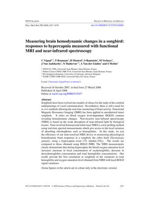
Positions of the input optical fiber F1 providing illumination and the optical fiber F2 collecting
transmitted light were chosen in order to probe the auditory regions of the telencephalon
(Field L, the caudo-medial Nidopallium NCM and the caudo-medial Mesopallium CMM
(figure 1(A))) with the best signal to noise ratio. We have developed a precise and reproducible
procedure for placing the optical fibers appropriately on the skin (Ramstein et al 2005). The
head of the bird is turned until the beak (rostral extremity) is perpendicular to the body
plane. This position allows us to define a stereotaxic origin point (0, 0, 0) defined by the
intersection of the vertical plane passing through the interaural line and the sagittal suture (the
4. 2460 C Vignal et al
Figure 1. (A) High-resolution magnetic resonance image of the head of a zebra finch (sagittal
image in the right hemisphere, 0.8 mm lateral to the sagittal suture). The positions of the ROIs
(Field L, caudo-medial nidopallium NCM and caudo-medial mesopallium CMM) are displayed.
The rostro-caudal positions of the input fiber F1 and the collecting fiber F2 of the NIRS setup are
shown according to the origin point (0, 0, 0) and the stereotaxic axes (X, Y, Z). Note that F1 and F2
are at 2 mm on the X axis. (B) Boundaries of the volume probed by the laser light that is projected
as 2D-ROI on the MR images to extract BOLD signal changes. The four panels show four sagittal
slices (slice 2: 0.5–1 mm from the midline, slice 4: 1.5–2 mm, slice 6: 2.5–3 mm, slice 8: 3.5–
4 mm) in one bird subject. These boundaries fit in a box with XYZ dimensions of 4 mm × 6 mm ×
3 mm centered on the two fibers.
5. Time-resolved NIRS and fMRI for probing hemodynamic changes in a songbird brain 2461
vena cerebralis dorsocaudalis) (figure 1(A)). The stereotaxic axes are chosen according to this
origin point. Previous works using post-mortem tissue (Ramstein et al 2005) allowed us to
define the coordinates of the auditory regions and to choose the positions of the two fibers
(F1 and F2) for optimal optical probing in the right hemisphere. These coordinates minimized
the absorption of light due to the sagittal venous sinus, the cerebellum, and the higher skull
thickness above the caudal part of the cerebellum. The head volume probed by the light
depends greatly on this positioning. Numerical simulations based on a steady-state analytical
closed-form Green’s function (Kienle and Patterson 1997) for a semi-infinite geometry showed
that the distance between the two fibers must be fixed around 5 mm to facilitate wide probing
of the auditory regions (Ramstein et al 2005). F1 was placed more rostrally on the right
hemisphere than F2 (figure 1(A)). The chosen coordinates (in millimetres) were F1 (2.0, 5.4,
−2.7) and F2 (2.0, 0.4, −0.3).
2.2.2. Determination of the volume probed by the laser light. In order to compare NIRS and
fMRI results, a near identical region of interest (ROI) must be considered. The boundaries of
the volume probed by the laser light are calculated in Ramstein et al (2005) using the absorption
coefficient µa = 0.083 mm−1 and the reduced scattering coefficient µs = 4.857 mm−1 which
were quantified during the baseline normocapnic period. Rough computations based on simple
models of light propagation in a homogeneous medium (Kienle and Patterson 1997) showed
that 90% of the collected light probed a tissue volume of 50 mm3. This volume fits in a box
with XYZ dimensions of 4 mm × 6 mm × 3 mm centered on the two fibers. Comparison with
previous work on post-mortem tissue (Ramstein et al 2005) showed that the tissue volume
probed by the light encompasses mainly the auditory regions. The same computations showed
that less than 1% of the collected light probed the vena cerebralis dorsocaudalis and that less
than 15% has probed the cerebellum. Although better light propagation models are needed, it
showed that the chosen fiber coordinates allowed probing the auditory regions non-invasively.
The boundaries of the volume probed by the laser light were projected on the MR images
to extract BOLD signal changes in the same ROI (figure 1(B)).
2.2.3. Optical setup and frame processing. The optical setup is described in Ramstein et al
(2005). It was composed of an ultrafast white laser (the light source) and a time-resolved
spectrometer (the detection system). The white laser was a supercontinuum obtained by
focusing amplified femtosecond laser pulses into pure water. The pulses (825 nm, 170 fs,
0.5 mJ, 1 kHz) were produced by a Ti:Sa chirped-pulse amplification laser chain (Coherent
Vitesse XT and BMI/Thales alpha 1000). The white light continuum (450 nm–950 nm) was
transmitted to an input optical fiber (core diameter 200 µm, numerical aperture 0.4, length
30 cm). Since the time-resolved spectrometer allows the detection of low light levels, very
low power level (1 mW) was transmitted through the bird’s head (Ramstein et al 2005). After
propagation through the bird’s head, the light was collected by a collecting optical fiber (same
model as the input optical fiber) toward the time-resolved spectrometer. This detection system
was composed of a polychromator (270M, Spex Jobin-Yvon) dispersing the light to ensure the
spectral analysis and a single shot streak camera (Hamamatsu Streakscope C4334) measuring
the time of propagation of the photons through tissues with a temporal resolution of 10 ps.
Each measure was a frame integrating 33 laser pulses due to the 33 ms CCD integration time
of the streak camera. Due to the jitter effect with 33 laser shots, the temporal resolution
was then around 18 ps. The spectro-temporal images had a spectral window extending from
668.0 nm to 844.6 nm on 640 pixels and a temporal window of 1.921 ns on 480 pixels.
The picosecond resolution of the time-of-flight of photons was used to probe deep tissues
6. 2462 C Vignal et al
(Ramstein et al 2005). The time-resolved transmittance (TRT) is the integral of the intensity
of the time-resolved signal for a time window excluding early arrived photons (slightly after
the maximum of the transmitted pulse). This time window was empirically chosen to optimize
the signal-to-noise ratio, where the signal is the variation of the TRT during the hypercapnia
event. This time window was chosen from 0.188 ns to the end of the recorded signal,
1.921 ns. The full spectrum was analyzed by 20 spectral windows.
The 640 pixel TRT variation spectrum was also fitted to the spectra of oxyhemoglobin
(HbO2) and deoxyhemoglobin (Hb) known in mammals (linear least-squares regression with
Matlab 7.1). The 640 simultaneous linear equations were solved by classic linear least-squares
procedure. This least-square fitting procedure was applied with the best estimate of the HbO2
and Hb extinction coefficient spectra (http://omlc.ogi.edu/spectra/hemoglobin/index.html).
The same procedure was applied to calculate the variations in concentration of HbO2 and
Hb. These concentration variations can be expressed using an absolute scale (µMol) because
our time-resolved detection system can measure the mean optical path through the bird’s
head thanks to the mean arrival time of photons. Indeed thanks to the mean arrival time of
photons ( t ) and the speed of light in tissues (v), it is possible to calculate the variation in the
TRT
log(1+ )
absorption coefficient ( µa in cm−1) with an absolute scale µa = v. t
TRT
, and then the
absolute variations in concentration of HbO2 and Hb (note that v. t is a mean pathlength).
The same time window used for the calculation of the TRT was used for the calculation of
the mean arrival time of photons. Assuming a mean refractive index of n = 1.4 (v = c/n)
as known in mammal tissue (Bolin et al 1989), the mean optical path length was found to
be 56 mm for an inter-fibers distance of 5.5 mm. Results were then filtered to get rid of the
high-frequency noise (Chebyshev filter, 120 samples time window (2 s)).
Differences between the TRT values were examined as for fMRI data, using an analysis
of variance (ANOVA) for repeated measures, with two factors: (1) the time points; (2) the
spectral points (repeated-measures ANOVA, p = 0.05, Statistica Software version 6.1). The
ANOVA was followed by a Fisher PLSD post-hoc test (p = 0.05).
2.3. Functional MRI measurements
2.3.1. Animal preparation. Anesthetized zebra finches were immobilized in a non-magnetic
lab-made head holder that enabled accurate positioning of the animals within the magnet. To
maintain optimal and stable physiological conditions during measurements, body temperature
and respiration were continuously monitored. Body temperature was monitored with a cloacal
temperature probe (SA-Instruments, Inc., New York, USA) and was maintained at 40.3 ±
0.3 ◦ C (mean ± SD) by a cotton jacket and a water-heated pad connected to an adjustable
heating pump (Neslab Instruments, ex111, Newington, CT, USA). Respiration rate and
amplitude were monitored with a small pneumatic sensor (SA-Instruments, Inc., New York,
USA) positioned under the bird.
2.3.2. fMRI setup. MR imaging was performed at 300 MHz on a 7 T horizontal bore
NMR microscope (MR Research Systems, MRRS, UK) with an actively shielded gradient-
insert (Magnex Scientific Ltd, Oxfordshire, UK) having an inner diameter of 80 mm and
a maximum gradient strength of 400 mT m−1. A Helmholtz (45 mm) antenna served for
transmitting the radio-frequency (RF) pulses and a circular RF surface antenna (15 mm) was
used for MR signal reception. Functional imaging was performed in the right hemisphere
(from midline to 4 mm lateral) with a T2∗-weighted multislice gradient-echo fast low angle
shot (GE-FLASH) sequence: TR 320 ms, TE 14 ms, acquisition matrix 128×62, FOV 25 mm,
8 sagittal slices, slice thickness 0.5 mm, temporal resolution 20 s, and spatial resolution
7. Time-resolved NIRS and fMRI for probing hemodynamic changes in a songbird brain 2463
195 × 195 µm2. Sagittal high-resolution imaging was performed in the same position as the
acquired functional slices with a T2-weighted spin-echo (SE) sequence: TR 2000 ms, TE
45 ms, acquisition matrix 256 × 128, FOV 25 mm, 8 sagittal slices, slice thickness 0.5 mm,
spatial resolution 98 × 98 µm2 and the number of averages 2. To limit the amount of data
to store, the MR measurements were subdivided into one normocapnic run (20 min) and one
hypercapnic run consisting of a 5 min of normocapnia, 5 min of hypercapnia and 10 min of
normocapnia rest period. We acquired 60 functional images during the run of 20 min.
2.3.3. Image processing. The boundaries of the volume probed by the laser light in NIRS
experiments were applied to the corresponding high-resolution MR images. This represents
a simple crop procedure that allowed matching the ROIs of both methods. An improvement
would be to use the photon measurement density function inside this volume; the resulting
ROIs would be weighted by the relative probability of a volume element to actually be crossed
by photons and to contribute toward the NIRS measurement (Mottin 2002, Ramstein et al
2005). The resulting brain ROIs were copied on all time point images of the fMRI experiment
and for each slice the mean signal intensity and area size of the selected 2D ROI were
calculated for the 60 time points. These mean signal intensities were subsequently expressed
as percent signal changes relative to the mean signal intensity of the 10 normocapnic time
points preceding the 5 min hypercapnic period. The percent signal changes for each time point
in the entire volume ROI spread over the eight sagittal slices—in resemblance to the NIRS
data—were calculated with a weighted average taking into account the different 2D ROI area
sizes in the eight slices. It means that the signal intensity over a 2D ROI in one slice was
weighted using the area size of this 2D ROI. Statistical analysis of these weighted averaged
percent signal changes was performed with SPSS (Statistical Package for Social Sciences,
release 12.0). Differences in the percent signal changes were statistically analyzed using an
analysis of variance (ANOVA) for repeated measures of the time points.
3. Results
3.1. NIRS measurements
Image processing of the four birds gives a mean temporal evolution of the time-resolved
transmittance (TRT) in 20 spectral windows (from 668.0 nm to 844.6 nm) (figure 2(A)). The
timing of the CO2 perturbation corresponding to the normoxic hypercapnia event is indicated
in figure 2. At t = 5 min the concentration of CO2 was increased to 7% and maintained
at this level for 5 min. At t = 10 min, the concentration was returned to the baseline level
of normocapnia. This defines three successive experimental time periods: normocapnia,
hypercapnia and rest.
The ANOVA for repeated measures using the 900 TRT values (one point per second and
15 min) as dependent factor demonstrates the existence of a significant effect of the spectral
window (p < 0.001, F = 417.8, df = 19, error = 63194), of the time period (p < 0.001,
F = 214.1, df = 2, error = 3326) and of the interaction of the spectral window and the
time period (p < 0.001, F = 161.6, df = 38, error = 63194). Figure 2(B) shows the mean
temporal evolution of the TRT in three spectral windows. The one-way ANOVA demonstrates
a significant effect for time points during hypercapnia in a spectral window of 676.8–
685.6 nm (figure 2(B); p < 0.001, F = 2.68, df = 499, error = 1500).
Figure 2(C) shows the mean spectrum of the TRT during normocapnia (mean on 300 TRT
normalized to one) and during the four last minutes of hypercapnia (240 TRT). From the 1st
to the 13th spectral window (668.0–676.8 nm to 774.0–782.8 nm), the TRT increase during
8. 2464 C Vignal et al
(A)
(B)
(C) (D)
Figure 2. (A) Mean temporal evolution (n = 4 birds) of the time-resolved transmittance (TRT)
in 20 spectral windows (Chebyshev time window with a 2 s length). From t = 5 min to t =
10 min the CO2 concentration was increased to 7%. (B) Mean temporal evolution of the TRT in
three spectral windows chosen for univariate analysis (∗∗∗: p < 0.001) (Chebyshev time window
with a 60 s length). (C) Mean spectrum of the TRT during normocapnia (mean on 300 TRT
normalized to 1) and during the last 4 min of hypercapnia (240 TRT). Error bars are standard
errors. From the 1st to the 13th spectral windows, the TRT increase during hypercapnia is
significant (∗∗∗: p < 0.001). (D) Fit of the TRT spectra variations induced by hypercapnia (last 4
min) to the spectra of oxyhemoglobin (HbO2) and deoxyhemoglobin (Hb).
hypercapnia is significant (post-hoc tests of the repeated measures ANOVA: Fisher PLSD,
p < 0.001) (figure 2(C)).
9. Time-resolved NIRS and fMRI for probing hemodynamic changes in a songbird brain 2465
Figure 3. Oxyhemoglobin (HbO2, continuous line) and deoxyhemoglobin (Hb, dashed line)
concentration variations during the normoxic hypercapnia (t = 5 to t = 10 min). Concentration
variations are shown with respect to the normocapnia period (t = 0 to t = 5 min).
A least-squares fitting procedure is applied with the oxyhemoglobin (HbO2) and
deoxyhemoglobin (Hb) extinction coefficient spectra known in mammals. Despite the
difference between the in vivo optical properties and the HbO2 and Hb extinction coefficient
spectra measured ex vivo, figure 2(D) shows that the TRT variation originates mainly from
the variations in concentration of HbO2 and Hb. The spectrum peak at 760 nm is significantly
measured (figure 2(D)) and the isobestic region around 800 nm is relatively stable despite
the increase of noise induced by the laser pump wavelength (825 nm, 170 fs). The
differences between fit and experimental spectrum come from tissue optics and complex
in vivo chemometrics (Mottin 2002). Despite the fact that the results about cytochrome aa
oxidation measurement seem unreliable (Plesnila et al 2002, Uludag et al 2004), we tested the
fit with the cytochrome extinction coefficient spectrum without success.
The HbO2 and Hb concentration variations throughout the successive time periods (5 min
normocapnia, 5 min hypercapnia and 5 min rest) were computed the same way in figure 3.
Figure 3 shows the variations of HbO2 and Hb, with a 4.6 µMol increase in HbO2 (with ±1.8
µMol on 4s time windows) and a 7.6 µMol-decrease in Hb (with ±1.4 µMol on 4s-time
window) during hypercapnia. HbO2 and Hb values come from linear unmixing based on
the NIRS measurements. These values show that the magnitude of the Hb decrease during
hypercapnia is higher than the magnitude of the HbO2 increase. These results are strengthened
by calculating the dimensionless ratio HbO2/ Hb = −0.61 and the quantity Hbdiff =
+12.2 µMol (with ±1.9 µMol for 4 s time window) with Hbdiff = HbO2 − Hb. Note that
the difference between Hbdiff standard deviation (SD) (1.9 µMol) and theoretical Hbdiff
SD (2.3 µMol) that can be calculated using HbO2 SD and of Hb SD, comes from the partial
correlation between HbO2 and Hb due to the linear unmixing procedure. Therefore the
total hemoglobin concentration HbTotal (HbTotal = HbO2 + Hb) appears to decrease during
hypercapnia, but this slight decrease of around 3 µMol ± 2.6 µMol was not detected using
the TRT variation at 800 nm (figures 2(B) and (C)). It should be noted that this slight decrease
remains almost stable during the rest period.
10. 2466 C Vignal et al
(A) (B)
Figure 4. (A) Mean temporal evolution of the percent blood oxygen level-dependent (BOLD)
signal change (n = 4 birds). From t = 5 min to t = 10 min the CO2 concentration was increased to
7% (∗∗∗: p < 0.001). (B) Mean percent BOLD signal change of the 2D ROI in the eight sagittal
slices, averaged over the last 3 min of hypercapnia. ANOVA demonstrates no significant effect of
the lateral position in the brain. Error bars are standard errors.
Consequently these results show a significant increase in oxygen saturation level
HbO2/HbTotal (StO2) while HbTotal decreases slightly through the normoxic hypercapnia event.
3.2. Functional MRI measurements
The mean temporal evolution of the percent BOLD signal changes calculated for the four birds
is displayed in figure 4(A). The timing of the CO2 perturbation corresponding to hypercapnia
is indicated.
Repeated measures ANOVA with the percent BOLD signal changes of the four birds as
dependent variables demonstrates the existence of a significant effect for time points during
hypercapnia (p < 0.001, F = 7.365, df = 58, error = 174). Post-hoc comparisons show that
these significant differences exist between time points of the two experimental time periods,
i.e. normocapnia (t = 0 to t = 5 min) and a subset of the hypercapnia images. We can conclude
that fMRI allows the detection of a hypercapnia-induced increase of blood oxygenation.
To investigate the effect of the lateral position in the brain (restricted to the eight sagittal
slices from midline to 4 mm lateral), we calculated for each slice the mean percent BOLD
signal change of the 2D ROI acquired during the last 3 min of hypercapnia (during which
a plateau was reached). These data are displayed in figure 4(B). A one-way ANOVA with
these mean percent BOLD signal change as dependent variable and the sagittal slice position
as independent factor demonstrates no significant effect on the mean percent BOLD signal
change for lateral position in the brain during both normocapnia (p = 0.085, F = 2.086,
df = 7), and hypercapnia (p = 0.901, F = 0.387, df = 7).
4. Discussion
The present study was aimed at testing the capacity of our ultrafast time-resolved NIRS design
to monitor hemodynamic changes in the brain of a songbird and comparing BOLD fMRI
and time-resolved NIRS within an identical paradigm. To the best of our knowledge, our
results provide the first correlation in songbirds of the variations in total hemoglobin (HbTotal)
and oxygen saturation level (StO2) obtained from NIRS with local BOLD signal variation
11. Time-resolved NIRS and fMRI for probing hemodynamic changes in a songbird brain 2467
measured with fMRI. Our NIRS results clearly demonstrate that during hypercapnia StO2
increases (increase in HbO2, decrease in Hb and HbTotal). These variations can be related to
the BOLD signal increase detected by fMRI. These results provide the basis of the in vivo
and non-invasive study in songbirds of the activation of auditory brain regions in response to
acoustic stimuli.
4.1. BOLD fMRI and NIRS signal changes during normoxic hypercapnia in songbirds
The contrast obtained with BOLD fMRI depends on the magnetic susceptibility difference
between oxygenated and deoxygenated hemoglobin. Magnetic susceptibility is the ability to
produce an internal magnetic field in response to an applied magnetic field. Whereas HbO2
is diamagnetic, Hb is a paramagnetic contrast agent that disturbs the local signals. A BOLD
signal increase thus indicates an increase in regional blood oxygenation originating from
variations in CBF, CBV and StO2. In our experiments, fMRI detected a significant BOLD
signal change reflecting an increase of blood oxygenation during the hypercapnia period and
the NIRS measurement detected a hypercapnia-induced increase of StO2 while the measured
HbTotal reflecting CBV slightly decreases.
Previous studies in mammals reported that hypercapnia induces an increase in CBF
(Reivich 1964, Duong et al 2001) associated with a small rising CBV (Grubb et al 1974,
Mandeville et al 1998, Ito et al 2003). Optical tomography (Bluestone et al 2004) and
fMRI (Lee et al 2001, Dutka et al 2002, Wu et al 2002) have confirmed these results: the
hypercapnia-induced CBF increase without change in oxygen consumption causes an increase
in StO2 (Dutka et al 2002). Because it provides overall information on CBF, CBV and StO2,
the fMRI BOLD signal increase measured during the hypercapnia period of our experiment is
in accordance with the expected BOLD response. In contrast, NIRS allows one to spectrally
quantify distinctively deoxyhemoglobin and oxyhemoglobin, then StO2 and HbTotal linked to
CBV. Whereas the increase of StO2 in response to hypercapnia measured by NIRS corresponds
to the standard oximetric response to a hypercapnic challenge, we measured a slight decrease
in HbTotal. The particularities of avian respiratory and cardiovascular systems (Sturkie 1986)
imply that circulatory adjustments in response to gas concentration modifications in birds might
differ from mammals. Moreover, an altered hematocrit could explain that the rise in overall
CBV is not accompanied by an increase of local CBV expressed by HbTotal (Kleinschmidt
et al 1996). Thus, it remains to be tested whether this slight decrease in HbTotal is explained
either by some particularities of the bird physiology or by our protocol. Indeed, 7% CO2 with
normoxia could be insufficient to induce a drastic vasodilatation in the songbird brain.
4.2. Differential detection sensitivities of fMRI and NIRS methods
Because the slight decrease in HbTotal was not detected using the TRT variations, one
hypothesis to explore is that the hypercapnia-induced HbTotal variation is below the detection
sensitivity of our NIRS design as previously mentioned in other NIRS techniques (Rostrup
et al 2002). Some authors have underlined the relative contribution of extracerebral
tissue such as skin, skull and cerebral spinal fluid (CSF) to the NIRS signal (Montcel et
al 2005) which could be a source of less CO2 reactivity. In our experimental design,
we used the time-resolution of NIRS in order to probe deep cerebral tissues and to get
the lowest effect from extracerebral tissue. To raise more precisely that question, NIRS
should be compared with other methods allowing the assessment of CBV in birds such
as MRI using contrast agents (Mandeville et al 1998) or ultrasound contrast densitometry
(Klaessens et al 2005). Moreover, it has been suggested that using a broad range of
12. 2468 C Vignal et al
wavelengths would be good for a reliable hemoglobin NIRS measurement (Strangman et al
2002, Ramstein et al 2005). Our NIRS design using a broadband white laser source would
allow such spectral investigations.
The chemometric analysis (by linear least-squares regression) shows that the variation
of transmittance originates mainly from the variation of oxygen saturation (figure 2(D)). In
conclusion, according to picosecond time-of-flight and chemometric spectral analysis, it is
clear that the normoxic 7% hypercapnia induces a significant increase of local StO2 in the
zebra finch brain.
The BOLD signal reflects the effect of paramagnetic deoxygenated hemoglobin upon the
magnetic field experienced by the protons in the surrounding water molecules in several spaces
including the water within the blood vessels and exterior to the blood vessels, while NIRS
measures the modifications of absorption linked only to the vascular bed. A previous study
(Toronov et al 2003) already showed that the BOLD signal does not necessarily correspond
to an increase in total blood volume, which is consistent with our results. The relationship
between these two physiological processes certainly needs to be further investigated.
4.3. fMRI and NIRS as methods for the in vivo investigation of songbird brain
As fMRI has been shown to be able to discriminate auditory induced activation in the songbird
brain (Van Meir et al 2005, Boumans et al 2007, 2008, Voss et al 2007), further investigations
are needed to raise the following question: could NIRS be used as an alternative or a
complementary in vivo method to probe acoustically induced brain activity in the songbird
brain? In the present work, we compared fMRI and time-resolved NIRS using a hypercapnia
protocol that evoked hemodynamic changes throughout the brain. Indeed, the lateral position
in the brain did not affect the BOLD signal change during hypercapnia. Our data show that
in these conditions both methods detected a significant hemodynamic change with a nearly
comparable signal-to-noise ratio. It remains thus to be tested whether our time-resolved NIRS
design could allow us to detect signals from highly localized brain regions as fMRI does.
Acknowledgments
We thank Colette Bouchut, Hugues Guillet de Chatellus, Pierre Laporte, Sabine Palle and
Nicolas Verjat for their help during NIRS experiments. The NIRS experiments were supported
by a grant of the French Agence Nationale de la Recherche (ANR, Birds’ voices project).
NM is supported by the Institut Universitaire de France. CV is supported by a Young
Investigator Sabbatical of the Jean Monnet University. The fMRI experiments were supported
by grants from the Research Foundation—Flanders (FWO-Flanders, project Nr G.0420.02),
by Concerted Research Actions (GOA funding) from the University of Antwerp, and by
European Network of Excellence Centra DIMI (Diagnostic Molecular Imaging; LSHB-
CT-2005-512146) and EMIL (European Molecular Laboratories; LSHC-CT-2004-503569)]
to AVdL. TB is research assistant of the Research Foundation—Flanders (FWO–Flanders,
Belgium). Collaboration between the labs was funded by a grant ‘Partenariat Hubert Curien
(Programme Tournesol)’.
References
Bluestone Y, Stewart M, Lasker J, Abdoulaev G S and Hielscher A H 2004 Three-dimensional optical tomographic
brain imaging in small animals: part 1. Hypercapnia and part 2. Unilateral carotid J. Biomed. Opt. 9 1046–73
13. Time-resolved NIRS and fMRI for probing hemodynamic changes in a songbird brain 2469
Bolin F P, Preuss L E, Taylor C and Ference R J 1989 Refractive index of some mammalian tissues using a fiber optic
cladding method Appl. Opt. 28 2297–303
Boumans T∗, Theunissen F E∗, Poirier C and Van der Linden A 2007 Neural representation of spectral and
temporal features of song in the auditory forebrain of zebra finches as revealed by functional MRI Eur. J.
Neurosci. 26 2613–26 (∗These authors have contributed equally to this work)
Boumans T, Vignal C, Smolders A, Sijbers J, Verhoye M, Van Audekerke J, Mathevon N and Van der Linden A 2008
Functional magnetic resonance imaging in zebra finch discerns the neural substrate involved in segregation of
conspecific song from background noise J. Neurophysiol. 99 931–8
Duong T Q, Iadecola C and Kim S G 2001 Effect of hyperoxia, hypercapnia and hypoxia on cerebral interstitial
oxygen tension and cerebral blood flow Magn. Reson. Med. 45 61–70
Dutka M V, Scanley B E, Does M D and Gore J C 2002 Changes in CBF-BOLD coupling detected by MRI during
and after repeated transient hypercapnia in rat Magn. Reson. Med. 48 262–70
Grubb R L J, Raichle M E, Eichling J O and Ter-Pogossian M M 1974 The effects of changes in PaCO2 on cerebral
blood volume, blood flow, and vascular mean transit time Stroke 5 630–9
Ito H, Kanno I, Takahashi K, Ibaraki M and Miura S 2003 Changes in human cerebral blood flow and cerebral blood
volume during hypercapnia and hypocapnia measured by positron emission tomography J. Cereb. Blood Flow
Metab. 23 665–70
Ito H, Takahashi K, Hatakawa J, Kim S G and Kanno I 2001 Changes in human regional cerebral blood flow and
cerebral blood volume during visual stimulation measured by positron emission tomography J. Cereb. Blood
Flow Metab. 21 608–12
Kienle A and Patterson M S 1997 Improved solutions of the steady state and the time-resolved diffusion equations
for reflectance from a semi-infinite turbid medium J. Opt. Soc. Am. A 14 246–54
Klaessens J H, Hopman J C, van Wijk M C, Djien Liemb K and Thijssen J M 2005 Assessment of local changes of
cerebral perfusion and blood concentration by near infrared spectroscopy and ultrasound contrast densitometry
Brain Dev. 27 406–14
Kleinschmidt A, Obrig H, Requardt M, Merboldt K D, Dirnagl U, Villringer A and Frahm J 1996 Simultaneous
recording of cerebral blood oxygenation changes during human brain activation by magnetic resonance
J. Cereb. Blood Flow Metab. 16 817–26
Lee S P, Duong T Q, Yang G, Ladecola C and Kim S G 2001 Relative changes of cerebral arterial and venous blood
volumes during increased cerebral blood flow: implications for BOLD fMRI Magn. Reson. Med. 45 791–800
Mandeville J B et al 1998 Dynamic functionnal imaging of relative cerebral blood volume during rat forepaw
stimulation Magn. Reson. Med. 39 615–24
Martin C, Jones M, Martindale J and Mayhew J 2006 Haemodynamic and neural responses to hypercapnia in the
awake rat Eur. J. Neurosci. 24 2601–10
Mehta A D, Jung J C, Flusberg B A and Schnitzer M J 2004 Fiber optic in vivo imaging in the mammalian nervous
system Curr. Opin. Neurobiol. 14 617–28
Montcel B, Chabrier R and Poulet P 2005 Detection of cortical activation using time-resolved diffuse optical methods
Appl. Opt. 44 1942–7
Mottin S 2002 Method and device for differential spectrophotometry of non-clear media by means of spectro-temporal
imaging in counting mode. WO/2004/013617, European Patent EP1525452, United States Patent 7265826
Obrig H et al 2000 Near-infrared spectroscopy: does it function in functional activation studies of the adult brain?
Int. J. Psychophysiol. 35 125–42
Obrig H and Villringer A 2003 Beyond the visible – imaging the human brain with light J. Cereb. Blood Flow
Metab. 23 1–18
Ogawa S, Lee T M, Kay A R and Tank D W 1990 Brain magnetic resonance imaging with contrast dependent on
blood oxygenation Proc. Natl. Acad. Sci. USA 87 9868–72
Plesnila N, Putz C, Rinecker M, Wiezorrek J, Schleinkofer L, Goetz A E and Kuebler W M 2002 Measurement
of absolute values of hemoglobin oxygenation in the brain of small rodents by near infrared reflection
spectrophotometry J. Neurosci. Methods 114 107–17
Ramstein S, Vignal C, Mathevon N and Mottin S 2005 In-vivo and non-invasive measurement of songbird head’s
optical properties Appl. Opt. 44 6197–204
Reivich M 1964 Arterial pCO2 and cerebral hemodynamic Am. J. Physiol. 206 25–35
Rostrup E, Law I, Pott F, Ide K and Knudsen G M 2002 Cerebral hemodynamics measured with simultaneous PET
and near-infrared spectroscopy in humans Brain Res. 954 183–93
Siegel A M, Culver J P, Mandeville J B and Boas D A 2003 Temporal comparison of functionnal brain imaging with
diffuse optical tomography and fMRI during rat forepaw stimulation Phys. Med. Biol. 48 1391–403
Strangman G, Culver J P, Thompson J H and Boas D A 2002 A quantitative comparison of simultaneous BOLD fMRI
and NIRS recordings during functional brain activation Neuroimage 17 719–31
14. 2470 C Vignal et al
Sturkie P D 1986 Avian Physiology 4th edn (NewYork: Springer)
Toronov V, Walker S, Gupta R, Choi J H, Gratton E, Hueber D and Webba A 2003 The roles of
changes in deoxyhemoglobin concentration and regional cerebral blood volume in the fMRI BOLD signal
Neuroimage 19 1521–31
Uludag K, Steinbrink J, Kohl-Bareis M, Wenzel R, Villringer A and Obrig H 2004 Cytochrome-c-oxidase redox
changes during visual stimulation measured by near-infrared spectroscopy cannot be explained by a mere cross
talk artefact Neuroimage 22 109–19
Van Meir V et al 2005 Spatiotemporal properties of the BOLD response in the songbirds’ auditory circuit during a
variety of listening tasks Neuroimage 25 1242–55
Voss H U, Tabelow K, Polzehl J, Tchernichovski O, Maul K K, Salgado-Commissariat D, Ballon D and Helekar S A
2007 Functional MRI of the zebra finch brain during song stimulation suggests a lateralized response topography
Proc. Natl. Acad. Sci. USA 104 10667–72
Wu G, Luo F, Li Z, Zhao X and Li S J 2002 Transient relationships among BOLD, CBV, and CBF changes in rat
brain as detected by functional MRI Magn. Reson. Med. 48 987–93