Introduction
• Every compoundthat is present in the nature has a
property to absorb ,transmit or reflect light
(electromagnetic radiation) at a certain wavelength.
• This property of the compound is measured
quantitatively by using spectrophotometric techniques.
• Spectrophotometry is a technique which deals with the
measurement of the interaction of light with materials.
• When light falls on a material that can be
reflected,transmitted,scattered or absorbed and at the
same time material on which light has fallen can emit
absorbed light with different frequency.
• This is due to the gained energy from the light
(electroluminescence) or due to its temperature.
3
4.
4
• UV Visiblespectrophotometry is a
basic technique to analyze the
samples based on the application of
the beer –lambert law.
• In biochemistry and molecular
biology ,spectrophotometric
analysis is essential for determining
biomolecule concentration of a
solution and is employed for
determining the concentration of
RNA,DNA and protein.
• The principleof a UV-spectrophotometer is
based on the interaction between light and
matter and how the absorption of ultraviolet or
visible light by a substance produces a
spectrum.
• The principle revolves around the fundamental
concept that substances selectevily absorb or
transmit light at specific wavelength.
• The absorbtion or transmission behaviour is
governed by the chemical structure and
composition of the substance.
• Spectrophotometer exploit this principle by
measuring the intensity of light before and after
interacting with a sample,allowing scientist to
determine its absorbtion or transmission
6
.
8
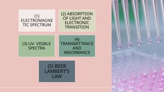
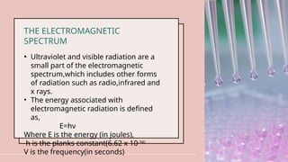
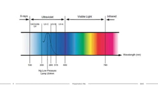
THE ELECTROMAGNETIC
SPECTRUM
• Ultravioletand visible radiation are a
small part of the electromagnetic
spectrum,which includes other forms
of radiation such as radio,infrared and
x rays.
• The energy associated with
electromagnetic radiation is defined
as,
E=hv
Where E is the energy (in joules),
h is the planks constant(6.62 x 10⁻³⁴)
V is the frequency(in seconds)
.
10
(2) ABSORPTION OFLIGHT AND ELECTRONIC
TRANSITIONS
• In UV-Vis spectroscopy, a sample is exposed to
light with wavelengths in the UV (200–400 nm)
and visible (400–700 nm) regions. If the energy of
the light matches the energy required for an
electronic transition in the molecule, absorption
occurs.
• Electronic Transitions: Different types of electronic
transitions can occur in a molecule, depending on
its structure:
• π π*
→ transitions: Occur in molecules with
conjugated double bonds.
• n π*
→ transitions: Occur in molecules with lone
pairs (non-bonding electrons) and π bonds.
• d-d transitions and charge transfer transitions:
11.
.
11
(3) UV VISIBLESPECTRA OR
ABSORBTION SPECTRUM
• When radiation interacts with matter,several
processes can be occurred including
reflection,scattering,absorbance,fluorescence or
phosphorescence and photochemical reactions
(absorbance and bond breaking).
• When measuring samples to determine their uv
visible spectrum absorbance is measured.
• The spectrometer measures the absorbance of
the sample across a range of wavelengths.
• The resulting absorption spectrum shows peaks
at wavelength where molecules absorbs light,
with each peak corresponding to a specific
electronic transition.
12.
.
12
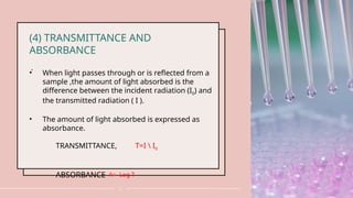
(4) TRANSMITTANCE AND
ABSORBANCE
•When light passes through or is reflected from a
sample ,the amount of light absorbed is the
difference between the incident radiation (I0) and
the transmitted radiation ( I ).
• The amount of light absorbed is expressed as
absorbance.
TRANSMITTANCE, T=I I0
ABSORBANCE A= -Log T
13.
.
13
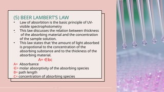
(5) BEER LAMBERT’SLAW
• Law of absorbtion is the basic principle of UV-
visible spectrophotometry
• This law discusses the relation between thickness
of the absorbing material and the concentration
of the sample solution.
• This law states that ‘the amount of light absorbed
is propotional to the concentration of the
absorbing substance and to the thickness of the
absorbing material.
A= bc
∈
A= Absorbance
∈= molar absorptivity of the absorbing species
B= path length
C= concentration of absorbing species
.
17
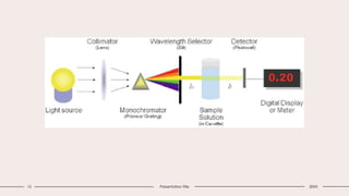
(1) LIGHT SOURCE
•Tungsten filament lamps and
Hydrogen-Deuterium lamps are most
widely used and suitable light source
as they cover the whole UV region.
• Tungsten filament lamps are rich in
red radiations; more specifically they
emit the radiations of 375 nm, while
the intensity of Hydrogen-Deuterium
lamps falls below 375 nm.
18.
.
18
(2)MONOCHROMATOR
• Monochromators generallyis composed of
prisms and slits.
• Most of the spectrophotometers are double
beam spectrophotometers.
• The radiation emitted from the primary source
is dispersed with the help of rotating prisms.
• The various wavelengths of the light source
which are separated by the prism are then
selected by the slits such the rotation of the
prism results in a series of continuously
increasing wavelength to pass through the
slits for recording purpose
• The beam selected by the slit is
monochromatic and further divided into two
beams with the help of another prism.
19.
.
19
(3)Sample and referencecells
• One of the two divided beams is passed
through the sample solution and
second beam is passes through the
reference solution.
• Both sample and reference solution are
contained in the cells.
• These cells are made of either silica or
quartz.
• Glass can't be used for the cells as it
also absorbs light in the UV region.
20.
.
20
(4)DETECTOR
• Generally twophotocells serve the
purpose of detector in UV spectroscopy.
• One of the photocell receives the beam
from sample cell and second detector
receives the beamfrom the reference.
• The intensity of the radiation from the
reference cell is stronger than the beam
of sample cell.
• This results in the generation of
pulsating or alternating currents in the
photocells.
23 Presentation title20XX
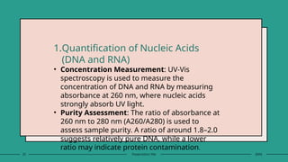
1.Quantification of Nucleic Acids
(DNA and RNA)
• Concentration Measurement: UV-Vis
spectroscopy is used to measure the
concentration of DNA and RNA by measuring
absorbance at 260 nm, where nucleic acids
strongly absorb UV light.
• Purity Assessment: The ratio of absorbance at
260 nm to 280 nm (A260/A280) is used to
assess sample purity. A ratio of around 1.8–2.0
suggests relatively pure DNA, while a lower
ratio may indicate protein contamination.
24.
24 Presentation title20XX
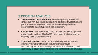
2.PROTIEN ANALYSIS
• Concentration Determination: Proteins typically absorb UV
light at 280 nm due to aromatic amino acids like tryptophan and
tyrosine. Measuring absorbance at this wavelength allows
researchers to quantify protein concentration.
• Purity Check: The A260/A280 ratio can also be used for protein
purity checks, with an A260/A280 ratio closer to 0.6 indicating
relatively pure protein solutions.
• Structural Studies: UV-Vis can provide insights into protein
secondary structure through circular dichroism (CD)
spectroscopy in the far-UV range, an extension of UV-Vis used
specifically to study protein folding and conformational changes.
25.
25 Presentation title20XX
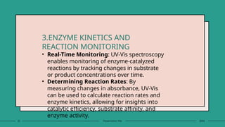
3.ENZYME KINETICS AND
REACTION MONITORING
• Real-Time Monitoring: UV-Vis spectroscopy
enables monitoring of enzyme-catalyzed
reactions by tracking changes in substrate
or product concentrations over time.
• Determining Reaction Rates: By
measuring changes in absorbance, UV-Vis
can be used to calculate reaction rates and
enzyme kinetics, allowing for insights into
catalytic efficiency, substrate affinity, and
enzyme activity.
26.
26 Presentation title20XX
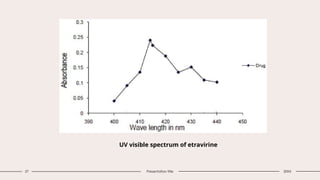
4.PHARMACEUTICAL ANALYSIS
• UV Visible spectroscopy has been widely
used technique in thedetermination of drug
concentration in pharmaceutical analysis.
• For example ,this technique is used in the
determination of etravirine in bulk and
pharmaceutical formulations.This is acting
as an antiviral drug and it showed the
maximum absorbance at 414 nm by
reacting with NaOH and naphthaquinone.
28 Presentation title20XX
(5)Interaction of human serum and
gold nanoparticles
• A case study has been conducted on combined uv
visible spectroscopy and chemometrics to determine
the interaction of human serum (HAS) and gold
nanoparticles(AuNPs).
• The data which has been recovered from the uv visible
spectroscopy and chemometrics about protein
interaction with nanoparticles were applied to
thermodynamics ,kinetic and structural parameters to
establish the evolution of protein nano conjugate.
29.
29 Presentation title20XX
Interaction of human serum albumin with citrate capped gold
nanoparticles
31 Presentation title20XX
The main limitations of UV-Visible spectroscopy include:
1. Limited Sensitivity: Less effective for detecting low-
concentration samples compared to other techniques
2. Lack of Specificity: Overlapping spectra can make it
difficult to distinguish between similar compounds in
mixtures.
3. Limited Structural Information: Provides minimal
detail about molecular structure.
4. Sensitivity to Solvent Effects: Different solvents can
alter absorption peaks, complicating analysis.
5. Interference from Particulates: Turbidity or
particulates can cause light scattering, affecting
accuracy.
32.
32 Presentation title20XX
6. Only Detects Compounds with Chromophores: Limited to
molecules with UV/visible-absorbing groups.
7. Path Length and Concentration Constraints: Deviations
occur at high concentrations or long path lengths
8. Photodegradation: Sensitive samples may degrade under
prolonged UV exposure.
9. Instrumental Limitations: Baseline drift and noise can
impact precision, especially at low absorbance.
10. Environmental Sensitivity: Changes in pH and
temperature can affect absorbance.These limitations often
require complementary methods for more detailed analysis.