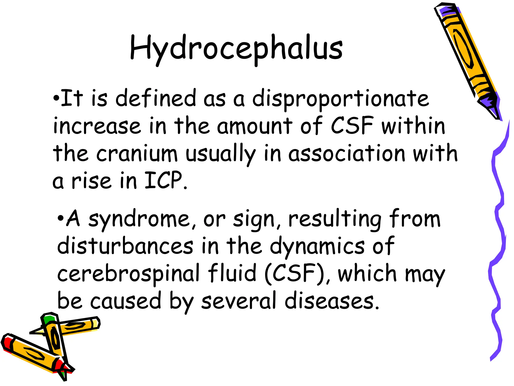
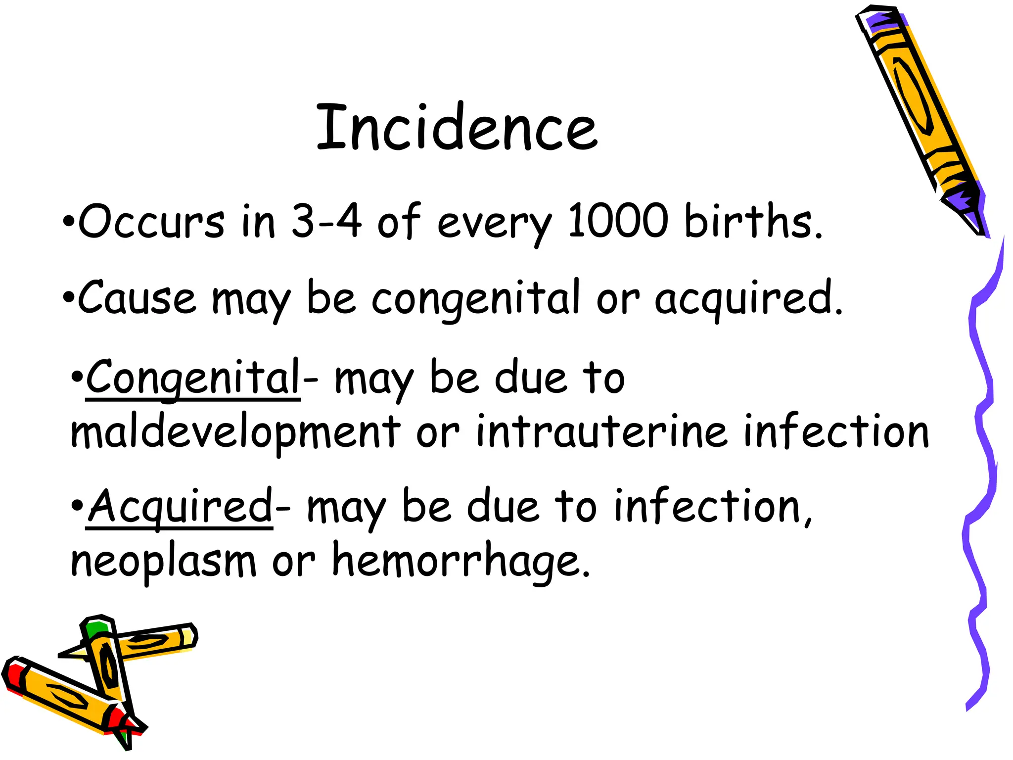
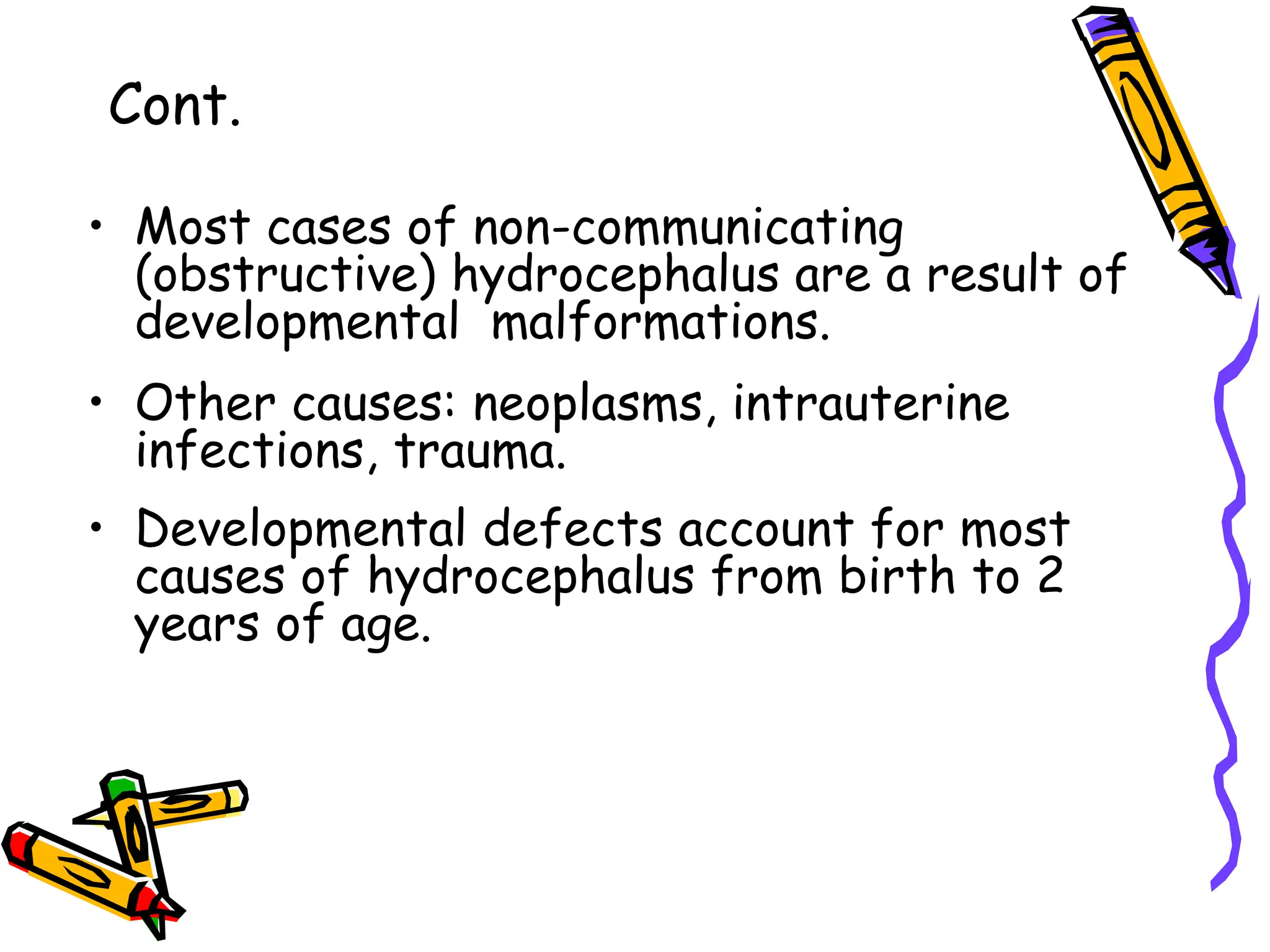
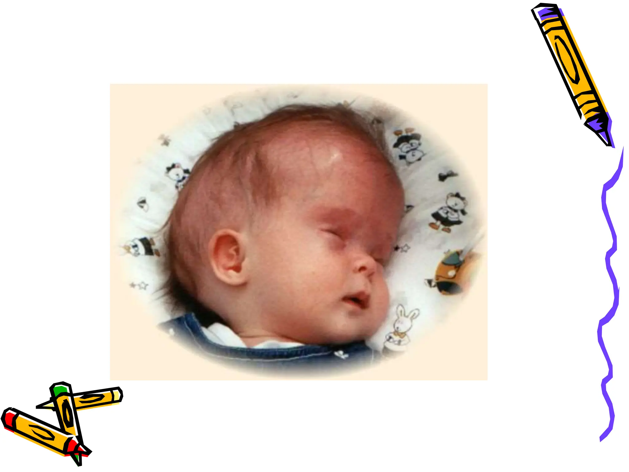
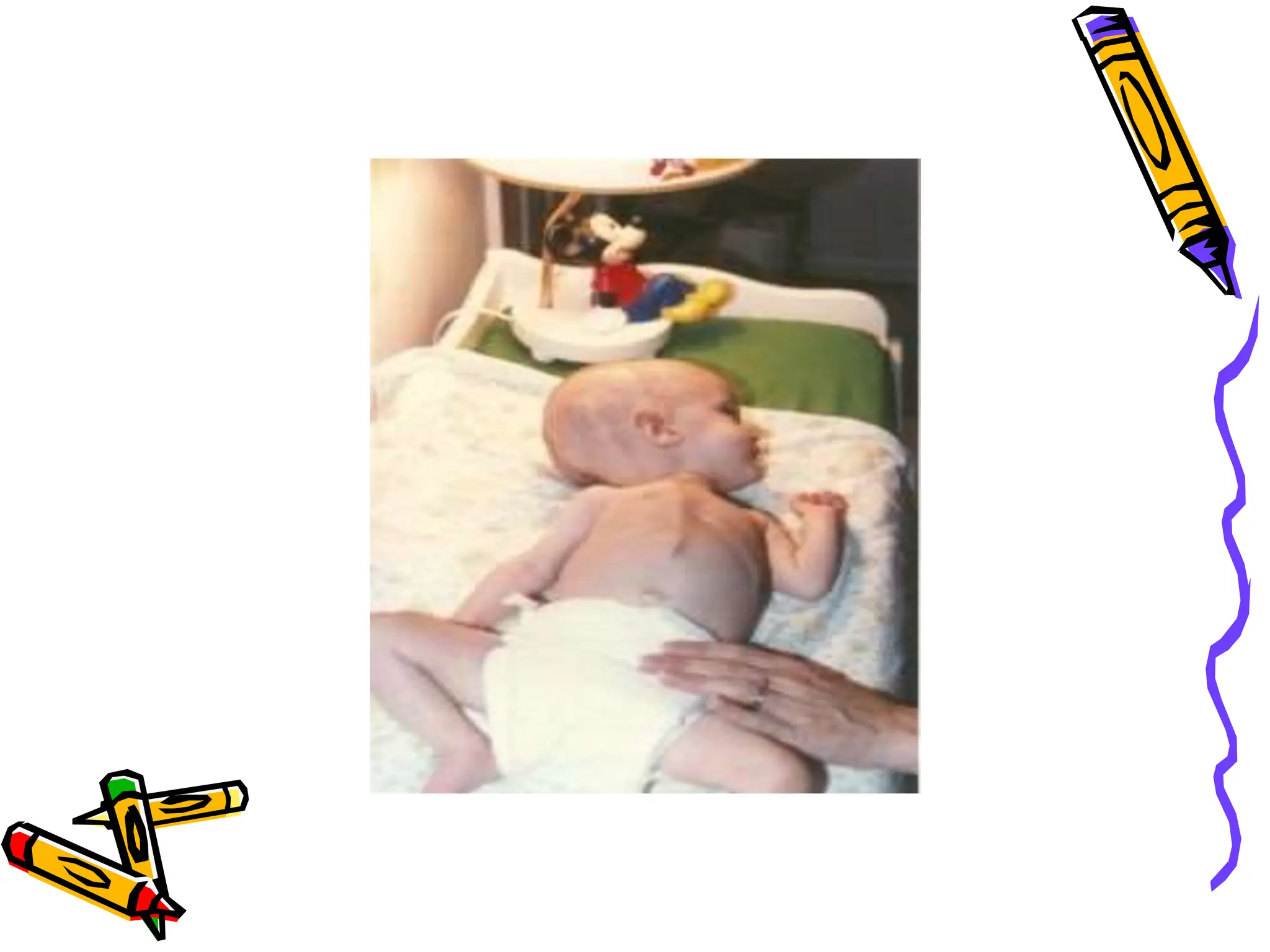
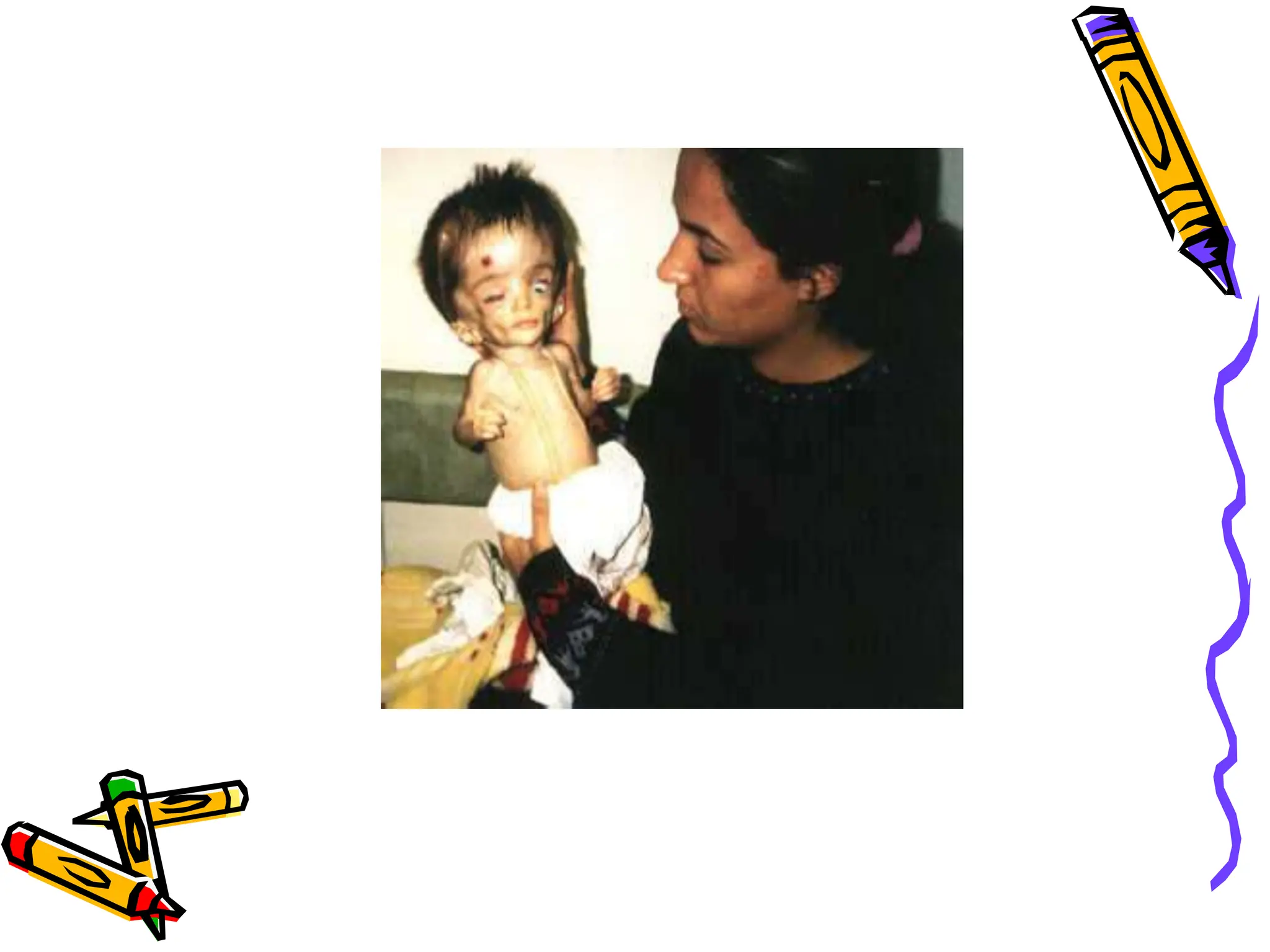
SCHOOL OF CLINICAL SCIENCES
MAKAMBO-MAKENI
Department of pathology
Lecturer: DR. ABUBAKAR KANU
Topic: hydrocephalus
Outline:
Definition
Incidence
Types
Classification
Pathophysiology
mechanism of fluid in balance
Common defect of the cranium
Clinical manifestation
Diagnosis
Hydrocephalus
•It is defined as a disproportionate
increase in the amount of CSF within
the cranium usually in association with
a rise in ICP.
•A syndrome, or sign, resulting from
disturbances in the dynamics of
cerebrospinal fluid (CSF), which may
be caused by several diseases.
Incidence
•Occurs in 3-4 of every 1000 births.
•Cause may be congenital or acquired.
•Congenital- may be due to
maldevelopment or intrauterine infection
•Acquired- may be due to infection,
neoplasm or hemorrhage.
Pathophysiology
•CSF is formed by two mechanisms:
-Secretion by the choroid plexus,
-Lymphatic-like drainage by the
extracellular fluid in brain.
•CSF circulates thru ventricular system
and is absorbed within subarachnoid
spaces by unknown mechanism.
Mechanisms of Fluid
Imbalance
• Hydrocephalus results from:
• 1. Impaired absorption of CSF within
the subarachnoid space
(communicating hydrocephalus), or
• 2. Obstruction to the flow of CSF
through the ventricular system (non-
communicating hydrocephalus)
Mechanisms of fluid
imbalance
• Both lead to increase accumulation of
CSF in the ventricles!
• Ventricles become dilated and
compress the brain.
• When this happens before cranial
sutures are closed, skull enlarges.
• In children <10-12, previously closed
sutures may open.
• Most cases of non-communicating
(obstructive) hydrocephalus are a result of
developmental malformations.
• Other causes: neoplasms, intrauterine
infections, trauma.
• Developmental defects account for most
causes of hydrocephalus from birth to 2
years of age.
Cont.
Common Defects
• Arnold-Chiari Malformation (ACM)
– Type 2 malformation of brain seen most
exclusively with myelomeningocele, is
characterized by herniation of a small
cerebellum, medulla, pons, and fourth
ventricle into the cervical spinal canal
through an enlarged foramen magnum.
Clinical manifestations
• Clinical picture depends on acuity of
onset and presence of preexisting
structural lesions.
Infancy
• Head grows at alarming rate with
hydrocephalus.
– First signs- bulging of fontanels without
head enlargement.
– Tense, bulging, non-pulsatile anterior
fontanel
– Dilated scalp veins, esp. when crying
– Thin skull bones with separated sutures
(cracked pot sounds on percussion)
Infancy
• Protruding forehead or bossing.
• Depressed eyes or setting-sun eyes (eyes
rotating or downward with sclera visible
above pupil)
• Pupils sluggish with unequal response to
light
• Irritability, lethargy, feeds poorly,
changes in LOC, arching of back
(opisthotonos), lower extremity spasticity.
• May cry when picked up or rocked; quiets
when allowed to lay still.
Infancy
• Swallowing difficulties, stridor,
apnea, aspiration, respiratory
difficulties and arm weakness may
indicate brain stem compression.
• If hydrocephalus progresses,
difficulty sucking and feeding, and a
high-pitched shrill cry results. (lower
brain stem dysfunction)
Infancy
• Emesis, somnolence, seizures, and
cardiopulmonary distress ensues and
hydrocephalus progresses.
• Severely affected infants may not
survive neonatal period.
Childhood
• Signs and symptoms caused by
increased ICP.
• Manifestations caused by posterior
neoplasms and aqueduct stenosis,
manifestations associated with
space-occupying lesions.
Childhood
• Headache on awakening with improvement
following emesis or sitting up.
• Papilledema (swelling of optic disc DT
obstruction), strabismus, and
extrapyramidal tract signs such as ataxia
• Irritability, lethargy, apathy, confusion,
and often incoherent
Childhood
• Dandy-Walker syndrome- congenital
defect-late onset.
– Obstruction of foramen of Luschka and
Magendie
– Bulging occiput, nystagmus, ataxia,
cranial nerve palsies
– Female predominance (3:1)
– Absence or occlusion of ventricles
Diagnostic Evaluation
• Antenatal- fetal ultrasound as early as 14
weeks
• Infancy- based on head circumference
crosses one or more grid lines on the
infant growth chart within a 4 week period
and there are progressive neuro signs.
• CT and
• MRI to localize site of obstruction; reveal
large ventricles
THANK YOU!!
ANY QUESTIONS!!

HYDROCEPHALY.ppt

  • 1.
    SCHOOL OF CLINICALSCIENCES MAKAMBO-MAKENI Department of pathology Lecturer: DR. ABUBAKAR KANU Topic: hydrocephalus
  • 2.
    Outline: Definition Incidence Types Classification Pathophysiology mechanism of fluidin balance Common defect of the cranium Clinical manifestation Diagnosis
  • 3.
    Hydrocephalus •It is definedas a disproportionate increase in the amount of CSF within the cranium usually in association with a rise in ICP. •A syndrome, or sign, resulting from disturbances in the dynamics of cerebrospinal fluid (CSF), which may be caused by several diseases.
  • 5.
    Incidence •Occurs in 3-4of every 1000 births. •Cause may be congenital or acquired. •Congenital- may be due to maldevelopment or intrauterine infection •Acquired- may be due to infection, neoplasm or hemorrhage.
  • 6.
    Pathophysiology •CSF is formedby two mechanisms: -Secretion by the choroid plexus, -Lymphatic-like drainage by the extracellular fluid in brain. •CSF circulates thru ventricular system and is absorbed within subarachnoid spaces by unknown mechanism.
  • 8.
    Mechanisms of Fluid Imbalance •Hydrocephalus results from: • 1. Impaired absorption of CSF within the subarachnoid space (communicating hydrocephalus), or • 2. Obstruction to the flow of CSF through the ventricular system (non- communicating hydrocephalus)
  • 9.
    Mechanisms of fluid imbalance •Both lead to increase accumulation of CSF in the ventricles! • Ventricles become dilated and compress the brain. • When this happens before cranial sutures are closed, skull enlarges. • In children <10-12, previously closed sutures may open.
  • 10.
    • Most casesof non-communicating (obstructive) hydrocephalus are a result of developmental malformations. • Other causes: neoplasms, intrauterine infections, trauma. • Developmental defects account for most causes of hydrocephalus from birth to 2 years of age. Cont.
  • 11.
    Common Defects • Arnold-ChiariMalformation (ACM) – Type 2 malformation of brain seen most exclusively with myelomeningocele, is characterized by herniation of a small cerebellum, medulla, pons, and fourth ventricle into the cervical spinal canal through an enlarged foramen magnum.
  • 13.
    Clinical manifestations • Clinicalpicture depends on acuity of onset and presence of preexisting structural lesions.
  • 14.
    Infancy • Head growsat alarming rate with hydrocephalus. – First signs- bulging of fontanels without head enlargement. – Tense, bulging, non-pulsatile anterior fontanel – Dilated scalp veins, esp. when crying – Thin skull bones with separated sutures (cracked pot sounds on percussion)
  • 18.
    Infancy • Protruding foreheador bossing. • Depressed eyes or setting-sun eyes (eyes rotating or downward with sclera visible above pupil) • Pupils sluggish with unequal response to light • Irritability, lethargy, feeds poorly, changes in LOC, arching of back (opisthotonos), lower extremity spasticity. • May cry when picked up or rocked; quiets when allowed to lay still.
  • 19.
    Infancy • Swallowing difficulties,stridor, apnea, aspiration, respiratory difficulties and arm weakness may indicate brain stem compression. • If hydrocephalus progresses, difficulty sucking and feeding, and a high-pitched shrill cry results. (lower brain stem dysfunction)
  • 20.
    Infancy • Emesis, somnolence,seizures, and cardiopulmonary distress ensues and hydrocephalus progresses. • Severely affected infants may not survive neonatal period.
  • 22.
    Childhood • Signs andsymptoms caused by increased ICP. • Manifestations caused by posterior neoplasms and aqueduct stenosis, manifestations associated with space-occupying lesions.
  • 23.
    Childhood • Headache onawakening with improvement following emesis or sitting up. • Papilledema (swelling of optic disc DT obstruction), strabismus, and extrapyramidal tract signs such as ataxia • Irritability, lethargy, apathy, confusion, and often incoherent
  • 24.
    Childhood • Dandy-Walker syndrome-congenital defect-late onset. – Obstruction of foramen of Luschka and Magendie – Bulging occiput, nystagmus, ataxia, cranial nerve palsies – Female predominance (3:1) – Absence or occlusion of ventricles
  • 25.
    Diagnostic Evaluation • Antenatal-fetal ultrasound as early as 14 weeks • Infancy- based on head circumference crosses one or more grid lines on the infant growth chart within a 4 week period and there are progressive neuro signs. • CT and • MRI to localize site of obstruction; reveal large ventricles
  • 26.