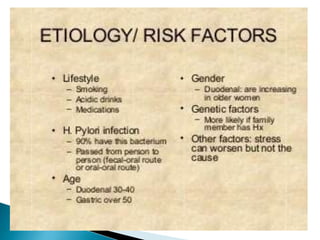
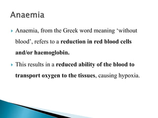
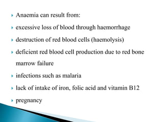
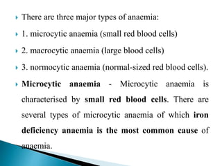
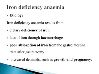
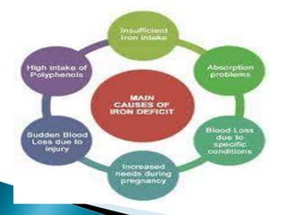
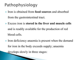
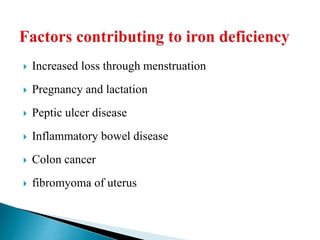
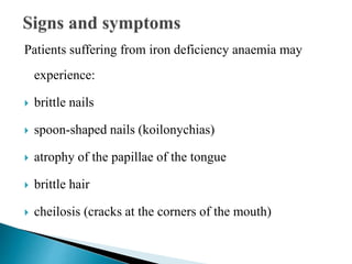
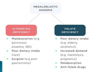
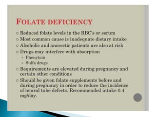
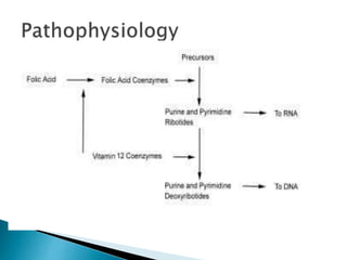
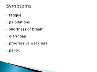
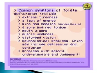
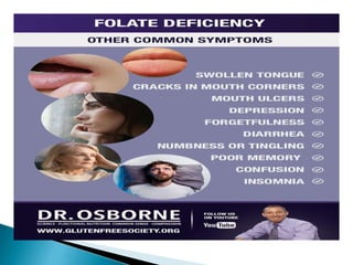
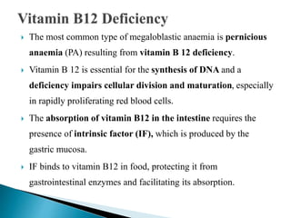
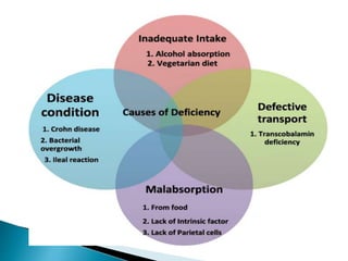
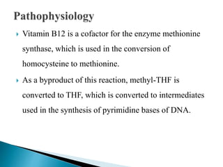
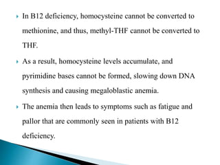
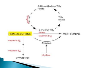
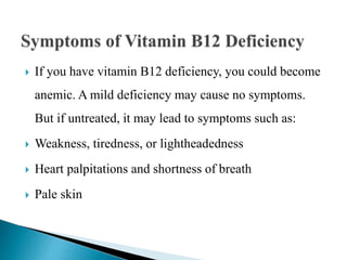
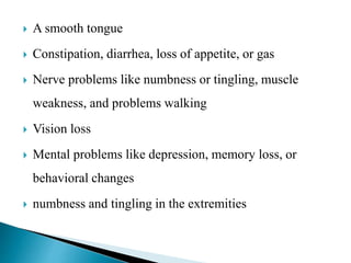
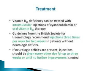
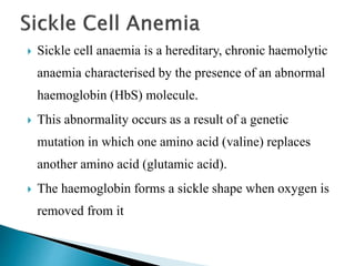
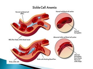
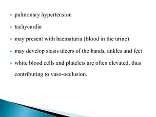
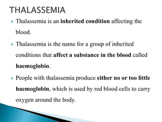
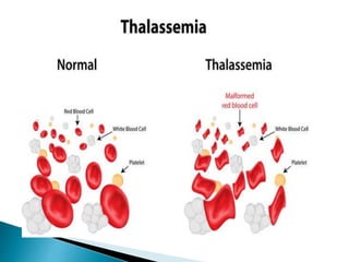
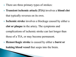
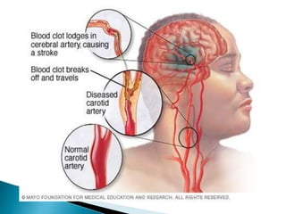
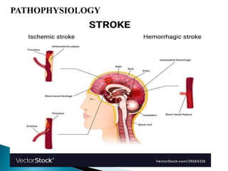
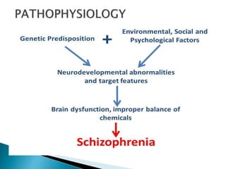
2. Peptic ulcer is the term used to define the
development of an ulcer in the lower part of the
oesophagus, the stomach or the duodenum.
Duodenal ulcers are more common than gastric
ulcers.
The ulcer develops as a result of exposure of the
gastrointestinal epithelium to the acidic gastric
secretions of the stomach.
3. Etiology
The causes of peptic ulcers include:
infection from Helicobacter pylori (a Gram-negative
bacterium)
excessive consumption of alcohol and cigarette smoking
excessive gastric secretions as a result of stress
excessive use of non-steroidal anti-inflammatory drugs
(NSAIDs) and aspirin derivatives
excessive consumption of caffeine
familial history of peptic ulcer.
4.
5.
6.
7. Mucus lines the digestive tract and acts as a barrier
against the acidic gastric secretions.
Too little mucus production coupled with too much
acid production will leave the digestive tract
vulnerable to acid erosion and ulceration.
Erosion of the mucosal lining may result in the
formation of a fistula.
8. The fistula allows the acidic gastric contents to leak
out into the peritoneum, resulting in peritonitis.
Stress, caffeine, cigarette smoking and alcohol
consumption increase acid production.
Medications such as NSAIDs and aspirin inhibit
prostaglandins, which protect the mucosal lining.
H. pylori bacterial infection leads to death of the
mucosal epithelial cells of the stomach and
duodenum.
9. The bacteria release toxins and enzymes that reduce
the efficiency of mucus in protecting the mucosal
lining of the gastrointestinal tract.
10.
11.
12.
13.
14.
15.
16.
17. Anaemia, from the Greek word meaning ‘without
blood’, refers to a reduction in red blood cells
and/or haemoglobin.
This results in a reduced ability of the blood to
transport oxygen to the tissues, causing hypoxia.
18. Anaemia can result from:
excessive loss of blood through haemorrhage
destruction of red blood cells (haemolysis)
deficient red blood cell production due to red bone
marrow failure
infections such as malaria
lack of intake of iron, folic acid and vitamin B12
pregnancy
19. There are three major types of anaemia:
1. microcytic anaemia (small red blood cells)
2. macrocytic anaemia (large blood cells)
3. normocytic anaemia (normal-sized red blood cells).
Microcytic anaemia - Microcytic anaemia is
characterised by small red blood cells. There are
several types of microcytic anaemia of which iron
deficiency anaemia is the most common cause of
anaemia.
20. Etiology
Iron deficiency anaemia results from:
dietary deficiency of iron
loss of iron through haemorrhage
poor absorption of iron from the gastrointestinal
tract after gastrectomy
increased demands, such as growth and pregnancy.
21.
22. Iron is obtained from food sources and absorbed
from the gastrointestinal tract.
Excess iron is stored in the liver and muscle cells
and is readily available for the production of red
blood cells.
Iron deficiency anaemia is present when the demand
for iron in the body exceeds supply; anaemia
develops slowly in three stages:
23. 1. The body’s iron stores are depleted; however,
erythropoiesis continues normally.
2. Iron transportation to bone marrow is
diminished, resulting in deficiency in red cell
production.
3. The number of microcytic red blood cells
increases in the circulation, replacing the normal
mature red blood cells.
24.
25. Increased loss through menstruation
Pregnancy and lactation
Peptic ulcer disease
Inflammatory bowel disease
Colon cancer
fibromyoma of uterus
26. Patients suffering from iron deficiency anaemia may
experience:
brittle nails
spoon-shaped nails (koilonychias)
atrophy of the papillae of the tongue
brittle hair
cheilosis (cracks at the corners of the mouth)
27. dizziness – due to lack of oxygen supply to the brain
hypoxia
breathlessness – physiological compensation resulting
from the lack of oxygen
loss of appetite, which may be due to a sore mouth.
30. Macrocytic anaemia is also termed megaloblastic
anaemia.
It is characterised by defective deoxyribonucleic acid
(DNA) synthesis resulting in the production of
unusually large stem cells (macrocytes) in the
circulation.
32. Folic acid (folate) is an essential vitamin for the
production and maturation of red blood cells.
Folate is obtained from the diet and is absorbed from the
jejunum and stored in the liver.
It is found in leafy vegetables, fruit, cereals and meat.
41. The most common type of megaloblastic anaemia is pernicious
anaemia (PA) resulting from vitamin B 12 deficiency.
Vitamin B 12 is essential for the synthesis of DNA and a
deficiency impairs cellular division and maturation, especially
in rapidly proliferating red blood cells.
The absorption of vitamin B12 in the intestine requires the
presence of intrinsic factor (IF), which is produced by the
gastric mucosa.
IF binds to vitamin B12 in food, protecting it from
gastrointestinal enzymes and facilitating its absorption.
42. Autoimmune disorders
Malabsorption due to inflammatory disease such as Crohn’s
disease
Dietary insufficiency
total gastrectomy, partial gastrectomy or gastrojejunostomy
gastric lesions
carcinoma of the stomach
alcohol abuse
43.
44. Vitamin B12 is a cofactor for the enzyme methionine
synthase, which is used in the conversion of
homocysteine to methionine.
As a byproduct of this reaction, methyl-THF is
converted to THF, which is converted to intermediates
used in the synthesis of pyrimidine bases of DNA.
45. In B12 deficiency, homocysteine cannot be converted to
methionine, and thus, methyl-THF cannot be converted to
THF.
As a result, homocysteine levels accumulate, and
pyrimidine bases cannot be formed, slowing down DNA
synthesis and causing megaloblastic anemia.
The anemia then leads to symptoms such as fatigue and
pallor that are commonly seen in patients with B12
deficiency.
46.
47. If you have vitamin B12 deficiency, you could become
anemic. A mild deficiency may cause no symptoms.
But if untreated, it may lead to symptoms such as:
Weakness, tiredness, or lightheadedness
Heart palpitations and shortness of breath
Pale skin
48. A smooth tongue
Constipation, diarrhea, loss of appetite, or gas
Nerve problems like numbness or tingling, muscle
weakness, and problems walking
Vision loss
Mental problems like depression, memory loss, or
behavioral changes
numbness and tingling in the extremities
49.
50. Sickle cell anaemia is a hereditary, chronic haemolytic
anaemia characterised by the presence of an abnormal
haemoglobin (HbS) molecule.
This abnormality occurs as a result of a genetic
mutation in which one amino acid (valine) replaces
another amino acid (glutamic acid).
The haemoglobin forms a sickle shape when oxygen is
removed from it
51.
52. The cause of the sickle shape is the deoxygenation of
the haemoglobin.
When the haemoglobin is fully saturated with oxygen,
the red blood cell has the normal shape but this changes
to the sickle shape as the oxygen content is reduced.
Sickled red blood cells are stiff and cannot change shape
as normal red blood cells do when they pass through
capillaries.
53. As a result, the sickled red blood cells obstruct blood flow,
causing vascular obstruction, pain and tissue ischaemia.
Sickling is not permanent; most sickled red blood cells
regain their normal shape once they are saturated with
oxygen.
However, repeated sickling causes loss of elasticity of the
cell membrane and over time the cells fail to return to the
normal shape when oxygen concentration increases.
The weakened red blood cells are haemolysed and removed
from the circulation.
54. Symptoms
pain and swelling caused by occluded blood vessels
affecting the hands and feet
abdominal pain if the abdominal blood vessels are
occluded
increased incidence of infection such as osteomyelitis
55. pulmonary hypertension
tachycardia
may present with haematuria (blood in the urine)
may develop stasis ulcers of the hands, ankles and feet
white blood cells and platelets are often elevated, thus
contributing to vaso-occlusion.
56.
57.
58. Thalassemia is an inherited condition affecting the
blood.
Thalassemia is the name for a group of inherited
conditions that affect a substance in the blood called
haemoglobin.
People with thalassemia produce either no or too little
haemoglobin, which is used by red blood cells to carry
oxygen around the body.
59. Thalassemia is caused by mutations in the DNA of cells
that make hemoglobin — the substance in red blood
cells that carries oxygen throughout your body.
The mutations associated with thalassemia are passed
from parents to children.
Hemoglobin molecules are made of chains called alpha
and beta chains that can be affected by mutations.
60.
61. In thalassemia, the production of either the alpha or beta chains
are reduced, resulting in either alpha-thalassemia or beta-
thalassemia.
In alpha-thalassemia, the severity of thalassemia you have
depends on the number of gene mutations you inherit from
your parents. The more mutated genes, the more severe your
thalassemia.
In beta-thalassemia, the severity of thalassemia you have
depends on which part of the hemoglobin molecule is affected.
62. Alpha-thalassemia
Four genes are involved in making the alpha hemoglobin chain.
You get two from each of your parents. If you inherit:
One mutated gene, you'll have no signs or symptoms of
thalassemia. But you are a carrier of the disease and can pass it on
to your children.
Two mutated genes, your thalassemia signs and symptoms will
be mild. This condition might be called alpha-thalassemia trait.
Three mutated genes, your signs and symptoms will be
moderate to severe.
63. Beta-thalassemia
Two genes are involved in making the beta hemoglobin
chain. You get one from each of your parents. If you inherit:
One mutated gene, you'll have mild signs and symptoms.
This condition is called thalassemia minor or beta-
thalassemia.
Two mutated genes, your signs and symptoms will be
moderate to severe. This condition is called thalassemia
major, or Cooley anemia.
64.
65.
66.
67.
68.
69.
70. Hemophilia is a rare disorder in which the blood
doesn't clot in the typical way because it doesn't have
enough blood-clotting proteins (clotting factors).
A disorder in which blood doesn't clot normally.
When blood can't clot properly, excessive bleeding
(external and internal) occurs after any injury or
damage.
71.
72. Hemophilia is caused by a mutation or change, in one
of the genes, that provides instructions for making the
clotting factor proteins needed to form a blood clot.
73. TYPES –
Congenital hemophilia
Hemophilia is usually inherited, meaning a person is
born with the disorder (congenital). Congenital
hemophilia is classified by the type of clotting factor
that's low.
The most common type is hemophilia A, associated
with a low level of factor 8 The next most common type
is hemophilia B, associated with a low level of factor 9.
74.
75. Acquired hemophilia
Some people develop hemophilia with no family history of the
disorder. This is called acquired hemophilia.
Acquired hemophilia is a variety of the condition that occurs
when a person's immune system attacks clotting factor 8 or 9 in
the blood. It can be associated with:
Pregnancy
Autoimmune conditions
Cancer
Multiple sclerosis
Drug reactions
76.
77.
78.
79. SEIZURE - A seizure is a sudden, uncontrolled
electrical disturbance in the brain. It can cause
changes in your behaviour, movements or feelings,
and in levels of consciousness.
Having two or more seizures at least 24 hours apart
that aren't brought on by an identifiable cause is
generally considered to be epilepsy.
80. STATUS EPILEPTICUS
A seizure that lasts longer than 5 minutes, or
having more than 1 seizure within a 5 minutes
period, without returning to a normal level of
consciousness between episodes is called status
epilepticus. This is a medical emergency that may
lead to permanent brain damage or death.
81. Epilepsy is a central nervous system (neurological)
disorder in which brain activity becomes abnormal,
causing seizures or periods of unusual behaviour,
sensations and sometimes loss of awareness.
It is characterized by recurrent seizures, which are brief
episodes of involuntary movement that may involve a
part of the body (partial) or the entire body (generalized)
and are sometimes accompanied by loss of
consciousness and control of bowel or bladder
function.
82. Seizure episodes are a result of excessive
electrical discharges in a group of brain
cells.
Epilepsy is a disorder of the brain
characterized by repeated seizures.
A seizure is usually defined as a sudden
alteration of behaviour due to a temporary
change in the electrical functioning of the
86. Focal, or Partial Seizures
Primary generalized seizure
Focal seizures are also called partial seizures since they
begin in one area of the brain. They can be caused by any
type of focal injury that leaves scar tangles. Medical history
or MRI will identify a cause (such as trauma, stroke or
meningitis) in about half of the people who have focal
seizures.
87. Focal seizures can start in one part of the brain and
spread to other areas, causing symptoms that are mild or
severe, depending on how much of the brain becomes
involved.
Generalized-onset Seizures
Generalized-onset seizures are surges of abnormal nerve
discharges throughout the cortex of the brain more or less
at the same time.
The most common cause is an imbalance in the excitatory
and inhibitory impulses in the brain.
88. Absence Seizures (“Petit Mal Seizures”)
Childhood absence epilepsy shows up as brief
staring episodes in children, usually starting between
ages four and six.
Myoclonic seizures
Myoclonic seizures consist of sudden body or limb
jerks that can involve the arms, head and neck.
89. The spasms occur on both sides of the body in clusters,
especially in the morning. When these seizures develop in
adolescence along with tonic-clonic seizures, they are part
of a syndrome called juvenile myoclonic epilepsy.
Tonic and Atonic Seizures (“Drop Attacks”)
Some people, usually those with multiple brain injuries
and intellectual disability, have tonic seizures consisting
of sudden stiffness in the arms and body, which can
cause falls and injuries.
90.
91. Given that the basic mechanism of neuronal
excitability is the action potential, a hyperexcitable
state can result from increased excitatory synaptic
neurotransmission, decreased inhibitory
neurotransmission, an alteration in voltage-gated ion
channels, or an alteration of intra- or extra-cellular
ion concentrations in favour of membrane
depolarization.
92.
93.
94.
95. Parkinson’s disease (PD) is a disease of the brain that
mainly occurs over the age of 50 years of age with
increasing prevalence over the age of 85 years.
It is a progressive degenerative disease which means
that there is a continual loss of neurons and their
function.
Parkinson’s disease (PD) is a common
neurodegenerative disorder.
96. It primarily affects part of the basal ganglia known as
the substantia nigra which is an important area for
controlling voluntary movement.
In Parkinson’s disease the nerves that use dopamine as
a neurotransmitter (dopaminergic neurons) start to die
and this leads to the signs and symptoms of
Parkinson’s disease.
97.
98. The cause of PD is not known, but a number of
genetic risk factors have now been characterized, as
well as several genes which cause rare familial
forms of PD.
The movement disorder arises due to the loss of
dopaminergic neurons of the substantia nigra pars
compacta, with the pathological hallmark being
intracellular aggregates of α-synuclein, in the form of
Lewy bodies and Lewy neurites.
99. Cigarette smoking
Caffeine
Pesticides, herbicides, and heavy metals
Genetics
NEUROPATHOLOGY OF PARKINSON’S
DISEASE
Macroscopically, the brain in idiopathic PD is often
unremarkable with mild atrophy of the frontal cortex.
100. The main distinctive morphological change in the PD
brain is observed in transverse sections of the
brainstem, where almost all cases present with loss of
the darkly pigmented area in the substantia nigra
pars compacta (SNpc) and locus coeruleus.
This pigmentation loss directly correlates with the
death of dopaminergic (DA) neuromelanin-
containing neurons in the SNpc and noradrenergic
neurons in the locus coeruleus
101. The pathological hallmark of PD is the presence of
abnormal cytoplasmic deposits within neuronal cell
bodies which are immunoreactive for the protein α-
synuclein.
These pathological protein aggregates are called Lewy
bodies (LBs) and are often accompanied by dystrophic
neurites (Lewy neurites)
102.
103.
104. slow movements (bradykinesia)
tremors (initially in the hand but also seen in the limbs,
head, face and jaw.
muscle stiffness and rigidity
tendency to walk forward on the toes with small,
shuffling steps
changes in balance
105. stooped posture
confusion
Depression
difficulty with fine motor functions, e.g. writing and
eating
‘mask’-like face
general weakness and muscle fatigue.
106.
107.
108.
109.
110.
111. Stroke occurs when the blood supply to part of the
brain is interrupted or reduced, preventing brain
tissue from getting oxygen and nutrients. Brain cells
begin to die in minutes.
A stroke occurs when a blood vessel in the brain
ruptures and bleeds, or when there’s a blockage in the
blood supply to the brain.
112. There are three primary types of strokes:
Transient ischemic attack (TIA) involves a blood clot
that typically reverses on its own.
Ischemic stroke involves a blockage caused by either a
clot or plaque in the artery. The symptoms and
complications of ischemic stroke can last longer than
those of a TIA, or may become permanent.
Hemorrhagic stroke is caused by either a burst or
leaking blood vessel that seeps into the brain.
121. Alzheimer's disease is a progressive neurologic
disorder that causes the brain to shrink and brain
cells to die. Alzheimer's disease is the most common
cause of dementia — a continuous decline in thinking,
behavioural and social skills that affects a person's
ability to function independently.
The early signs of the disease include forgetting recent
events or conversations.
122. As the disease progresses, a person with Alzheimer's
disease will develop severe memory impairment and
lose the ability to carry out everyday tasks.
Medications may temporarily improve or slow
progression of symptoms.
There is no treatment that cures Alzheimer's disease or
alters the disease process in the brain.
123.
124.
125.
126.
127. The amygdala is part of the limbic system that plays a major
role in the processing and memorizing of emotional
reactions.
Neuropathologically, AD brains are characterized by the
presence of extracellular fibrillar amyloid beta peptide
(Aβ) in amyloid plaques, intraneuronal neurofibrillary
tangles consisting of aggregated hyperphosphorylated
tau, and elevated brain levels of soluble Aβ oligomers.
128. Plaques and neurofibrillary tangles are observed mostly
in the cerebral cortex, but subcortical regions such as
nucleus basalis, thalamus, locus coeruleus and raphe
nuclei are also affected
The patients with AD present neuropsychiatric
symptoms, such as hallucinations, delusions,
paranoia, anxiety, agitation, and affective
disturbances during the course of their illness
129.
130.
131.
132. Depression is a mood disorder that causes a
persistent feeling of sadness and loss of interest.
Also called major depressive disorder or clinical
depression, it affects how you feel, think and behave
and can lead to a variety of emotional and physical
problems.
133. Abuse - Physical, sexual, or emotional abuse
Age
Certain medications. Some drugs, such as
isotretinoin (used to treat acne), the antiviral drug
interferon-alpha, and corticosteroids, can increase your
risk of depression.
Conflict
134. Death or a loss
Gender
Genes
Other personal problems
Serious illnesses
135.
136.
137.
138.
139.
140. Feelings of sadness, tearfulness, emptiness or hopelessness
Angry outbursts, irritability or frustration, even over small
matters
Loss of interest or pleasure in most or all normal activities,
such as sex, hobbies or sports
Sleep disturbances, including insomnia or sleeping too
much
Tiredness and lack of energy, so even small tasks take extra
effort
141. Reduced appetite and weight loss or increased cravings for food
and weight gain
Anxiety, agitation or restlessness
Slowed thinking, speaking or body movements
Feelings of worthlessness or guilt, fixating on past failures or
self-blame
Trouble thinking, concentrating, making decisions and
remembering things
Frequent or recurrent thoughts of death, suicidal thoughts,
suicide attempts or suicide
Unexplained physical problems, such as back pain or headaches
142.
143.
144.
145.
146.
147. Schizophrenia is a serious mental disorder in which
people interpret reality abnormally.
Schizophrenia may result in some combination of
hallucinations, delusions, and extremely disordered
thinking and behaviour that impairs daily
functioning, and can be disabling.
148. The exact causes of schizophrenia are unknown.
Research suggests a combination of physical, genetic,
psychological and environmental factors can make a
person more likely to develop the condition.
Genetics
Brain development
Neurotransmitters - dopamine and serotonin.
Pregnancy and birth complications
155. Diseases that result in too much sugar in the blood (high
blood glucose).
A disease in which the body does not control the
amount of glucose (a type of sugar) in the blood and
the kidneys make a large amount of urine.
This disease occurs when the body
does not make enough insulin or
does not use it properly.
Diabetes mellitus (DM) is a disease of inadequate
control of blood levels of glucose.
156. Diabetes mellitus (DM) is a metabolic disease, involving
inappropriately elevated blood glucose levels.
A blood sugar level less than 140 mg/dL (7.8 mmol/L) is
normal.
Prediabetes is a serious health condition where blood sugar
levels are higher than normal, but not high enough yet to be
diagnosed as type 2 diabetes.
A reading of more than 200 mg/dL (11.1 mmol/L) after two
hours indicates diabetes.
A reading between 140 and 199 mg/dL (7.8 mmol/L and 11.0
mmol/L) indicates prediabetes.
157. Prediabtes Symptoms
Increased thirst.
Frequent urination.
Increased hunger.
Fatigue.
Blurred vision.
Numbness or tingling in the feet or hands.
Frequent infections.
Slow-healing sores.
158. In the islets of Langerhans in the pancreas, there are two
main subclasses of endocrine cells: insulin-producing beta
cells and glucagon secreting alpha cells.
Beta and alpha cells are continually changing their levels of
hormone secretions based on the glucose environment.
Without the balance between insulin and glucagon, the
glucose levels become inappropriately skewed.
In the case of DM, insulin is either absent and/or has
impaired action (insulin resistance), and thus leads to
hyperglycemia.
159.
160.
161. Insulin dependent diabetes mellitus (IDDM), also known as type
1 diabetes, usually starts before 15 years of age, but can occur in
adults also.
T1DM is characterized by the destruction of beta cells in the
pancreas, typically secondary to an autoimmune process. The result
is the absolute destruction of beta cells, and consequentially, insulin
is absent or extremely low.
so it used to be called juvenile diabetes.
Most of the type 1 DM in which, a T-cell mediated autoimmune
attack leads to the loss of B cells.
162. T2DM or Non-insulin dependent diabetes mellitus
or adult/maturity onset DM - involves a more
insidious onset where an imbalance between insulin
levels and insulin sensitivity causes a functional deficit
of insulin. Insulin resistance is multifactorial but
commonly develops from obesity and aging.
Or excessive inappropriate glucagon secretion.
It is attributed to typical genetic makeup, familial
history, obesity and defects in insulin receptor.
163. Diabetes can occur temporarily during pregnancy and it occurs
in 2% to 10% of all pregnancies.
Significant hormonal changes in pregnancy can lead to blood
sugar elevation in genetically predisposed individual.
Gestational diabetes usually resolves once the baby is born.
35% to 60% of women with gestational diabetes will develop
into type II DM.
172. Too little thyroid hormone from an underactive thyroid gland is called
hypothyroidism.
Body’s metabolism is slowed.
Symptoms –
Fatigue
Foggy
Dry skin
Constipation
Feeling cold
Fluid retention
Muscle and joint aches
depression
173. Too much of thyroid hormone from an overactive thyroid
gland.
Symptoms –
Tremor
Nervousness
Fast heart rate
Fatigue
Increase in bowel movments
Unintentional weight loss
174. It is inflammation of the thyroid. It occurs when the body’s
immune system makes antibodies that attack the thyroid.
Causes – Autoimmune disease like type I DM and RA,
genetics, viral or bacterial infection.
Two types - postpartus thyroiditis & Hashimoto’s diagnosis
postpartus thyroiditis - inflammation of the thyroid after
giving birth.
Hashimoto’s diagnosis – testing the level of TSH.
175. It is an autoimmune disorder that occurs when the body’s
immune system mistakenly attacks the thyroid gland.
This can cause the gland to overproduce the hormone
responsible for regulating metabolism.
THYROID NODULES
Nodules are lumps or abnormal masses within the thyroid.
Nodules can be caused by benign cysts, benign tumors or
cancer.
176. Papillary thyroid cancer
Medullary thyroid cancer
Follicular thyroid cancer
GOITER
Enlargement of the thyroid gland.
Colloid goiter
Non-toxic goiter
Toxic nodular goiter