
Recommended
More Related Content
Similar to Carbohydrate & lipid Metabolism_food Sci.pdf
Similar to Carbohydrate & lipid Metabolism_food Sci.pdf (20)
More from EmmanuelSimonMarino
Recently uploaded
Recently uploaded (20)
Carbohydrate & lipid Metabolism_food Sci.pdf
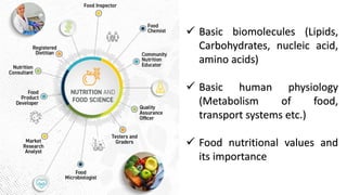
- 1. ✓ Basic biomolecules (Lipids, Carbohydrates, nucleic acid, amino acids) ✓ Basic human physiology (Metabolism of food, transport systems etc.) ✓ Food nutritional values and its importance
- 2. Syllabus • Unit I: Enzymology • Unit II: Molecular aspect of transport • Unit III: Metabolism of carbohydrate • Unit IV: Metabolism of Protein
- 3. What is a metabolic pathway? • A metabolic pathway is a series of chemical reactions that takes a starting molecule and modifies it, step-by- step, through a series of metabolic intermediates, eventually yielding a final product. • It is irreversible and committed to the first step. • All metabolic pathways are regulated and occurs at specific cellular location in eukaryotes.
- 4. Two types of metabolic pathways Anabolic pathways are those that require energy to synthesize larger molecules. Catabolic pathways are those that generate energy by breaking down larger molecules.
- 8. Glucose occupies central position in metabolism • The complete oxidation of glucose to carbon dioxide and water proceeds with a standard free- energy change of 2,840 kJ/mol. • By storing glucose as a high molecular weight polymer such as starch or glycogen, a cell can stockpile large quantities of hexose units while maintaining a relatively low cytosolic osmolarity. When energy demands increase, glucose can be released from these intracellular storage polymers and used to produce ATP either aerobically or anaerobically.
- 9. Glucose occupies central position in metabolism • Many tissues can also use fat or protein as an energy source but others, such as the brain and red blood cells, can only use glucose. • Glucose is stored in the body as glycogen. The liver is an important storage site for glycogen.
- 11. Glycolysis
- 12. Glycolysis = E.M. Pathway History of Glycolysis Breakdown of glucose in yeast cells Otto Warburg Hans von Euler-Chelpin Gustav Embden Otto Meyerhof Breakdown of glucose in muscle cells In 1930,
- 13. History of Glycolysis Breakdown of glucose in yeast cells Otto Warburg Hans von Euler-Chelpin Gustav Embden Otto Meyerhof Breakdown of glucose in muscle cells In 1930, Glycolysis = E.M. Pathway
- 14. Location: Cytoplasmic fraction of cell “Glycolysis is defined as the sequence of reactions converting glucose (or glycogen) to pyruvate or lactate, with the production of ATP.” It can occur in absence of oxygen (anerobic) and have the end-product Lactate It can occur in presence of oxygen (aerobic) and have the end-product Pyruvate Glycolysis is a major pathway for ATP synthesis in tissues lacking mitochondria, e.g. erythrocytes, cornea, lens etc. Glycolysis is very essential for brain which is dependent on glucose for energy. The glucose in brain has to undergo glycolysis before it is oxidized to CO2 and H2O. Glycolysis has two major phases: Preparatory phase and Payoff phase
- 16. Irreversible
- 17. Bisphosphate
- 18. Bisphosphate ➢ In a bisphosphate compound, the two phosphate moieties are not attached to each other, but rather are bonded at different places in the compound. Diphosphate ➢ in a diphosphate compound, the two phosphate moieties are attached to each other
- 24. Important chemical transformations 1. Degradation of the carbon skeleton of glucose to yield pyruvate. 2. Phosphorylation of ADP to ATP by high-energy phosphate compounds formed during glycolysis 3. Transfer of a hydride ion to NAD⁺ forming NADH.
- 25. ATP Formation Coupled to Glycolysis Glucose + 2NAD + 2ADP + 2Pi 2 pyruvate + 2NADH + 2H + 2ATP + 2H2O ΔG°= -85kJ/mol ➢ Glycolysis is tightly regulated in coordination with other energy-yielding pathways to assure a steady supply of ATP. ➢ Hexokinase, PFK-1, and pyruvate kinase are all subject to allosteric regulation that controls the flow of carbon through the pathway and maintains constant levels of metabolic intermediates.
- 27. Regulation of Glycolysis 1. Regulation of hexokinase 2. Regulation of phosphofructokinase -1 (PFK-1) 3. Regulation of Pyruvate kinase
- 30. Hexokinase Hexokinase Regulation of Glycolysis Glucose Glucose-6-phosphate ATP ADP Hexokinase Insulin Allows glucose in cell 1. Hexokinase regulation Induces Hexokinase
- 31. Hexokinase Regulation of Glycolysis Glucose Glucose-6-phosphate ATP ADP Hexokinase 1. Hexokinase regulation
- 32. Regulation of Glycolysis Fructose-6- phosphate Fructose 1-6 bisphosphate ATP ADP RATE LIMITING STEP 2. PFK-1 regulation Glucose PFK-1 ADP AMP Pi Fructose- 2- phosphate Induces Hexokinase
- 33. Regulation of Glycolysis Fructose-6- phosphate Fructose 1-6 bisphosphate ATP ADP RATE LIMITING STEP 2. PFK-1 regulation Glucose PFK-1 ADP AMP Pi Fructose- 2- phosphate ATP
- 34. Regulation of Glycolysis PEP Pyruvate ATP ADP 3. Pyruvate kinase regulation Fructose 1-6 bisphosphate Pyruvate kinase Induces pyruvate kinase
- 35. Regulation of Glycolysis PEP Pyruvate ATP ADP 3. Pyruvate kinase regulation Fructose 1-6 bisphosphate Pyruvate kinase ATP
- 39. Lactic acid formation ➢ Glycolysis in the erythrocytes leads to lactate production, since mitochondria the centers for aerobic oxidation—are absent. Brain, retina, skin, renal medulla and gastrointestinal tract derive most of their energy from glycolysis. ➢ Mild forms of lactic acidosis are associated with strenuous exercise, shock, respiratory diseases, cancers, etc. ➢ Severe forms of lactic acidosis are observed due to impairment/collapse of circulatory system which is often encountered in myocardial infarction, pulmonary embolism, uncontrolled hemorrhage and severe shock.
- 40. Ethanol formation ➢ Yeast, other microorganisms and some plants ferment glucose to ethanol and CO2.
- 41. RAPAPORT- LEUBERING CYCLE ➢ This is a supplementary pathway to glycolysis is in the erythrocytes of humans and other mammals. ➢ About 15-25% of the glucose converts to lactate in erythrocytes goes via 2,3-BPG synthesis. Significance of 2,3-BPG 1. It is a shunt pathway of glycolysis to dissipate or waste the energy not needed by erythrocytes. 2. It combines with Hb and reduces its affinity with oxygen. Therefore, in the presence of 2,3-BPG, oxyhemoglobin unloads more oxygen to the tissues. ➢ Increase in erythrocyte 2,3-BPG is observed in hypoxic condition, high altitude, anemic condition and fetal tissues.
- 42. RAPAPORT- LEUBERING CYCLE 2-3, BPG No more profitable ATP production
- 43. Conversion of pyruvate to acetyl-CoA PDH complex
- 44. Conversion of pyruvate to acetyl-CoA PDH complex Insulin Induces PDH complex ADP AMP
- 45. Conversion of pyruvate to acetyl-CoA PDH complex Insulin ADP AMP ATP
- 46. Location: mitochondrial matrix Citric acid cycle Also called Tricarboxylic acid (TCA) cycle or Krebs cycle. About 65-70% of the ATP is synthesized in Krebs cycle. Citric acid cycle essentially involves the oxidation of acetyl CoA to CO2 and H2O. This cycle utilizes about two thirds of total oxygen consumed by the body. It is a central metabolic pathway that connects almost all metabolic pathway.
- 47. Discovered by Hans Adolf Kreb in 1937 Citric acid cycle
- 49. Offering Acetyl to Citrate Cister Is O K !!! you Sico Silly Funny Man Oxaloacetate Acetyl CoA Citrate Cis-Aconitate Isocitrate Oxalo-succinate α-Ketoglutarate Succinyl CoA Succinate Fumarate Malate
- 50. Acetyl CoA + 3 NAD+ + FAD + GDP + Pi + 2H2O 2CO2 + 3NADH + 3H+ + FADH2 + GTP + CoA
- 51. Major anaplerotic reactions of TCA
- 52. Role of vitamins in TCA cycle Thiamine (vitamin B1) as a coenzyme (TPP) for α-ketoglutarate dehydrogenase Riboflavin (vitamin B2) as a coenzyme (FAD) for succinate dehydrogenase. Niacin (vitamin B3) as NAD works as electron acceptor for isocitrate dehydrogenase, α-ketoglutarate dehydrogenase and malate dehydrogenase. Pantothenic acid (vitamin B5) as coenzyme A attached to active carboxylic acid residues i.e. acetyl CoA, succinyl CoA.
- 55. Regulation of TCA 1. Citrate synthase: Inhibited by ATP, NADH, Acetyl CoA and Succinyl CoA Activated by ADP 2. Isocitrate dehydrogenase: Inhibited by ATP and NADH Activated by ADP 3. α-Ketoglutarate dehydrogenase: Inhibited by NADH and Succinyl CoA Activated by ADP ➢ Availability of ADP is very important for the citric acid cycle to proceed. This is due to the fact that unless sufficient levels of ADP are available, oxidation (coupled with phosphorylation of ADP to ATP) of NADH and FADH2 through electron transport chain stops.
- 56. Location: mitochondrial inner membrane Electron Transport Chain Discovered by Eugene Kennedy and Albert Lehninger in 1948
- 57. Location: mitochondrial inner membrane Electron Transport Chain
- 59. Cell plasma Matrix Inner membrane Outer membrane NADH dehydrogenase Complex I Complex II Succinate dehydrogenase Complex III Cytochrome reductase Complex IV Cytochrome C Oxidase Coenzyme-Q Cytochrome C ATP synthase
- 60. Cell plasma Matrix IM OM NADH dehydrogenase Complex I Complex II Succinate dehydrogenase Complex III Cytochrome reductase Complex IV Cytochrome C Oxidase Coenzyme-Q Cytochrome C ATP synthase IMS
- 61. ▪ Called as NADH dehydrogenase complex or NADH:ubiquinone oxido- reductase. ▪ Contains flavin mononucleotides (FMN) and six iron sulphate (Fe-S) complexes. Complex I Complex II • Called as succinate dehydrogenase complex. • The only membrane bound protein of TCA cycle. Complex III • Called as Cytochrome reductase or Q-cytochrome C oxidoreductase. • It is having cytochrome C1 and cytochrome B. Complex IV • Celled as Cytochrome C Oxidase. • It has heme and copper units. Coenzyme-Q Cytochrome C ATP synthase
- 62. FMN Cell plasma Matrix IM OM Coenzyme-Q IMS Complex I NADH NAD⁺ H⁺ FMN Fe-S e¯ e¯ H⁺ H⁺ H⁺
- 63. FMN Cell plasma Matrix IM OM Coenzyme-Q IMS Complex I NADH NAD⁺ H⁺ FMN Fe-S e¯ e¯ H⁺ H⁺ H⁺
- 64. FMN Cell plasma Matrix IM OM Coenzyme-Q IMS Complex I NADH NAD⁺ H⁺ FMN Fe-S e¯ e¯ Fe²⁺ to Fe3⁺ H⁺ H⁺ H⁺
- 65. FMN Cell plasma Matrix IM OM Coenzyme-Q IMS Complex I NADH NAD⁺ H⁺ FMN Fe-S e¯ e¯ Fe²⁺ to Fe3⁺ H⁺ H⁺ H⁺
- 66. FMN Cell plasma Matrix IM OM Coenzyme-Q IMS Complex I NADH NAD⁺ H⁺ FMN Fe-S e¯ e¯ Fe²⁺ to Fe3⁺ H⁺ H⁺ H⁺ H⁺ H⁺
- 67. FMN Cell plasma Matrix IM OM Coenzyme-Q IMS Complex I NADH NAD⁺ H⁺ FMN Fe-S Fe²⁺ to Fe3⁺ H⁺ H⁺ H⁺ QH2
- 68. FMN Cell plasma Matrix Complex II Coenzyme-Q QH2 succinate Fumarate FADH2 Fe-S
- 69. FMN Cell plasma Matrix Complex II Coenzyme-Q QH2 succinate Fumarate FADH2 e¯ e¯ FAD⁺ H⁺ H⁺ Fe-S
- 70. FMN Cell plasma Matrix Complex II Coenzyme-Q QH2 succinate Fumarate FADH2 e¯ e¯ FAD⁺ H⁺ H⁺ Fe-S
- 71. FMN Cell plasma Matrix Complex II Coenzyme-Q QH2 succinate Fumarate FADH2 e¯ e¯ FAD⁺ H⁺ H⁺ Fe-S H⁺ H⁺
- 72. FMN Cell plasma Matrix Complex II Complex III Coenzyme-Q QH2 succinate Fumarate FADH2 e¯ e¯ FAD⁺ H⁺ H⁺ Fe-S Fe-S Cyt c1 Cyt b H⁺ H⁺
- 73. FMN Cell plasma Matrix Complex II Complex III Coenzyme-Q QH2 succinate Fumarate FADH2 e¯ e¯ FAD⁺ Fe-S Fe-S Cyt c1 Cyt b H⁺ H⁺ H⁺ H⁺
- 74. FMN Cell plasma Matrix Complex II Complex III Coenzyme-Q QH2 succinate Fumarate FADH2 e¯ e¯ FAD⁺ Fe-S Fe-S Cyt c1 Cyt b H⁺ H⁺ H⁺ H⁺
- 75. FMN Cell plasma Matrix Complex II Complex III Coenzyme-Q QH2 succinate Fumarate FADH2 e¯ e¯ FAD⁺ Fe-S Fe-S Cyt c1 Cyt b H⁺ H⁺ H⁺ H⁺
- 76. FMN Cell plasma Matrix Complex II Complex III Coenzyme-Q Cytochrome C QH2 succinate Fumarate FADH2 e¯ e¯ FAD⁺ Fe-S Fe-S Cyt c1 Cyt b H⁺ H⁺ H⁺ H⁺
- 77. FMN Cell plasma Matrix Complex II Complex III Coenzyme-Q Cytochrome C succinate Fumarate FADH2 e¯ e¯ FAD⁺ Fe-S Fe-S Cyt c1 Cyt b H⁺ H⁺ H⁺ H⁺ e¯ e¯
- 78. FMN Cell plasma Matrix Complex II Complex III Coenzyme-Q Cytochrome C succinate Fumarate FADH2 e¯ e¯ FAD⁺ Fe-S Fe-S Cyt c1 Cyt b H⁺ H⁺ H⁺ H⁺ e¯ e¯
- 79. FMN Cell plasma Matrix Complex II Complex III Coenzyme-Q Cytochrome C succinate Fumarate FADH2 e¯ e¯ FAD⁺ Fe-S Fe-S Cyt c1 Cyt b H⁺ H⁺ H⁺ H⁺ e¯ e¯
- 80. FMN Cell plasma Matrix Complex II Complex III Complex IV Coenzyme-Q Cytochrome C QH2 succinate Fumarate FADH2 e¯ e¯ FAD⁺ Fe-S Fe-S Cyt c1 Cyt b H⁺ H⁺ H⁺ H⁺ e¯ e¯ Cu-a Cu-b Cyt a Cyt a3
- 81. FMN Cell plasma Matrix Complex II Complex III Complex IV Coenzyme-Q Cytochrome C QH2 succinate Fumarate FADH2 e¯ e¯ FAD⁺ Fe-S Fe-S Cyt c1 Cyt b H⁺ H⁺ H⁺ H⁺ e¯ e¯ Cu-a Cu-b Cyt a Cyt a3
- 82. FMN Cell plasma Matrix Complex II Complex III Complex IV Coenzyme-Q Cytochrome C QH2 succinate Fumarate FADH2 e¯ e¯ FAD⁺ Fe-S Fe-S Cyt c1 Cyt b H⁺ H⁺ H⁺ H⁺ e¯ e¯ Cu-a Cu-b Cyt a Cyt a3
- 83. FMN Cell plasma Matrix Complex II Complex III Complex IV Coenzyme-Q Cytochrome C QH2 succinate Fumarate FADH2 e¯ e¯ FAD⁺ Fe-S Fe-S Cyt c1 Cyt b H⁺ H⁺ H⁺ H⁺ e¯ e¯ Cu-a Cu-b Cyt a Cyt a3
- 84. FMN Cell plasma Matrix Complex II Complex III Complex IV Coenzyme-Q Cytochrome C QH2 succinate Fumarate FADH2 e¯ e¯ FAD⁺ Fe-S Fe-S Cyt c1 Cyt b H⁺ H⁺ H⁺ H⁺ e¯ e¯ Cu-a Cu-b Cyt a Cyt a3
- 85. FMN Cell plasma Matrix Complex II Complex III Complex IV Coenzyme-Q Cytochrome C QH2 succinate Fumarate FADH2 e¯ e¯ FAD⁺ Fe-S Fe-S Cyt c1 Cyt b H⁺ H⁺ H⁺ H⁺ e¯ e¯ Cu-a Cu-b Cyt a Cyt a3 H⁺ H⁺ H⁺ H⁺
- 86. H⁺ H⁺ FMN Cell plasma Matrix Complex II Complex III Complex IV Coenzyme-Q Cytochrome C QH2 succinate Fumarate FADH2 e¯ e¯ FAD⁺ Fe-S Fe-S Cyt c1 Cyt b H⁺ H⁺ H⁺ H⁺ e¯ e¯ Cu-a Cu-b Cyt a Cyt a3 H⁺ H⁺
- 87. FMN Cell plasma Matrix Complex II Complex III Complex IV Coenzyme-Q Cytochrome C QH2 succinate Fumarate FADH2 e¯ FAD⁺ Fe-S Fe-S Cyt c1 Cyt b H⁺ H⁺ H⁺ H⁺ e¯ Cu-a Cu-b Cyt a Cyt a3 H⁺ H⁺
- 88. FMN Cell plasma Matrix Complex II Complex III Complex IV Coenzyme-Q Cytochrome C QH2 succinate Fumarate FADH2 FAD⁺ Fe-S Fe-S Cyt c1 Cyt b H⁺ H⁺ H⁺ H⁺ Cu-a Cu-b Cyt a Cyt a3 H⁺ H⁺
- 89. FMN Cell plasma Matrix Complex II Complex III Complex IV Coenzyme-Q Cytochrome C QH2 succinate Fumarate FADH2 FAD⁺ Fe-S Fe-S Cyt c1 Cyt b H⁺ H⁺ H⁺ H⁺ Cu-a Cu-b Cyt a Cyt a3 H⁺ H⁺
- 90. Cell plasma Matrix Inner membrane Outer membrane NADH dehydrogenase Complex I Complex II Succinate dehydrogenase Complex III Cytochrome reductase Complex IV Cytochrome C Oxidase Coenzyme-Q Cytochrome C ATP synthase H⁺ H⁺ H⁺ H⁺ H⁺ H⁺ H⁺ H⁺ H⁺ H⁺
- 91. Cell plasma Matrix Inner membrane Outer membrane NADH dehydrogenase Complex I Complex II Succinate dehydrogenase Complex III Cytochrome reductase Complex IV Cytochrome C Oxidase Coenzyme-Q Cytochrome C ATP synthase H⁺ H⁺ H⁺ H⁺ H⁺ H⁺ H⁺ H⁺ H⁺ H⁺
- 93. ATP synthase
- 94. ATP synthase
- 95. ATP synthase
- 97. ATP calculation Glycolysis 2 ATP 2 NADH (3 via ATP G3P shuttle or 5 ATP via Malate-Aspartate shuttle) 4 H⁺ ion = 1 ATP Pyruvate dehydrogenase complex 2 NADH (5 ATP) TCA 2 GTP (2 ATP) 6 NADH (15 ATP) 2 FADH2 (3 ATP)
- 98. Major anaplerotic reactions of TCA
- 99. Gluconeogenesis
- 100. Gluconeogenesis ▪ Mostly takes place in cytosol of liver cells (1 gm glucose is synthesized everyday) Importance of gluconeogenesis • Brain and central nervous system, erythrocytes, testes and kidney medulla are dependent on glucose for continuous supply of energy. • Glucose is the only source that supplies energy to the skeletal muscle, under anaerobic conditions. • In fasting even more than a day, gluconeogenesis must occur to meet the basal requirements of the body for glucose and to maintain the intermediates of citric acid cycle. • Certain metabolites produced in the tissue accumulate in the blood, e.g. lactate, glycerol, propionate etc. Gluconeogenesis effectively clears them from the blood.
- 101. Gluconeogenesis 2 Pyruvate + 4ATP + 2GTP + 2NADH + 2H⁺ + 4H2O glucose + 4ADP + 2GDP + 6Pi + 2NAD⁺
- 102. Gluconeogenesis It’s an expensive but important process because ....... ➢ Citric Acid Cycle Intermediates and Many Amino Acids Are Glucogenic. ➢ The amino group of these amino acid can be removed in liver mitochondria and the carbon skeleton enters the gluconeogenesis pathway. ➢ No net conversion of fatty acids to glucose occurs in mammalian cells. Rather, it converts to acetyl CoA that can not be used as precursor for gluconeogenesis.
- 103. Gluconeogenesis Gluconeogenesis from lactate (Cori cycle)
- 104. Lipid metabolism Why should fat be the fuel reserve of the body? • Triacylglycerols (TG) are highly concentrated form of energy, yielding 9 Cal/g, in contrast to carbohydrates and proteins that produce only 4 Cal/g. • The triacylglycerols are non-polar and hydrophobic in nature, hence stored in pure form without any association with water (anhydrous form).
- 105. ➢ Breakdown of lipid is by hormone sensitive TG lipase enzyme. ➢ Hormones like epinephrine (most effective), norepinephrine, glucagon, thyroxine, ACTH etc.— enhance the activity of adenylate cyclase and, thus, increase lipolysis. ➢ On the other hand, insulin decreases cAMP levels and thereby inactivates lipase. Caffeine promotes lipolysis by increasing cAMP levels through its inhibition on phosphodiesterase activity.
- 106. Fate of glycerol • The adipose tissue lacks the enzyme glycerol kinase, hence glycerol produced in lipolysis cannot be phosphorylated here. • It is transported to liver where it is activated to glycerol 3-phosphate. • The latter may be used for the synthesis of triacylglycerols and phospholipids. • Glycerol 3-phosphate may also enter glycolysis by getting converted to dihydroxyacetone phosphate
- 107. Fate of free fatty acids • The fatty acids released in the adipocytes enter the circulation and are transported in a bound form to albumin. • The free fatty acids enter various tissues and are utilized for the energy. • About 95% of the energy obtained from fat comes from the oxidation of fatty acids. • Certain tissues, however, cannot oxidize fatty acids, e.g. brain, erythrocytes.
- 108. The process involves three stages 1. Activation of fatty acids occurring in the cytosol 2. Transport of fatty acids into mitochondria 3. β-Oxidation proper in the mitochondrial matrix. FATTY ACID OXIDATION • The fatty acids in the body are mostly oxidized by β-oxidation. • β-Oxidation may be defined as the oxidation of fatty acids on the β-carbon atom. • This results in the sequential removal of two carbon fragment, acetyl CoA.
- 109. 1. Activation of fatty acids occurring in the cytosol • Fatty acids are activated to acyl CoA by thiokinases or acyl CoA synthetases. • Fatty acid reacts with ATP to form acyladenylate which then combines with coenzyme A to produce acyl CoA. • The immediate elimination of PPi makes this reaction totally irreversible.
- 110. • The inner mitochondrial membrane is impermeable to fatty acids. • A specialized carnitine carrier system (carnitine shuttle) operates to transport activated fatty acids from cytosol to the mitochondria. 2. Transport of acyl CoA into mitochondria
- 111. This occurs into four stages 1. Acyl group of acyl CoA is transferred to carnitine catalyzed by carnitine acyltransferase I (present on the outer surface of inner mitochondrial membrane). 2. Transport of acyl CoA into mitochondria
- 112. This occurs into four stages 2. The acyl-carnitine is transported across the membrane to mitochondrial matrix by a specific carrier protein. 2. Transport of acyl CoA into mitochondria
- 113. This occurs into four stages 3. Carnitine acyl transferase II (found on the inner surface of inner mitochondrial membrane) converts acyl-carnitine to acyl CoA. 2. Transport of acyl CoA into mitochondria
- 114. This occurs into four stages 4. The carnitine released returns to cytosol for reuse. 2. Transport of acyl CoA into mitochondria
- 115. Inhibitors of carnitine shuttle • Malonyl CoA inhibits carnitine acyl transferase. • So, when fatty acid synthesis is happening this pathway is inhibited 2. Transport of acyl CoA into mitochondria Carnitine shuttle
- 116. Each cycle of β-oxidation, liberating two carbon unit- acetyl CoA, occurs in a sequence of four reactions 1. Oxidation : Acyl CoA undergoes dehydrogenation by an FAD-dependent flavoenzyme, acyl CoA dehydrogenase. A double bond is formed between α and β-carbons 3. β- Oxidation proper
- 117. Each cycle of β-oxidation, liberating two carbon unit- acetyl CoA, occurs in a sequence of four reactions 2. Hydration : Enoyl CoA hydratase brings about the hydration of the double bond to form β-hydroxyacyl CoA. 3. β- Oxidation proper
- 118. Each cycle of β-oxidation, liberating two carbon unit- acetyl CoA, occurs in a sequence of four reactions 3. Oxidation : β-Hydroxyacyl CoA dehydrogenase catalyzes the second oxidation and generates NADH. The product formed is β-ketoacyl CoA. 3. β- Oxidation proper
- 119. Each cycle of β-oxidation, liberating two carbon unit- acetyl CoA, occurs in a sequence of four reactions 4. Cleavage : The final reaction in β-oxidation is the liberation of 2 carbon fragment, acetyl CoA from acyl CoA. This occurs by a thiolytic cleavage catalysed by β- ketoacyl CoA thiolase (or simply thiolase). 3. β- Oxidation proper
- 121. Important notes • Unsaturated fat generates lesser energy than saturated fats • Diseases like SIDS, Methylmalonic acidemia are result of deficiencies that hinder the fatty acid metabolism
- 122. Ketone bodies
- 123. Ketogenesis • The synthesis of ketone bodies occurs in the liver. • The enzymes for ketone body synthesis are located in the mitochondrial matrix.
- 124. Utilization of ketone bodies • They are sources of energy for the peripheral tissues such as skeletal muscle, cardiac muscle, renal cortex etc. • During prolonged starvation, ketone bodies are the major fuel source for the brain and other parts of central nervous system. • The ability of the brain to utilize fatty acids for energy is very limited. The ketone bodies can meet 50-70% of the brain’s energy needs. • This is an adaptation for the survival of the organism during the periods of food deprivation.