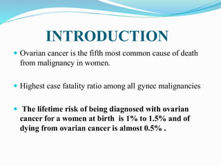
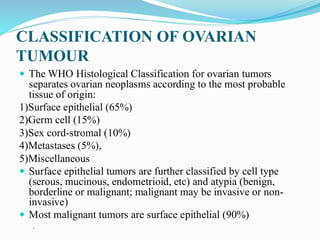
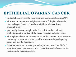
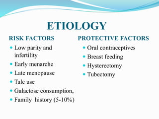
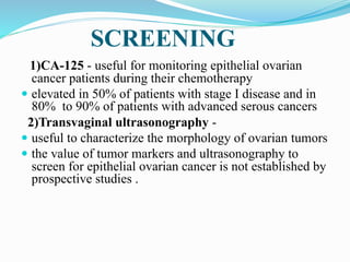
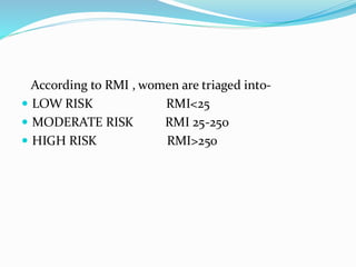
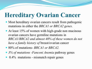
Dr. SHRADHA JAI N
INTRODUCTION
 Ovarian cancer is the fifth most common cause of death
from malignancy in women.
 Highest case fatality ratio among all gynec malignancies
 The lifetime risk of being diagnosed with ovarian
cancer for a women at birth is 1% to 1.5% and of
dying from ovarian cancer is almost 0.5% .
CLASSIFICATION OF OVARIAN
TUMOUR
 The WHO Histological Classification for ovarian tumors
separates ovarian neoplasms according to the most probable
tissue of origin:
1)Surface epithelial (65%)
2)Germ cell (15%)
3)Sex cord-stromal (10%)
4)Metastases (5%),
5)Miscellaneous
 Surface epithelial tumors are further classified by cell type
(serous, mucinous, endometrioid, etc) and atypia (benign,
borderline or malignant; malignant may be invasive or non-
invasive)
 Most malignant tumors are surface epithelial (90%)

EPITHELIAL OVARIAN CANCER
 Epithelial cancers are the most common ovarian malignancy.(90%)
 Most serous carcinomas originate from the fallopian tube while
other subtypes (clear cell, endometrioid) are derived from
endometriosis.
 previously it was thought to be derived from the coelomic
epithelium on the surface of the ovary / ovarian inclusion cysts.
 Most epithelial ovarian cancers are sporadic, but up to one-quarter of
cases may be associated with germline mutations in predisposing
genes and may be hereditary.
 Hereditary ovarian cancers, particularly those caused by BRCA1
mutations, occur at a younger age, typically about 10 years earlier
than sporadic ovarian cancers.
CLASSIFICATION OF EPITHELIAL
OVARIAN TUMOUR
 IV. Clear-cell (5%) Müllerian
A. Benign
B. Borderline
C. Malignant
 V. Brenner (less than 1%) Transitional
A. Benign
B. Borderline (proliferating)
C. Malignant
CLASSIFICATION OF EPITHELIAL
OVARIAN TUMOUR
 VI. Mixed (less than 1%) epithelial Mixed
 A. Benign
 B. Borderline
 C. Malignant
 VII. Undifferentiated (less than 1%) May be
anaplastic
 VIII. Unclassified (less than 1%)
SEROUS EPITHELIAL CANCER
 Resemble tubal secretory cells
 Psammoma bodies (made up of concentric rings of
calcifications.)
 Many high-grade serous carcinomas of the ovary arise
from the fimbrial end of the fallopian tube rather than
from the ovary
 Serous epithelial ovarian cancers can be separated into
two distinct groups—
Type I
Type II
TYPE 1 TYPE 2
 serous borderline tumors
 low-grade serous
carcinoma
 Genetically stable,
 KRAS or BRAF mutation
 may have precursor
lesions in the fimbria of
the fallopian tube
 advanced stage at
diagnosis
 genetically unstable
 p53 mutation
 rapidly growing, highly
aggressive
BORDERLINE SEROUS TUMOUR
 Tumours of low malignant potentrial
 Confined to ovary for longer periods
 Predominantly in pre-menopuasal women
 Good prognosis
 Age- 30-50 yrs (average age- 46 yrs)
 10% of all serous ovarian tumour fall in borderline
category
 40% serous borderline tumour spread beyond ovary
 May harbour foci of stromal microinvasion
DIAGONOSTIC CRITERIA
1)Epithelial hyperplasia-
- Pseudostratification
- Tufting
- Cribriform
- Micropapillary architecture
2)Mild nuclear atypia and mild increased mitotic
activity
3)Detached cell clusters
4)Absence of destructive stromal invasion
IMPLANTS
NON INVASIVE INVASIVE
 Pappilary proliferation of
atypical cells involve
peritoneal surface and
form smooth invaginations
 90%
 usually confined to the
abdominal cavity and may
be seen in pelvis,omentum,
and adjacent tissues,
including lymph nodes, but
spread outside the
abdominal cavity is rare.
 Resemble well differentiated serous
carcinoma
 characterized by atypical cells forming
irregular glands with sharp borders.
 10%
 higher likelihood of progression, in the
peritoneal cavity, which can ultimately
lead to intestinal obstruction caused by
associated fibrosis and death
SEROUS CARCINOMA
 In malignant serous tumors, stromal invasion is
present .
 Low-grade serous adenocarcinomas- papillary and
glandular structures predominate
 High-grade neoplasms -solid sheets of cells
nuclear pleomorphism,
high mitotic activity
 Laminated, calcified psammoma bodies are found in
80% of serous carcinomas.
 Serous psammocarcinoma - rare variant of serous
carcinoma characterized by massive psammoma body
formation and low-grade cytologic features.
 Patients with serous psammocarcinoma have a
protracted clinical course and a relatively favorable
prognosis
 clinical course more closely resembles that of high-
stage, progressive serous borderline tumor than serous
carcinoma.
MUCINOUS TUMOUR
 These cystic ovarian tumors have loculi lined with
mucin-secreting epithelium.
 lining epithelial cells contain intracytoplasmic mucin
and resemble those of endocervix,gastric pylorus, or
intestine.
 They represent about 8% to 10% of epithelial ovarian
tumors.
BORDERLINE MUCINOUS
TUMOUR
 difficult to diagnose
 Well-differentiated mucinous epithelium may be seen
immediately adjacent to a poorly differentiated focus.
 It is important to take multiple sections from many
areas in the mucinous tumor to identify the most
malignant alteration
MUCINOUS CARCINOMA
 Bilateral in 8% to 10% of cases.
 These lesions are confined to the ovary in 95% to 98% of
cases
 Most ovarian mucinous carcinomas contain enteric-type
cells.
 Primary ovarian neoplasms rarely metastasize to the
mucosa of the bowel, although they commonly involve the
serosa, whereas gastrointestinal lesions frequently involve
the ovary by direct extension or lymphatic spread
PSEUDOMYXOMA
PERITONI
 Pseudomyxoma peritonei is a clinical term used to
describe the finding of abundant mucoid or gelatinous
material in the pelvis and abdominal cavity surrounded by
fibrous tissue.
 It is most commonly secondary to a well-differentiated
appendiceal mucinous neoplasm or other gastrointestinal
primary; rarely, mucinous tumors arising in an ovarian
mature teratoma are associated with pseudomyxoma
peritonei
ENDOMETROID TUMOURS
 6% to 8% of epithelial tumors.
 Endometrioid neoplasia includes all the benign
demonstrations of endometriosis.
 The malignant potential of endometriosis is very low
 Borderline Endometrioid Tumors- wide morphologic
spectrum.
 Tumors may resemble an endometrial polyp or
complex endometrial hyperplasia with glandular
crowding.
 Some borderline endometrioid tumors have a prominent
fibromatous component. In such cases, the term
adenofibroma is used to describe them
 Endometrioid Carcinomas- characterized by a
markedly complex glandular pattern with all the
potential variations of epithelia found in the uterus
 Endometrioid carcinoma of the ovary is associated in
15% to 20% of the cases with carcinoma of the
endometrium.
CLEAR CELL TUMOUR
 Several basic histologic patterns are present in the clear cell
adenocarcinoma (i.e.,tubulocystic, papillary, reticular, and
solid)
 made up of clear and hobnail cells
 clear cells have - abundant clear or vacuolated cytoplasm
-hyperchromatic irregular nucle
-nucleoli of various sizes
 mixed clear cell and endometrioid carcinoma may occur
 The clear cell carcinoma seen in the ovary is
histologically identical to that seen in the uterus or vagina
of the young patient who has been exposed to
diethylstilbestrol (DES) in utero.
 Almost invariably high-grade nuclei are identified. Hence,
clear cell carcinomas are not graded
Transitional (Brenner) Tumors
Borderline Brenner Tumors
Proliferative Brenner tumors were previously subclassified
as -
 Proliferating tumors (those tumors that resemble low-
grade papillary urothelial carcinoma of the urinary
bladder)
 Borderline tumors (those tumors that resemble high-grade
papillary urothelial carcinoma),
 These groups of tumors are now classified as borderline
Brenner tumors
Malignant Brenner Tumors- These rare tumors are defined
as benign or borderline Brenner tumors coexisting with
invasive transitional cell carcinoma.
Transitional Cell Carcinoma- these are primary ovarian
carcinoma resembling transitional cell carcinoma of the
urinary bladder without a recognizable Brenner tumor.
 They are a variant of high-grade serous carcinoma and do
not represent a distinct epithelial subtype of carcinoma
 Unlike malignant Brenner tumors, these tumors are more
frequently diagnosed in an advanced stage
Clinical Features
 More than 80% of epithelial ovarian cancers are found in
postmenopausal women.
 The peak incidence of invasive epithelial ovarian cancer is at 56
to 60 years of age.
 About 30% of ovarian neoplasms in postmenopausal women are
malignant, whereas only about 7% of ovarian epithelial tumors in
premenopausal patients are malignant
 The average age of patients with borderline tumors is
approximately 46 years
 Less than 1% of epithelial ovarian cancers occur before the age of
20 years, with two-thirds of ovarian malignancies in such patients
being germ cell tumors.
ETIOLOGY
RISK FACTORS PROTECTIVE FACTORS
 Low parity and
infertility
 Early menarche
 Late menopause
 Talc use
 Galactose consumption,
 Family history (5-10%)
 Oral contraceptives
 Breast feeding
 Hysterectomy
 Tubectomy
PREVENTION
1)Because parity is inversely related to the risk of ovarian
cancer, having at least one child is protective for the
disease, with a risk reduction of 0.3 to 0.4.
2) Oral contraceptive use -reduces the risk
 Women who use oral contraceptives for 5 or more years
reduce their relative risk to 0.5
 Women who had two children and used oral
contraceptives for 5 or more years have a relative risk of
ovarian cancer as low as 0.3, or a 70% reduction
 only documented method of chemoprevention for ovarian
cancer
3)Prophylactic salpingo-oophorectomy -significantly
reduces, but does not totally eliminate the risk.
(peritoneal carcinomas can occur in 2% to 3% of women
even after prophylactic bilateral salpingo-oophorectomy)
 performance of a prophylactic salpingo-oophorectomy
reduced the risk of BRCA-related gynecologic cancer by
96%
 The risk of developing subsequent breast cancer may be
reduced by 50% to 80% in BRCA carriers.
SCREENING
1)CA-125 - useful for monitoring epithelial ovarian
cancer patients during their chemotherapy
 elevated in 50% of patients with stage I disease and in
80% to 90% of patients with advanced serous cancers
2)Transvaginal ultrasonography -
 useful to characterize the morphology of ovarian tumors
 the value of tumor markers and ultrasonography to
screen for epithelial ovarian cancer is not established by
prospective studies .
3) CA125 + TVS-
 The use of transvaginal ultrasound alone or in
combination with CA-125 for early ovarian cancer
detection has not proven effective in average-risk women
to decrease mortality from ovarian cancer.
 Instead there are harms to this strategy, including false-
positive results and more invasive procedures being
performed such as surgery
The U.S. Preventative Services Task Force (USPSTF)
recommends against screening asymptomatic women for
ovarian cancer.
 The USPSTF concluded that there is adequate evidence that
there is no mortality benefit to routine screening for ovarian
cancer with TVS or single-threshold serum CA-125 testing
and that the harms of such screening are at least moderate.
 There is no effective strategy for early detection of
ovarian cancer that cause reduction in ovarian cancer
mortality.
United Kingdom Collaborative Trial of Ovarian
Cancer Screening (UKCTOCS)
 has not shown a significant reduction of ovarian
cancer mortality with multimodal screening when
compared with no screening.
 Multimodal screening with the ROCA was superior to
using fixed CA-125 cut-off .
 The ROCA (risk of ovarian cancer algorithm)-
 uses serially measured CA-125 and mathematical
modeling to determine whether an increase in levels
should prompt referral for ultrasound (score>1%-
women are subjected for TVS)
 has a better predictive performance for detecting
ovarian cancer than using a fixed CA-125 value cut-
off.
US-based Prostate, Lung, Colorectal, and Ovarian
(PLOC)Cancer Screening Trial-
 Showed that using a baseline and annual measurement of CA-
125 with a cut-off (35 U/mL) in combination with pelvic
ultrasound as a way for early assessment does not result in
decreased ovarian cancer mortality
 This large trial showed that screening women at average risk
with annual TVS provided no mortality benefit when compared
with no screening.
 There is no evidence to demonstrate a benefit of screening
high-risk women, even with active investigation
RISK OF MALIGNANCY INDEX(RMI)
 RMI= U X M X CA 125
 U=USG SCORE - 1 point for each- Multilocularity
-Metastasis
-Ascitis
-Bilateral
-Solid areas
 M –menopausal score -1 point-pre-menopausal
 3-postmenopausal
 CA125- value in U/ml
According to RMI , women are triaged into-
 LOW RISK RMI<25
 MODERATE RISK RMI 25-250
 HIGH RISK RMI>250
Hereditary Ovarian Cancer
 Most hereditary ovarian cancers result from pathogenic
mutations in either the BRCA1 or BRCA2 genes.
 At least 15% of women with high-grade non mucinous
ovarian cancers have germline mutations in
BRCA1/BRCA2 and almost 40% of these women do not
have a family history of breast/ovarian cancer
 80% of mutations- BRCA1 or BRCA2.
 3% of mutations -Fanconi Anemia pathway genes
 0.4% mutations - mismatch repair genes
 The mutations are inherited in an autosomal dominant
fashion, and therefore a full pedigree analysis is essential
when taking a family history.
 All women with high-grade invasive ovarian cancers
should be offered genetic testing, even if they do not have
a family history of breast/ovarian cancer.
 Hereditary ovarian cancers occur in women who are
approximately 10 years younger than those with non-
hereditary tumors
 A woman with a first- or second-degree relative who had
premenopausal ovarian cancer may have a higher probability of
being a BRCA carrier.
 Women with BRCA germline mutations tend to be diagnosed at a
younger age than women with sporadic ovarian cancers.
 In women with BRCA1 mutations, less than 2% to 3% of carriers
develop ovarian cancer by the age of 40 which increases to 10% to
21% by the age of 50.
 In women with BRCA2 mutations, less than 3% of carriers develop
ovarian cancer by the age of 50 and most develop after age 50.
 Most BRCA ovarian cancers are high-grade serous carcinomas
GENETICS
1) HEREDITORY BREAST OVARIAN CANCER(BOC
)SYNDROME-
 80-95% Cases
 BRCA1 and BRCA2 gene mutations are seen
 Present at early age
2)Lynch Syndrome or HNPCC- which
multiple adenocarcinomas, involves a combination of
colon cancer and endometrial or ovarian cancer and other
malignancies of the gastrointestinal and genitourinary
systems .
 Autosomal dominant
 The chief gene mutations - MSH2, MLH1, MSH6, and
PMS2 or the EPCAM gene.
 These women appear to have at least three times the
relative risk of the general population.
 Life time Risk of C/A endometrium- 50%
 Risk of C/A ovary- 12%
Management of Women at High
Risk for Ovarian Cancer
Recommendations-
 1. Genetic counseling is recommended for all women with
ovarian/fallopian tube/peritoneal epithelial cancer and for
individuals who have a personal or family history of breast
cancer or ovarian cancer.
 2. Women who wish to preserve their reproductive capacity can
undergo screening by TVS every 6 months or CA-125
measurement and this may be a reasonable short-term
surveillance strategy for women at high risk of ovarian cancer
starting at age 30 to 35 years until they decide to pursue
definitive risk-reducing bilateral salpingo-oophorectomy.
3. Oral contraceptives should be recommended to young women
before they are ready to start a family.
4. Women who do not wish to maintain their fertility or who have
completed their families should be recommended to undergo
prophylactic bilateral salpingo oophorectomy after the age of
35, but by age 40 years.
 Typically, risk-reducing salpingo-oophorectomy is
recommended at age 35 to 40 years for BRCA1 carriers with
the highest lifetime risk of ovarian cancer, whereas women
with BRCA2 may consider delaying until age 40 to 45 years
because of later onset of ovarian cancer.
5. Women with BRCA mutations or who carry another
deleterious mutation that is predisposing to breast cancer
should be offered risk-reducing bilateral mastectomy.
6. For a risk-reducing bilateral salpingo-ophorectomy, all
tissue from the ovaries and fallopian tubes should be
removed
7. For women aged 25 to 29 years with known BRCA
mutations, recommended breast cancer surveillance includes
-
clinical breast examination every 6 to 12 months and annual
radiographic evaluation (preferably magnetic resonance
imaging [MRI], with contrast)
 . For women aged 30 years and older with known BRCA
mutations or other actionable breast cancer mutations,
recommended breast cancer surveillance includes-
annual mammography and annual breast MRI with contrast,
often alternating every 6 months.
8. Women with a personal or family history of breast or
ovarian cancer but who do not have a documented mutation
in BRCA1 or BRCA2 or other hereditary breast- or ovarian-
cancer–associated genes should be managed based on their
family history.
 9. Women with a documented Lynch syndrome (HNPCC)
should undergo periodic colonoscopy every 1 to 2 years,
starting at age 20 to 25 years or 2 to 5 years before the earliest
colon cancer diagnosis in the family, depending on which is
earlier.
 Endometrial biopsy every 1 to 2 years starting at age 30 to 35
years, is recommended, prophylactic hysterectomy, and
bilateral salpingo-oophorectomy is a risk-reducing option after
the completion of childbearing.
 In general, risk-reducing hysterectomy and
salpingooophorectomy should be discussed with patients by
their early to mid-40s
SYMPTOMS
EARLY STAGE DISEASE-
 Irregular meneses.(pre-menopausal)
 Urinary frequency
 Constipation
 Lower abdominal distention, dyspareunia
Acute symptoms –
 Pain secondary to rupture or torsion,
 shortness of breath (malignant pleural effusion)
 symptoms of a bowel obstruction
 ADVANCED STAGE DISEASE-patients have symptoms
related to the presence of ascites, omental metastases, or
bowel metastases.
 Abdominal distention
 Bloating
 Constipation
 Nausea
 Anorexia
 Irregular or heavy menses(pre-menopausal)
 Vaginal bleeding (postmenopausal women)- If the ovarian
tumors are estrogen producing
Ovarian cancer symptom index
 By Goff et al
 symptoms associated with ovarian cancer, when present
for less than 1 year and occurring longer than 12 days a
month, were pelvic/abdominal pain, urinary
frequency/urgency, increased abdominal size or bloating,
and difficulty eating or feeling full .
 The index had a sensitivity of 56.7% for early ovarian
cancer and 79.5% for advanced-stage disease
SIGNS
 The most important sign of epithelial ovarian cancer is the
presence of a pelvic mass on physical examination.
 A solid, irregular, fixed pelvic mass is highly suggestive of an
ovarian malignancy.
 If an upper abdominal mass or ascites is present, the diagnosis
of ovarian cancer is almost certain.
 Palpable pelvic mass in these patients should be considered
potentially malignant, a situation that was referred to as the
postmenopausal palpable ovary syndrome
 only about 3% of palpable masses measuring less than 5
cm in postmenopausal women are malignant
 Atypical glandular cells on cervical pathology may signal
an upper genital tract malignancy.
 Rarely present with signs of a paraneoplastic syndrome,
which may include disseminated intravascular
coagulation, polyneuritis, dermatomyositis, hemolytic
anemia, and cerebellar degeneration
DIAGONOSIS
 The diagnosis of an ovarian cancer require surgical
exploration by a gynecologic oncologist, which may be by
an exploratory laparotomy or a minimally invasive
technique such as laparoscopy or a robotic platform
 For a postmenopausal patient with an adnexal mass and a
very high serum CA-125 level (>200 U/mL), there is a 96%
positive predictive value for malignancy.
 Ultrasonographic signs of malignancy include an adnexal
pelvic mass with areas of complexity, such as irregular
borders, multiple echogenic patterns within the mass, and
dense multiple irregular septae.
Differential Diagnosis
 Ovarian epithelial cancers must be differentiated from
benign neoplasms and functional cysts of the ovaries.
pelvic inflammatory disease
endometriosis
pedunculated uterine leiomyomata
 Nongynecologic causes -
 inflammatory or neoplastic colonic mass
 Gastrointestinal and breast cancers are the most common
nongenital malignancies that metastasize to the ovary
Patterns of Spread
 Ovarian epithelial cancers spread primarily by exfoliation of
cells into the peritoneal cavity, lymphatic dissemination, and
hematogenous spread.
1)Transcoelomic-
 The most common and earliest mode of dissemination of
ovarian epithelial cancer is by exfoliation of cells that
implant along the surfaces of the peritoneal cavity.
2)Lymphatic
 Lymphatic dissemination to the pelvic and para-aortic lymph
nodes is common, particularly in advanced-stage disease
 78% of patients with stage III disease have metastases
to the pelvic lymph nodes .
 The rate of para-aortic lymph nodes positive for
metastasis -
 18% in stage I
 20% in stage II
 42% in stage III
 67% in stage IV
3)Hematogenous-
 Hematogenous dissemination at the time of diagnosis is
uncommon.
 Spread to vital organ parenchyma, such as the lungs and
liver, occurs in only about 2% to 3% of patients.
FIGO Staging for Carcinoma Ovary
 Stage I- Growth Limited to the Ovaries
 IA- Growth limited to one ovary
No ascites containing malignant cells
No tumor on the external surface
Capsule intact
 IB -Growth limited to both ovaries
No ascites containing malignant cells
No tumor on the external surfaces;
Capsules intact
 IC- Tumor limited to one or both ovaries or fallopian
tubes with any of the following:
IC1- Surgical spill
IC2- Capsule ruptured before surgery
or tumor on ovarian surface
IC3 - Malignant cells in the ascites
or peritoneal washings
 Stage II Growth involving one or both ovaries or
fallopian tubes with pelvic extension (below the
pelvic brim)
 IIA- Extension and/or metastases to the uterus and/or
fallopian tubes
 IIB -Extension to other pelvic tissues
 Stage III- Tumor involving one or both ovaries with cytologically
or histologically confirmed spread to the peritoneum outside the
pelvis and/or metastasis to the retroperitoneal lymph nodes
 IIIA - Positive retroperitoneal lymph nodes
and/or microscopic metastasis beyond pelvis
 IIIA1(i) Positive retroperitoneal lymph nodes with metastasis up to
10 mm in greatest dimension
 IIIA1(ii) Positive retroperitoneal lymph nodes with metastasis more
than 10 mm in greatest dimension
 IIIA2 Microscopic extrapelvic (above the pelvic brim) peritoneal
involvement, with or without positive retroperitoneal lymph
nodes
 IIIB Macroscopic peritoneal metastasis beyond pelvis up to 2 cm
in greatest dimension, with or without positive retroperitoneal
nodes
 IIIC Macroscopic peritoneal metastasis beyond pelvis more than
2 cm in greatest dimension (includes extension of tumor to
capsule of liver and spleen without parenchymal involvement of
either organ), with or without positive retroperitoneal nodes
 IV Distant metastasis (excludes peritoneal metastasis)
 IVA -Pleural effusion is present, there must be positive
cytologic test results
 IVB- Parenchymal metastases and metastases to extra-
abdominal organs
 Dense adhesions with histologically proven tumor cells
justify upgrading to stage II.
 Transmural bowel infiltration or umbilical deposit are
stage IVB.
MANAGEMENT
1) Pre-operative evaluation
2) Staging laparotomy
3)Cytoreduction surgery
4)chemotherapy
5) harmone therapy
6) radiotherapy
PRE-OPERATIVE EVALUATION
 Assesing fitness for surgery
 Fitness for anesthesia
 To identify extent of disease
 To detect primary site
 Routine investigations- CBC, Urine analysis, Blood
grouping and Rh typing,screening for infectious
disease,liver and renal function test
 ECG
 Chest X-ray
 Ultrasound-
 to identify origin
 to differentiate benign from malignant
tumour(multiloculation, papillary projections, thick
septations, increased vascularity, solid areas)
 Ascitis
 Omental caking
 Para-aortic and pelvic lymph node enlargement
 Liver/spleen involvement
 Tumour markers-
 CT/MRI
 Barium enema / colonoscopy- to rule out colon cancer..
 Upper GI endoscopy – in presence of upper GI symptoms
 Pap smear
 Endometrial sampling
 Bilateral diagnostic mammography- if there is any breast
mass
STAGING LAPAROTOMY
 Vertical abdominal incision is recommended to allow
adequate access to the upper abdomen
 The ovarian tumor should be removed intact, and a frozen
histologic section should be obtained.
Steps-
 1. Any free fluid, especially in the pelvic cul-de-sac, should
be submitted for cytologic evaluation.
 2. If no free fluid is present, peritoneal washings should be
performed by instilling and recovering 50 to 100 mL of
saline from the pelvic cul-de-sac, each paracolic gutter, and
beneath each hemidiaphragm.
 3. A systematic exploration of all the intra-abdominal
surfaces and viscera is performed, proceeding in a clockwise
fashion starting from cecum.
 4. Any suspicious areas or adhesions on the peritoneal
surfaces should be biopsied. If there is no evidence of
disease, multiple IP biopsies should be performed.
 5. The diaphragm should be sampled, either by biopsy or by
scraping with a tongue depressor.
6. The omentum should be resected from the transverse
colon, including from the hepatic and splenic flexure
of the colon and separated from its attachment to the
greater curvature of the stomach, including ligation of
the left and right gastroepiploic and short gastric
arteries, a procedure called supracolic omentectomy.
7. The retroperitoneal spaces are explored to evaluate
the pelvic and para-aortic lymph nodes.
 Any enlarged lymph nodes should be resected and
submitted for frozen section.
 If no metastases are present, pelvic
lymphadenectomy is performed.
 The para-aortic area should be palpated and lymph
nodes sampled if enlarged or suspicious
BORDERLINE TUMOUR
 The principal treatment of borderline tumors is surgical
resection of the primary tumor.
 Premenopausal patients who desire preservation of the
ovarian function may undergo a conservative operation, a
unilateral oophorectomy F/b removal of uterus and other
ovary after completion of child bearing.
 Pelvic and para-aortic sampling not needed.
STAGE 1 TREATMENT
 Primary treatment for stage I epithelial ovarian cancer is
surgical, and patients should undergo total abdominal
hysterectomy, bilateral salpingooophorectomy, and
surgical staging
 In certain circumstances, a unilateral salpingo-
oophorectomy may be performed as a part of fertility-
sparing surgery.
 Based on the findings at surgery and the pathologic
evaluation, patients stage I ovarian cancer can be grouped
into low-risk and high-risk categories
Low Risk High Risk
 Low grade High grade
 Intact capsule Tumor growth through capsule
 No surface excrescences Surface excrescences
 No ascites Ascites
 Negative peritoneal cytology Malignant cells in fluid
 Unruptured/ intraoperative rupture Preoperative rupture
 No dense adherence Dense adherence
 Diploid tumor Aneuploid tumor
Stage I Low Risk
 Total abdominal hysterectomy and bilateral salpingo-
oophorectomy are appropriate therapy
 The uterus and the contralateral ovary can be preserved in
women with stage IA, grade 1 to 2 disease who desire to
preserve fertility.
 They should be monitored carefully with routine periodic
pelvic examinations and determinations of serum CA- 125
levels.
 The other ovary and the uterus are removed at the
completion of childbearing.
Stage I High Risk
 Patients who have more poorly differentiated disease or
who have malignant cells either in ascites fluid or in
peritoneal washings, must undergo Total abdominal
hysterectomy and bilateral salpingo-oophorectomy and
complete surgical staging .
 Most patients require chemotherapy.
ADVANCED STAGE TREATMENT
 If the patient is medically stable, she should undergo
cytoreductive surgery to remove as much of the tumor and
its metastases as possible .
 The operation to remove the primary tumor and the
associated metastatic disease – debulking
 Includes TAH+BSO+ complete omentectomy and resection
of any metastatic lesions from the peritoneal surfaces or
from the intestines.
 Routine lymphadenectomy should be omitted in these
patients to reduce postoperative morbidity and
mortality(LION Study)
ADVANTAGES OF DEBULKYING
 Reduce the volume of ascites present, may reduce symptoms.
 Removal of intestinal metastases may restore adequate
intestinal function and improve the overall nutritional status of
the patient, thereby facilitating the patient’s ability to tolerate
subsequent chemotherapy
 Surgical removal of these bulky tumor may eliminate poorly
vascularised areas that could be relatively resistant to radiation
and chemotherapeutic treatment.
 Cytoreductive surgery reduce larger proportion of cells that are
in the “resting” phase (i.e., G0 cells, which are resistant to the
therapy).
 The principal goal of cytoreductive surgery is removal of
all of the primary cancer and, if possible, all metastatic
disease.
 If resection of all metastases is not feasible, the goal is to
reduce the tumor burden by resection of all individual
tumors to an optimal status (<1 cm of any single implant
of residual disease).
 The resectability of the metastatic tumor is usually
determined by the location of the disease
Optimal cytoreduction is difficult to achieve in the presence
of -
-Extensive disease on the diaphragm
-Parenchyma of the liver
-Base of the small Bowel mesentery
-Lesser omentum
-Porta hepatis
 Neoadjuvant chemotherapy is an option for these
patients
Neoadjuvant chemotherapy
 Two or three cycles of chemotherapy before cytoreductive
surgery may be helpful in patients with massive ascites and
large pleural effusions.
INDICATIONS-
 Very advanced (stages IIIC and IV) ovarian cancer
 poor performance status
 high perioperative risk
ADVANTAGE-
 The chemotherapy may decrease the effusions, improve the
patient’s performance status, and decrease postoperative
morbidity, particularly chest morbidity
Interval cytoreductive surgery
 Performed after three cycles of platinum-combination
chemotherapy (Neo-adjuvant chemotherapy )in patients
whose primary attempt at cytoreduction was suboptimal.
INDICATIONS-
 Advanced epithelial ovarian cancer
 High risk for surgery
 Co-morbidities(pleural effusion)
CHEMOTHERAPY
Stage I Epithelial Ovarian Cancer-
Early Stage, Low Risk-no adjuvant chemotherapy
Early Stage, High Risk- The recommendations are-
 Patients with high-grade, high-risk stage I epithelial ovarian
cancer should be offered adjuvant chemotherapy depending
on the patient’s overall health and medical comorbidities.
 Treatment with CP( carboplatin and paclitaxel) in
chemotherapy for three to six cycles is used in these
patients.
 single-agent carboplatin may be preferable for older women
and patients with other medical comorbidities
Advanced-Stage Epithelial Ovarian
Cancer-
 For the treatment of advanced-stage epithelial ovarian
cancer, the following treatment regimens should be
considered -
1)Combination chemotherapy or IV CP every 3 weeks or
IP carboplatin or cisplatin and paclitaxel are the
treatments of choice for patients with advanced disease.
 The advantages and disadvantages of the IV versus IP
routes of administration of these drugs should be
discussed with the patient
2) The recommended doses and schedule for IV
chemotherapy are-
 carboplatin (starting dose AUC = 5 to 6), and paclitaxel
(175 mg/m2), every 3 weeks for six cycles.
 Calvert formula-dose(mg )=Target AUC X GFR +25
 Alternatively, the dose-dense regimen of carboplatin
AUC 6 every 3 weeks for six cycles and weekly paclitaxel
80 mg/m2 could be used,
3)The recommended doses and schedule for IP
chemotherapy are
 Day 1- paclitaxel 135 mg/m2 IV over 24 hrs
 Day 2- carboplatin AUC 5(400 mg/m2) or cisplatin 75 to
100 mg/m2 IP
 Day8- paclitaxel 60 mg/m2 IP
 every 3 weeks for six cycles.
IP CHEMOTHERAPY
 IP chemotherapy is best suited to patients who have
optimally resected tumors, a good performance status, and
are in overall good health
 Given in low volume stage 3 disease after surgery
ADVANTAGES-
 more efficacious because provide high concentration of drug
in the site.
 Survival rate is more
DISADVANTAGES-
 More toxicity
 Catheter related problems
 Can not be used in large volume residual disease
4)Bevacizumab 7.5 to 15 mg/kg can be added to any of these IV or I
chemotherapy regimens.
5)In patients who cannot tolerate combination chemotherapy, single-
agent, IV administered carboplatin (AUC 5 to 6) can be given.
6)In patients who have a hypersensitivity to paclitaxel or carboplatin
an alternative active drug can be substituted (e.g., docetaxel,
nanoparticle paclitaxel, cisplatin).
In the case of carboplatin hypersensitivity, desensitization could be
attempted.
TREATMENT
ASSESSMENT
 Many patients who undergo optimal cytoreductive surgery
and subsequent chemotherapy for epithelial ovarian cancer
have no evidence of disease at the completion of treatment.
 Historically, a second-look surgery was performed to
evaluate these patients, but that was abandoned, as there was
no evidence of a meaningful benefit to patients
 It was done on those with no clinical evidence of persistant
disease to determine the disease status after chemotherapy to
acess if person has responded to a paerticular treatment.
Tumor Markers
1)CA-125 -is a surface glycoprotein associated with müllerian
epithelial tissues, and is elevated in about 80% of patients
with epithelial ovarian cancers, particularly those with non
mucinous tumors .
 The CA-125 level frequently falls below the normal range
after the initial surgical resection and one or two cycles of
chemotherapy.
 Srum CA-125 levels can be used during chemotherapy to
follow those patients whose levels were elevated at initial
diagnosis and at the initiation of therapy
.. The fall in the CA-125 correlates with response.
 Sensitivity- 50-74%(pre-menopausal)
 69-87%(postmenopausal)
 Specificity -26-92%(pre-menopausal)
 81-100%(post menopausal)
 After surgery and first line chemotherapy pt are followed
with CA-125 levels every 3 to 4 months.
 There are well-accepted Gynecologic Oncology
Intergroup (GCIG) criteria for CA-125 progression.
 Patients with an elevated CA-125 pre treatment and in the
normal range following the completion of treatment must
have a CA-125 greater than, or equal to, two times the
upper normal limit on two occasions at least 1 week apart
to be classified as having CA-125 progression
2)HE4-
 Elevated in women with serous epithelial cancer
 Comination of CA125+HE4 is superior to any other
marker.
3) CEAAND CA19-9-Elevated in mucinous cancer
4)Other-
 M-CSF
 OVX1
Other conditions in which CA125 is raised-
Normal postmenopausal women(2-3%)
Fibroid
PID
Endometriosis
Endometrial carcinoma
Breast, colon and lung cancers
Pancreatitis
peritonitis
Radiologic Assessment
 CT can- be used to assess the response in patients with
measurable lesions at the start of therapy.
 In patients who have no or minimal residual disease
following surgery, they may be useful in follow-up,
especially to document the site of recurrence in patients with
CA-125 progression.
 false-negative rate of a CT scan is about 45%
 MRI can be used as an alternative to CT in patients with
allergies to the contrast medium
 PET/CT has high diagnostic accuracy in patients with
recurrent ovarian cancer.
PROGNOSTIC FACTORS
 The outcome of treatment can be evaluated in the context
of prognostic factors, which can be grouped into
pathologic, biologic, and clinical factors
 PATHOLOGICAL FACTORS-
 1) Histologic type- clear cell –worst prognosis
 2) Histologic grade- pattern of differentiation
extent of cellular anaplasia
proportion of undifferentiated cells
CLINICAL FACTORS
 Stage of disease
 Extent of residual disease after primary surgery
 Volume of ascites,
 Patient age
 Performance status
OTHER
 capsular penetration
 surface excrescences
 Ploidy status
RECURRENT OVARIAN CANCER
 About 80% of patient devlope relapse after treatment.
 Recurrence- Disease free intervel of 6 months
 Persistant- Disease free intervel less than 6 months
 Platinum sensitive –recurrence after 6 months of
chemotherapy
 Platinum resistant –recurrence within 6 months of
chemotherapy
Second-Line Therapy
Secondary Cytoreductive Surgery-
 defined as an attempt at cytoreductive surgery in patients with
recurrent ovarian cancer or Cytoreductive surgery after
completion of first line chemotherapy
 Indications-Prolonged disease free intervel
 - Platinum sensitive recurrent ovarian cancer
 - no ascitis
 -good performance status
 -absence of peritoneal carcinomatosis
 -solitary/limited site of recurrence
 (when it is considered possible to resect all disease)
 Not suitable for- progressive disease on chemotherapy
(platinum refractory) or
platinum-resistant ovarian cancer
 secondary debulking should be restricted to patients who
have a disease-free interval of at least 12, but preferably
24 months or those in whom it is expected that all
macroscopic disease can be resected
 The role of secondary debulking surgery has been
addressed in the DESKTOP III trial.(randomisation is
done to sec cytoreductive surgery with chemo or chemo
alone)
Chemotherapy for Recurrent Ovarian Cancer
 Platinum-sensitive-Women who relapse later than 6 months
after primary chemotherapy
 usually receive further platinum-based chemotherapy with
response rates ranging from 27% to 65% a median survival of
12 to 24 months
Platinum-resistant- Patients who relapse within 6 months of
completing first-line chemotherapy
 have a median survival of 6 to 9 months and a 10% to 30%
likelihood of responding to chemotherapy.
 Patients who progress while on treatment are classified as
having Platinum-refractory disease.
 response rates to chemotherapy in patients with platinum-
refractory ovarian cancer are low—less than 20%
PLATINUM SENSITIVE DISEASE
1) Combination platinum plus paclitaxel chemotherapy
2) Paclitaxel therapy
 Data support the slight advantage of a second-line regimen
containing both paclitaxel and a platinum agent compared
with platinum-based therapy alone.
(ICON4 and AGO-OVAR-2.2 trials)
3) Carboplatin + gemcitabine / liposomal doxorubicin
4)Carboplatin +gemcitabine + bevacizumab
 Carboplatin, gemcitabine, and bevacizumab have been
shown in a phase III randomized control trial to be superior
to carboplatin and gemcitabine alone for platinum-sensitive
recurrent epithelial ovarian cancer.(OCEANS study)
 studies show that maintenance with bevacizumab can
improve the PFS and may improve the overall survival in a
subset of patients.
5) PARP Inhibitors-
 have been approved by FDA for maintenance therapy in
patients with platinum-sensitive disease following a partial
or complete response to platinum-based chemotherapy.
 The PFS is improved with these agents regardless of the
BRCA status, although the patients with BRCA
germline/somatic mutations have a greater benefit than
patients without mutations
(A)Niraparib- has been approved by the U.S. FDA for
maintenance therapy in patients with platinum-sensitive
disease following a partial or complete response to
platinum-based chemotherapy for both germline BRCA1 and
BRCA2 carriers and nongermline carriers.
(B)Olaparib- FDA approved for the treatment of patients with
gBRCA-mutated recurrent ovarian cancer who have received
three or more prior lines of chemotherapy.
 (C)Rucaparib- Rucaparib is approved for treatment of
BRCA-mutation–associated advanced ovarian cancer after
completion of treatment with two or more chemotherapy
regimens regardless of whether patients are platinum-
sensitive or resistant.
 D) Other PARP Inhibitors
 There are a number of other PARP inhibitors under
investigation.
Veliparib is being investigated in GOG-3005 in combination
with chemotherapy in the frontline and maintenance setting.
Platinum-Resistant and Refractory
Disease
 In platinum-refractory patients (i.e., those progressing on
treatment), response rates to second-line chemotherapy are less
than 10% and the median survival is short, around 3 to 5 months.
 The management of women who are platinum-resistant (i.e.,
progressing within 6 months of completion of chemotherapy) is
difficult and “non–cross-resistant agents” are selected, but there
does not appear to be one best treatment.
 Single agent therapy is typically used because combination
regimens are associated with more toxicity without any apparent
additional benefit
 There are a variety of potentially active drugs:
 Paclitaxel,
 Docetaxel,
 Topotecan,
 Liposomal doxorubicin,
 Gemcitabine,
 Oral etoposide,
 Tamoxifen, and
 Bevacizumab are the most frequently used.
 Other agents include vinorelbine and newer drugs such as
trabectedin.
 1)Taxanes-
 paclitaxel- shows objective responses in 20% to 30% in
phase II trials of women with platinum-resistant ovarian
cancer
 Weekly paclitaxel appears to be more active than 3-weekly
administration in patients with platinum-resistant ovarian
cancer.
 Side effects - alopecia in 62% to 100%,
neurotoxicity (any grade) in 5% to 42%
grade 3 to 4 leukopenia in 4% to 24% patients.
 Docetaxel -has some activity in these patients
2)Topotecan
 Topotecan is a relatively active second-line treatment for
patients with platinum sensitive and platinum-resistant
disease
 The predominant toxicity of topotecan is hematologic,
especially neutropenia, cause greater myelosuppression
than liposomal doxorubicin or paclitaxel and is observed in
49% to 76% of patients
 Weekly topotecan administered at a dose of 4 mg/m2 per
week for 3 weeks with a week off every month produced a
similar response with considerably less toxicity, and this
dose schedule is commonly used in the recurrent setting
3)Liposomal Doxorubicin
 Liposomal doxorubicin has activity in platinum- and
taxane-refractory disease.
 One of the most important side effects of liposomal
doxorubicin is the hand–foot syndrome, also known as
palmar-plantar erythrodysesthesia or acral erythema,
which occurs in 20% of patients who receive 50 mg/m2
every 4 weeks
 Severe stomatitis is reported in up to 10% of patients
4)Gemcitabine
 Gemcitabine is associated with response rates of 20% to
50%, with 15% to 30% in patients who are platinum-
resistant .
 The principal toxicity is myelosuppression.
5)Oral Etoposide
 The most common toxicities with oral etoposide are
myelosuppression and gastrointestinal issues: grade 4
neutropenia is observed in about one-fourth of patients, and
10% to 15% have severe nausea and vomiting
HARMONE THERAPY
 Tamoxifen is associated with CA-125 response rates of 15%
to 20% in small studies of patients with recurrent ovarian
cancer
 Aromatase inhibitors (e.g., letrozole, anastrozole, and
exemestane), which have activity in metastatic breast
cancer, are being studied in relapsed ovarian cancer .
 One of the principal advantages of this class of agents is its
very low toxicity
BEVACIZUMAB
 Bevacizumab was the first targeted drug to show significant
single-agent activity in ovarian cancer.
 It is a humanized MonAb that targets angiogenesis by
binding to VEGF-A, thereby blocking the interaction of
VEGF with its receptor.
 a number of studies reported using bevacizumab in patients
with platinum-sensitive and platinum-resistant ovarian
cancer with response rates ranging from 16% to 22% in
platinum-sensitive and refractory patients
SIDE EFFECTS
 The side effects of bevacizumab are well recognized -
 hypertension (most common )
 fatigue
 proteinuria
 gastrointestinal perforation (most concerning)or fistula
 vascular thrombosis
 central nervous system ischemia
 pulmonary hypertension
 bleeding and wound-healing complications.
RADIATION THERAPY
 Whole-abdominal radiation therapy was given as a
treatment for recurrent or persistent disease but is
associated with high morbidity and is not used.
 30% of patients receiving this treatment develop intestinal
obstruction, which necessitated exploratory surgery with
potential morbidity.
SURVIVAL
 The 5-year survival rate for carefully and properly staged
patients -
 stage I - 94%,
 stage II - 73%,
 stage III or IV - 28%
stage IIIA 41%,
stage IIIB 25%,
stage IIIC 23%,
stage IV 11%
 Survival of patients with borderline tumors is excellent
stage I lesions having a 98% 15-year survival .
 When all stages of borderline tumors are included, the 5-
year survival rate is about 86% to 90%
THANK
YOU

OVARIAN CNCER powerpoint presentation....

  • 1.
  • 2.
    INTRODUCTION  Ovarian canceris the fifth most common cause of death from malignancy in women.  Highest case fatality ratio among all gynec malignancies  The lifetime risk of being diagnosed with ovarian cancer for a women at birth is 1% to 1.5% and of dying from ovarian cancer is almost 0.5% .
  • 3.
    CLASSIFICATION OF OVARIAN TUMOUR The WHO Histological Classification for ovarian tumors separates ovarian neoplasms according to the most probable tissue of origin: 1)Surface epithelial (65%) 2)Germ cell (15%) 3)Sex cord-stromal (10%) 4)Metastases (5%), 5)Miscellaneous  Surface epithelial tumors are further classified by cell type (serous, mucinous, endometrioid, etc) and atypia (benign, borderline or malignant; malignant may be invasive or non- invasive)  Most malignant tumors are surface epithelial (90%) 
  • 4.
    EPITHELIAL OVARIAN CANCER Epithelial cancers are the most common ovarian malignancy.(90%)  Most serous carcinomas originate from the fallopian tube while other subtypes (clear cell, endometrioid) are derived from endometriosis.  previously it was thought to be derived from the coelomic epithelium on the surface of the ovary / ovarian inclusion cysts.  Most epithelial ovarian cancers are sporadic, but up to one-quarter of cases may be associated with germline mutations in predisposing genes and may be hereditary.  Hereditary ovarian cancers, particularly those caused by BRCA1 mutations, occur at a younger age, typically about 10 years earlier than sporadic ovarian cancers.
  • 5.
    CLASSIFICATION OF EPITHELIAL OVARIANTUMOUR  IV. Clear-cell (5%) Müllerian A. Benign B. Borderline C. Malignant  V. Brenner (less than 1%) Transitional A. Benign B. Borderline (proliferating) C. Malignant
  • 6.
    CLASSIFICATION OF EPITHELIAL OVARIANTUMOUR  VI. Mixed (less than 1%) epithelial Mixed  A. Benign  B. Borderline  C. Malignant  VII. Undifferentiated (less than 1%) May be anaplastic  VIII. Unclassified (less than 1%)
  • 7.
    SEROUS EPITHELIAL CANCER Resemble tubal secretory cells  Psammoma bodies (made up of concentric rings of calcifications.)  Many high-grade serous carcinomas of the ovary arise from the fimbrial end of the fallopian tube rather than from the ovary  Serous epithelial ovarian cancers can be separated into two distinct groups— Type I Type II
  • 8.
    TYPE 1 TYPE2  serous borderline tumors  low-grade serous carcinoma  Genetically stable,  KRAS or BRAF mutation  may have precursor lesions in the fimbria of the fallopian tube  advanced stage at diagnosis  genetically unstable  p53 mutation  rapidly growing, highly aggressive
  • 9.
    BORDERLINE SEROUS TUMOUR Tumours of low malignant potentrial  Confined to ovary for longer periods  Predominantly in pre-menopuasal women  Good prognosis  Age- 30-50 yrs (average age- 46 yrs)  10% of all serous ovarian tumour fall in borderline category  40% serous borderline tumour spread beyond ovary  May harbour foci of stromal microinvasion
  • 10.
    DIAGONOSTIC CRITERIA 1)Epithelial hyperplasia- -Pseudostratification - Tufting - Cribriform - Micropapillary architecture 2)Mild nuclear atypia and mild increased mitotic activity 3)Detached cell clusters 4)Absence of destructive stromal invasion
  • 11.
    IMPLANTS NON INVASIVE INVASIVE Pappilary proliferation of atypical cells involve peritoneal surface and form smooth invaginations  90%  usually confined to the abdominal cavity and may be seen in pelvis,omentum, and adjacent tissues, including lymph nodes, but spread outside the abdominal cavity is rare.  Resemble well differentiated serous carcinoma  characterized by atypical cells forming irregular glands with sharp borders.  10%  higher likelihood of progression, in the peritoneal cavity, which can ultimately lead to intestinal obstruction caused by associated fibrosis and death
  • 12.
    SEROUS CARCINOMA  Inmalignant serous tumors, stromal invasion is present .  Low-grade serous adenocarcinomas- papillary and glandular structures predominate  High-grade neoplasms -solid sheets of cells nuclear pleomorphism, high mitotic activity
  • 13.
     Laminated, calcifiedpsammoma bodies are found in 80% of serous carcinomas.  Serous psammocarcinoma - rare variant of serous carcinoma characterized by massive psammoma body formation and low-grade cytologic features.  Patients with serous psammocarcinoma have a protracted clinical course and a relatively favorable prognosis  clinical course more closely resembles that of high- stage, progressive serous borderline tumor than serous carcinoma.
  • 14.
    MUCINOUS TUMOUR  Thesecystic ovarian tumors have loculi lined with mucin-secreting epithelium.  lining epithelial cells contain intracytoplasmic mucin and resemble those of endocervix,gastric pylorus, or intestine.  They represent about 8% to 10% of epithelial ovarian tumors.
  • 15.
    BORDERLINE MUCINOUS TUMOUR  difficultto diagnose  Well-differentiated mucinous epithelium may be seen immediately adjacent to a poorly differentiated focus.  It is important to take multiple sections from many areas in the mucinous tumor to identify the most malignant alteration
  • 16.
    MUCINOUS CARCINOMA  Bilateralin 8% to 10% of cases.  These lesions are confined to the ovary in 95% to 98% of cases  Most ovarian mucinous carcinomas contain enteric-type cells.  Primary ovarian neoplasms rarely metastasize to the mucosa of the bowel, although they commonly involve the serosa, whereas gastrointestinal lesions frequently involve the ovary by direct extension or lymphatic spread
  • 17.
    PSEUDOMYXOMA PERITONI  Pseudomyxoma peritoneiis a clinical term used to describe the finding of abundant mucoid or gelatinous material in the pelvis and abdominal cavity surrounded by fibrous tissue.  It is most commonly secondary to a well-differentiated appendiceal mucinous neoplasm or other gastrointestinal primary; rarely, mucinous tumors arising in an ovarian mature teratoma are associated with pseudomyxoma peritonei
  • 18.
    ENDOMETROID TUMOURS  6%to 8% of epithelial tumors.  Endometrioid neoplasia includes all the benign demonstrations of endometriosis.  The malignant potential of endometriosis is very low  Borderline Endometrioid Tumors- wide morphologic spectrum.  Tumors may resemble an endometrial polyp or complex endometrial hyperplasia with glandular crowding.
  • 19.
     Some borderlineendometrioid tumors have a prominent fibromatous component. In such cases, the term adenofibroma is used to describe them  Endometrioid Carcinomas- characterized by a markedly complex glandular pattern with all the potential variations of epithelia found in the uterus  Endometrioid carcinoma of the ovary is associated in 15% to 20% of the cases with carcinoma of the endometrium.
  • 20.
    CLEAR CELL TUMOUR Several basic histologic patterns are present in the clear cell adenocarcinoma (i.e.,tubulocystic, papillary, reticular, and solid)  made up of clear and hobnail cells  clear cells have - abundant clear or vacuolated cytoplasm -hyperchromatic irregular nucle -nucleoli of various sizes  mixed clear cell and endometrioid carcinoma may occur
  • 21.
     The clearcell carcinoma seen in the ovary is histologically identical to that seen in the uterus or vagina of the young patient who has been exposed to diethylstilbestrol (DES) in utero.  Almost invariably high-grade nuclei are identified. Hence, clear cell carcinomas are not graded
  • 22.
    Transitional (Brenner) Tumors BorderlineBrenner Tumors Proliferative Brenner tumors were previously subclassified as -  Proliferating tumors (those tumors that resemble low- grade papillary urothelial carcinoma of the urinary bladder)  Borderline tumors (those tumors that resemble high-grade papillary urothelial carcinoma),  These groups of tumors are now classified as borderline Brenner tumors
  • 23.
    Malignant Brenner Tumors-These rare tumors are defined as benign or borderline Brenner tumors coexisting with invasive transitional cell carcinoma. Transitional Cell Carcinoma- these are primary ovarian carcinoma resembling transitional cell carcinoma of the urinary bladder without a recognizable Brenner tumor.  They are a variant of high-grade serous carcinoma and do not represent a distinct epithelial subtype of carcinoma  Unlike malignant Brenner tumors, these tumors are more frequently diagnosed in an advanced stage
  • 24.
    Clinical Features  Morethan 80% of epithelial ovarian cancers are found in postmenopausal women.  The peak incidence of invasive epithelial ovarian cancer is at 56 to 60 years of age.  About 30% of ovarian neoplasms in postmenopausal women are malignant, whereas only about 7% of ovarian epithelial tumors in premenopausal patients are malignant  The average age of patients with borderline tumors is approximately 46 years  Less than 1% of epithelial ovarian cancers occur before the age of 20 years, with two-thirds of ovarian malignancies in such patients being germ cell tumors.
  • 25.
    ETIOLOGY RISK FACTORS PROTECTIVEFACTORS  Low parity and infertility  Early menarche  Late menopause  Talc use  Galactose consumption,  Family history (5-10%)  Oral contraceptives  Breast feeding  Hysterectomy  Tubectomy
  • 26.
    PREVENTION 1)Because parity isinversely related to the risk of ovarian cancer, having at least one child is protective for the disease, with a risk reduction of 0.3 to 0.4. 2) Oral contraceptive use -reduces the risk  Women who use oral contraceptives for 5 or more years reduce their relative risk to 0.5  Women who had two children and used oral contraceptives for 5 or more years have a relative risk of ovarian cancer as low as 0.3, or a 70% reduction
  • 27.
     only documentedmethod of chemoprevention for ovarian cancer 3)Prophylactic salpingo-oophorectomy -significantly reduces, but does not totally eliminate the risk. (peritoneal carcinomas can occur in 2% to 3% of women even after prophylactic bilateral salpingo-oophorectomy)  performance of a prophylactic salpingo-oophorectomy reduced the risk of BRCA-related gynecologic cancer by 96%  The risk of developing subsequent breast cancer may be reduced by 50% to 80% in BRCA carriers.
  • 28.
    SCREENING 1)CA-125 - usefulfor monitoring epithelial ovarian cancer patients during their chemotherapy  elevated in 50% of patients with stage I disease and in 80% to 90% of patients with advanced serous cancers 2)Transvaginal ultrasonography -  useful to characterize the morphology of ovarian tumors  the value of tumor markers and ultrasonography to screen for epithelial ovarian cancer is not established by prospective studies .
  • 29.
    3) CA125 +TVS-  The use of transvaginal ultrasound alone or in combination with CA-125 for early ovarian cancer detection has not proven effective in average-risk women to decrease mortality from ovarian cancer.  Instead there are harms to this strategy, including false- positive results and more invasive procedures being performed such as surgery
  • 30.
    The U.S. PreventativeServices Task Force (USPSTF) recommends against screening asymptomatic women for ovarian cancer.  The USPSTF concluded that there is adequate evidence that there is no mortality benefit to routine screening for ovarian cancer with TVS or single-threshold serum CA-125 testing and that the harms of such screening are at least moderate.  There is no effective strategy for early detection of ovarian cancer that cause reduction in ovarian cancer mortality.
  • 31.
    United Kingdom CollaborativeTrial of Ovarian Cancer Screening (UKCTOCS)  has not shown a significant reduction of ovarian cancer mortality with multimodal screening when compared with no screening.  Multimodal screening with the ROCA was superior to using fixed CA-125 cut-off .
  • 32.
     The ROCA(risk of ovarian cancer algorithm)-  uses serially measured CA-125 and mathematical modeling to determine whether an increase in levels should prompt referral for ultrasound (score>1%- women are subjected for TVS)  has a better predictive performance for detecting ovarian cancer than using a fixed CA-125 value cut- off.
  • 33.
    US-based Prostate, Lung,Colorectal, and Ovarian (PLOC)Cancer Screening Trial-  Showed that using a baseline and annual measurement of CA- 125 with a cut-off (35 U/mL) in combination with pelvic ultrasound as a way for early assessment does not result in decreased ovarian cancer mortality  This large trial showed that screening women at average risk with annual TVS provided no mortality benefit when compared with no screening.  There is no evidence to demonstrate a benefit of screening high-risk women, even with active investigation
  • 34.
    RISK OF MALIGNANCYINDEX(RMI)  RMI= U X M X CA 125  U=USG SCORE - 1 point for each- Multilocularity -Metastasis -Ascitis -Bilateral -Solid areas  M –menopausal score -1 point-pre-menopausal  3-postmenopausal  CA125- value in U/ml
  • 35.
    According to RMI, women are triaged into-  LOW RISK RMI<25  MODERATE RISK RMI 25-250  HIGH RISK RMI>250
  • 36.
    Hereditary Ovarian Cancer Most hereditary ovarian cancers result from pathogenic mutations in either the BRCA1 or BRCA2 genes.  At least 15% of women with high-grade non mucinous ovarian cancers have germline mutations in BRCA1/BRCA2 and almost 40% of these women do not have a family history of breast/ovarian cancer  80% of mutations- BRCA1 or BRCA2.  3% of mutations -Fanconi Anemia pathway genes  0.4% mutations - mismatch repair genes
  • 37.
     The mutationsare inherited in an autosomal dominant fashion, and therefore a full pedigree analysis is essential when taking a family history.  All women with high-grade invasive ovarian cancers should be offered genetic testing, even if they do not have a family history of breast/ovarian cancer.  Hereditary ovarian cancers occur in women who are approximately 10 years younger than those with non- hereditary tumors
  • 38.
     A womanwith a first- or second-degree relative who had premenopausal ovarian cancer may have a higher probability of being a BRCA carrier.  Women with BRCA germline mutations tend to be diagnosed at a younger age than women with sporadic ovarian cancers.  In women with BRCA1 mutations, less than 2% to 3% of carriers develop ovarian cancer by the age of 40 which increases to 10% to 21% by the age of 50.  In women with BRCA2 mutations, less than 3% of carriers develop ovarian cancer by the age of 50 and most develop after age 50.  Most BRCA ovarian cancers are high-grade serous carcinomas
  • 39.
    GENETICS 1) HEREDITORY BREASTOVARIAN CANCER(BOC )SYNDROME-  80-95% Cases  BRCA1 and BRCA2 gene mutations are seen  Present at early age 2)Lynch Syndrome or HNPCC- which multiple adenocarcinomas, involves a combination of colon cancer and endometrial or ovarian cancer and other malignancies of the gastrointestinal and genitourinary systems .
  • 40.
     Autosomal dominant The chief gene mutations - MSH2, MLH1, MSH6, and PMS2 or the EPCAM gene.  These women appear to have at least three times the relative risk of the general population.  Life time Risk of C/A endometrium- 50%  Risk of C/A ovary- 12%
  • 41.
    Management of Womenat High Risk for Ovarian Cancer Recommendations-  1. Genetic counseling is recommended for all women with ovarian/fallopian tube/peritoneal epithelial cancer and for individuals who have a personal or family history of breast cancer or ovarian cancer.  2. Women who wish to preserve their reproductive capacity can undergo screening by TVS every 6 months or CA-125 measurement and this may be a reasonable short-term surveillance strategy for women at high risk of ovarian cancer starting at age 30 to 35 years until they decide to pursue definitive risk-reducing bilateral salpingo-oophorectomy.
  • 42.
    3. Oral contraceptivesshould be recommended to young women before they are ready to start a family. 4. Women who do not wish to maintain their fertility or who have completed their families should be recommended to undergo prophylactic bilateral salpingo oophorectomy after the age of 35, but by age 40 years.  Typically, risk-reducing salpingo-oophorectomy is recommended at age 35 to 40 years for BRCA1 carriers with the highest lifetime risk of ovarian cancer, whereas women with BRCA2 may consider delaying until age 40 to 45 years because of later onset of ovarian cancer.
  • 43.
    5. Women withBRCA mutations or who carry another deleterious mutation that is predisposing to breast cancer should be offered risk-reducing bilateral mastectomy. 6. For a risk-reducing bilateral salpingo-ophorectomy, all tissue from the ovaries and fallopian tubes should be removed 7. For women aged 25 to 29 years with known BRCA mutations, recommended breast cancer surveillance includes - clinical breast examination every 6 to 12 months and annual radiographic evaluation (preferably magnetic resonance imaging [MRI], with contrast)
  • 44.
     . Forwomen aged 30 years and older with known BRCA mutations or other actionable breast cancer mutations, recommended breast cancer surveillance includes- annual mammography and annual breast MRI with contrast, often alternating every 6 months. 8. Women with a personal or family history of breast or ovarian cancer but who do not have a documented mutation in BRCA1 or BRCA2 or other hereditary breast- or ovarian- cancer–associated genes should be managed based on their family history.
  • 45.
     9. Womenwith a documented Lynch syndrome (HNPCC) should undergo periodic colonoscopy every 1 to 2 years, starting at age 20 to 25 years or 2 to 5 years before the earliest colon cancer diagnosis in the family, depending on which is earlier.  Endometrial biopsy every 1 to 2 years starting at age 30 to 35 years, is recommended, prophylactic hysterectomy, and bilateral salpingo-oophorectomy is a risk-reducing option after the completion of childbearing.  In general, risk-reducing hysterectomy and salpingooophorectomy should be discussed with patients by their early to mid-40s
  • 46.
    SYMPTOMS EARLY STAGE DISEASE- Irregular meneses.(pre-menopausal)  Urinary frequency  Constipation  Lower abdominal distention, dyspareunia Acute symptoms –  Pain secondary to rupture or torsion,  shortness of breath (malignant pleural effusion)  symptoms of a bowel obstruction
  • 47.
     ADVANCED STAGEDISEASE-patients have symptoms related to the presence of ascites, omental metastases, or bowel metastases.  Abdominal distention  Bloating  Constipation  Nausea  Anorexia  Irregular or heavy menses(pre-menopausal)  Vaginal bleeding (postmenopausal women)- If the ovarian tumors are estrogen producing
  • 48.
    Ovarian cancer symptomindex  By Goff et al  symptoms associated with ovarian cancer, when present for less than 1 year and occurring longer than 12 days a month, were pelvic/abdominal pain, urinary frequency/urgency, increased abdominal size or bloating, and difficulty eating or feeling full .  The index had a sensitivity of 56.7% for early ovarian cancer and 79.5% for advanced-stage disease
  • 49.
    SIGNS  The mostimportant sign of epithelial ovarian cancer is the presence of a pelvic mass on physical examination.  A solid, irregular, fixed pelvic mass is highly suggestive of an ovarian malignancy.  If an upper abdominal mass or ascites is present, the diagnosis of ovarian cancer is almost certain.  Palpable pelvic mass in these patients should be considered potentially malignant, a situation that was referred to as the postmenopausal palpable ovary syndrome
  • 50.
     only about3% of palpable masses measuring less than 5 cm in postmenopausal women are malignant  Atypical glandular cells on cervical pathology may signal an upper genital tract malignancy.  Rarely present with signs of a paraneoplastic syndrome, which may include disseminated intravascular coagulation, polyneuritis, dermatomyositis, hemolytic anemia, and cerebellar degeneration
  • 51.
    DIAGONOSIS  The diagnosisof an ovarian cancer require surgical exploration by a gynecologic oncologist, which may be by an exploratory laparotomy or a minimally invasive technique such as laparoscopy or a robotic platform  For a postmenopausal patient with an adnexal mass and a very high serum CA-125 level (>200 U/mL), there is a 96% positive predictive value for malignancy.  Ultrasonographic signs of malignancy include an adnexal pelvic mass with areas of complexity, such as irregular borders, multiple echogenic patterns within the mass, and dense multiple irregular septae.
  • 52.
    Differential Diagnosis  Ovarianepithelial cancers must be differentiated from benign neoplasms and functional cysts of the ovaries. pelvic inflammatory disease endometriosis pedunculated uterine leiomyomata  Nongynecologic causes -  inflammatory or neoplastic colonic mass  Gastrointestinal and breast cancers are the most common nongenital malignancies that metastasize to the ovary
  • 53.
    Patterns of Spread Ovarian epithelial cancers spread primarily by exfoliation of cells into the peritoneal cavity, lymphatic dissemination, and hematogenous spread. 1)Transcoelomic-  The most common and earliest mode of dissemination of ovarian epithelial cancer is by exfoliation of cells that implant along the surfaces of the peritoneal cavity. 2)Lymphatic  Lymphatic dissemination to the pelvic and para-aortic lymph nodes is common, particularly in advanced-stage disease
  • 54.
     78% ofpatients with stage III disease have metastases to the pelvic lymph nodes .  The rate of para-aortic lymph nodes positive for metastasis -  18% in stage I  20% in stage II  42% in stage III  67% in stage IV
  • 55.
    3)Hematogenous-  Hematogenous disseminationat the time of diagnosis is uncommon.  Spread to vital organ parenchyma, such as the lungs and liver, occurs in only about 2% to 3% of patients.
  • 56.
    FIGO Staging forCarcinoma Ovary  Stage I- Growth Limited to the Ovaries  IA- Growth limited to one ovary No ascites containing malignant cells No tumor on the external surface Capsule intact  IB -Growth limited to both ovaries No ascites containing malignant cells No tumor on the external surfaces; Capsules intact
  • 57.
     IC- Tumorlimited to one or both ovaries or fallopian tubes with any of the following: IC1- Surgical spill IC2- Capsule ruptured before surgery or tumor on ovarian surface IC3 - Malignant cells in the ascites or peritoneal washings
  • 58.
     Stage IIGrowth involving one or both ovaries or fallopian tubes with pelvic extension (below the pelvic brim)  IIA- Extension and/or metastases to the uterus and/or fallopian tubes  IIB -Extension to other pelvic tissues
  • 59.
     Stage III-Tumor involving one or both ovaries with cytologically or histologically confirmed spread to the peritoneum outside the pelvis and/or metastasis to the retroperitoneal lymph nodes  IIIA - Positive retroperitoneal lymph nodes and/or microscopic metastasis beyond pelvis  IIIA1(i) Positive retroperitoneal lymph nodes with metastasis up to 10 mm in greatest dimension  IIIA1(ii) Positive retroperitoneal lymph nodes with metastasis more than 10 mm in greatest dimension
  • 60.
     IIIA2 Microscopicextrapelvic (above the pelvic brim) peritoneal involvement, with or without positive retroperitoneal lymph nodes  IIIB Macroscopic peritoneal metastasis beyond pelvis up to 2 cm in greatest dimension, with or without positive retroperitoneal nodes  IIIC Macroscopic peritoneal metastasis beyond pelvis more than 2 cm in greatest dimension (includes extension of tumor to capsule of liver and spleen without parenchymal involvement of either organ), with or without positive retroperitoneal nodes
  • 61.
     IV Distantmetastasis (excludes peritoneal metastasis)  IVA -Pleural effusion is present, there must be positive cytologic test results  IVB- Parenchymal metastases and metastases to extra- abdominal organs  Dense adhesions with histologically proven tumor cells justify upgrading to stage II.  Transmural bowel infiltration or umbilical deposit are stage IVB.
  • 62.
    MANAGEMENT 1) Pre-operative evaluation 2)Staging laparotomy 3)Cytoreduction surgery 4)chemotherapy 5) harmone therapy 6) radiotherapy
  • 63.
    PRE-OPERATIVE EVALUATION  Assesingfitness for surgery  Fitness for anesthesia  To identify extent of disease  To detect primary site  Routine investigations- CBC, Urine analysis, Blood grouping and Rh typing,screening for infectious disease,liver and renal function test  ECG  Chest X-ray
  • 64.
     Ultrasound-  toidentify origin  to differentiate benign from malignant tumour(multiloculation, papillary projections, thick septations, increased vascularity, solid areas)  Ascitis  Omental caking  Para-aortic and pelvic lymph node enlargement  Liver/spleen involvement
  • 65.
     Tumour markers- CT/MRI  Barium enema / colonoscopy- to rule out colon cancer..  Upper GI endoscopy – in presence of upper GI symptoms  Pap smear  Endometrial sampling  Bilateral diagnostic mammography- if there is any breast mass
  • 66.
    STAGING LAPAROTOMY  Verticalabdominal incision is recommended to allow adequate access to the upper abdomen  The ovarian tumor should be removed intact, and a frozen histologic section should be obtained. Steps-  1. Any free fluid, especially in the pelvic cul-de-sac, should be submitted for cytologic evaluation.  2. If no free fluid is present, peritoneal washings should be performed by instilling and recovering 50 to 100 mL of saline from the pelvic cul-de-sac, each paracolic gutter, and beneath each hemidiaphragm.
  • 67.
     3. Asystematic exploration of all the intra-abdominal surfaces and viscera is performed, proceeding in a clockwise fashion starting from cecum.  4. Any suspicious areas or adhesions on the peritoneal surfaces should be biopsied. If there is no evidence of disease, multiple IP biopsies should be performed.  5. The diaphragm should be sampled, either by biopsy or by scraping with a tongue depressor.
  • 68.
    6. The omentumshould be resected from the transverse colon, including from the hepatic and splenic flexure of the colon and separated from its attachment to the greater curvature of the stomach, including ligation of the left and right gastroepiploic and short gastric arteries, a procedure called supracolic omentectomy. 7. The retroperitoneal spaces are explored to evaluate the pelvic and para-aortic lymph nodes.
  • 69.
     Any enlargedlymph nodes should be resected and submitted for frozen section.  If no metastases are present, pelvic lymphadenectomy is performed.  The para-aortic area should be palpated and lymph nodes sampled if enlarged or suspicious
  • 70.
    BORDERLINE TUMOUR  Theprincipal treatment of borderline tumors is surgical resection of the primary tumor.  Premenopausal patients who desire preservation of the ovarian function may undergo a conservative operation, a unilateral oophorectomy F/b removal of uterus and other ovary after completion of child bearing.  Pelvic and para-aortic sampling not needed.
  • 71.
    STAGE 1 TREATMENT Primary treatment for stage I epithelial ovarian cancer is surgical, and patients should undergo total abdominal hysterectomy, bilateral salpingooophorectomy, and surgical staging  In certain circumstances, a unilateral salpingo- oophorectomy may be performed as a part of fertility- sparing surgery.  Based on the findings at surgery and the pathologic evaluation, patients stage I ovarian cancer can be grouped into low-risk and high-risk categories
  • 72.
    Low Risk HighRisk  Low grade High grade  Intact capsule Tumor growth through capsule  No surface excrescences Surface excrescences  No ascites Ascites  Negative peritoneal cytology Malignant cells in fluid  Unruptured/ intraoperative rupture Preoperative rupture  No dense adherence Dense adherence  Diploid tumor Aneuploid tumor
  • 73.
    Stage I LowRisk  Total abdominal hysterectomy and bilateral salpingo- oophorectomy are appropriate therapy  The uterus and the contralateral ovary can be preserved in women with stage IA, grade 1 to 2 disease who desire to preserve fertility.  They should be monitored carefully with routine periodic pelvic examinations and determinations of serum CA- 125 levels.  The other ovary and the uterus are removed at the completion of childbearing.
  • 74.
    Stage I HighRisk  Patients who have more poorly differentiated disease or who have malignant cells either in ascites fluid or in peritoneal washings, must undergo Total abdominal hysterectomy and bilateral salpingo-oophorectomy and complete surgical staging .  Most patients require chemotherapy.
  • 75.
    ADVANCED STAGE TREATMENT If the patient is medically stable, she should undergo cytoreductive surgery to remove as much of the tumor and its metastases as possible .  The operation to remove the primary tumor and the associated metastatic disease – debulking  Includes TAH+BSO+ complete omentectomy and resection of any metastatic lesions from the peritoneal surfaces or from the intestines.  Routine lymphadenectomy should be omitted in these patients to reduce postoperative morbidity and mortality(LION Study)
  • 76.
    ADVANTAGES OF DEBULKYING Reduce the volume of ascites present, may reduce symptoms.  Removal of intestinal metastases may restore adequate intestinal function and improve the overall nutritional status of the patient, thereby facilitating the patient’s ability to tolerate subsequent chemotherapy  Surgical removal of these bulky tumor may eliminate poorly vascularised areas that could be relatively resistant to radiation and chemotherapeutic treatment.  Cytoreductive surgery reduce larger proportion of cells that are in the “resting” phase (i.e., G0 cells, which are resistant to the therapy).
  • 77.
     The principalgoal of cytoreductive surgery is removal of all of the primary cancer and, if possible, all metastatic disease.  If resection of all metastases is not feasible, the goal is to reduce the tumor burden by resection of all individual tumors to an optimal status (<1 cm of any single implant of residual disease).  The resectability of the metastatic tumor is usually determined by the location of the disease
  • 78.
    Optimal cytoreduction isdifficult to achieve in the presence of - -Extensive disease on the diaphragm -Parenchyma of the liver -Base of the small Bowel mesentery -Lesser omentum -Porta hepatis  Neoadjuvant chemotherapy is an option for these patients
  • 79.
    Neoadjuvant chemotherapy  Twoor three cycles of chemotherapy before cytoreductive surgery may be helpful in patients with massive ascites and large pleural effusions. INDICATIONS-  Very advanced (stages IIIC and IV) ovarian cancer  poor performance status  high perioperative risk ADVANTAGE-  The chemotherapy may decrease the effusions, improve the patient’s performance status, and decrease postoperative morbidity, particularly chest morbidity
  • 80.
    Interval cytoreductive surgery Performed after three cycles of platinum-combination chemotherapy (Neo-adjuvant chemotherapy )in patients whose primary attempt at cytoreduction was suboptimal. INDICATIONS-  Advanced epithelial ovarian cancer  High risk for surgery  Co-morbidities(pleural effusion)
  • 81.
    CHEMOTHERAPY Stage I EpithelialOvarian Cancer- Early Stage, Low Risk-no adjuvant chemotherapy Early Stage, High Risk- The recommendations are-  Patients with high-grade, high-risk stage I epithelial ovarian cancer should be offered adjuvant chemotherapy depending on the patient’s overall health and medical comorbidities.  Treatment with CP( carboplatin and paclitaxel) in chemotherapy for three to six cycles is used in these patients.  single-agent carboplatin may be preferable for older women and patients with other medical comorbidities
  • 82.
    Advanced-Stage Epithelial Ovarian Cancer- For the treatment of advanced-stage epithelial ovarian cancer, the following treatment regimens should be considered - 1)Combination chemotherapy or IV CP every 3 weeks or IP carboplatin or cisplatin and paclitaxel are the treatments of choice for patients with advanced disease.  The advantages and disadvantages of the IV versus IP routes of administration of these drugs should be discussed with the patient
  • 83.
    2) The recommendeddoses and schedule for IV chemotherapy are-  carboplatin (starting dose AUC = 5 to 6), and paclitaxel (175 mg/m2), every 3 weeks for six cycles.  Calvert formula-dose(mg )=Target AUC X GFR +25  Alternatively, the dose-dense regimen of carboplatin AUC 6 every 3 weeks for six cycles and weekly paclitaxel 80 mg/m2 could be used,
  • 84.
    3)The recommended dosesand schedule for IP chemotherapy are  Day 1- paclitaxel 135 mg/m2 IV over 24 hrs  Day 2- carboplatin AUC 5(400 mg/m2) or cisplatin 75 to 100 mg/m2 IP  Day8- paclitaxel 60 mg/m2 IP  every 3 weeks for six cycles.
  • 85.
    IP CHEMOTHERAPY  IPchemotherapy is best suited to patients who have optimally resected tumors, a good performance status, and are in overall good health  Given in low volume stage 3 disease after surgery ADVANTAGES-  more efficacious because provide high concentration of drug in the site.  Survival rate is more DISADVANTAGES-  More toxicity  Catheter related problems  Can not be used in large volume residual disease
  • 86.
    4)Bevacizumab 7.5 to15 mg/kg can be added to any of these IV or I chemotherapy regimens. 5)In patients who cannot tolerate combination chemotherapy, single- agent, IV administered carboplatin (AUC 5 to 6) can be given. 6)In patients who have a hypersensitivity to paclitaxel or carboplatin an alternative active drug can be substituted (e.g., docetaxel, nanoparticle paclitaxel, cisplatin). In the case of carboplatin hypersensitivity, desensitization could be attempted.
  • 89.
    TREATMENT ASSESSMENT  Many patientswho undergo optimal cytoreductive surgery and subsequent chemotherapy for epithelial ovarian cancer have no evidence of disease at the completion of treatment.  Historically, a second-look surgery was performed to evaluate these patients, but that was abandoned, as there was no evidence of a meaningful benefit to patients  It was done on those with no clinical evidence of persistant disease to determine the disease status after chemotherapy to acess if person has responded to a paerticular treatment.
  • 90.
    Tumor Markers 1)CA-125 -isa surface glycoprotein associated with müllerian epithelial tissues, and is elevated in about 80% of patients with epithelial ovarian cancers, particularly those with non mucinous tumors .  The CA-125 level frequently falls below the normal range after the initial surgical resection and one or two cycles of chemotherapy.  Srum CA-125 levels can be used during chemotherapy to follow those patients whose levels were elevated at initial diagnosis and at the initiation of therapy
  • 91.
    .. The fallin the CA-125 correlates with response.  Sensitivity- 50-74%(pre-menopausal)  69-87%(postmenopausal)  Specificity -26-92%(pre-menopausal)  81-100%(post menopausal)  After surgery and first line chemotherapy pt are followed with CA-125 levels every 3 to 4 months.
  • 92.
     There arewell-accepted Gynecologic Oncology Intergroup (GCIG) criteria for CA-125 progression.  Patients with an elevated CA-125 pre treatment and in the normal range following the completion of treatment must have a CA-125 greater than, or equal to, two times the upper normal limit on two occasions at least 1 week apart to be classified as having CA-125 progression
  • 93.
    2)HE4-  Elevated inwomen with serous epithelial cancer  Comination of CA125+HE4 is superior to any other marker. 3) CEAAND CA19-9-Elevated in mucinous cancer 4)Other-  M-CSF  OVX1
  • 94.
    Other conditions inwhich CA125 is raised- Normal postmenopausal women(2-3%) Fibroid PID Endometriosis Endometrial carcinoma Breast, colon and lung cancers Pancreatitis peritonitis
  • 95.
    Radiologic Assessment  CTcan- be used to assess the response in patients with measurable lesions at the start of therapy.  In patients who have no or minimal residual disease following surgery, they may be useful in follow-up, especially to document the site of recurrence in patients with CA-125 progression.  false-negative rate of a CT scan is about 45%  MRI can be used as an alternative to CT in patients with allergies to the contrast medium  PET/CT has high diagnostic accuracy in patients with recurrent ovarian cancer.
  • 96.
    PROGNOSTIC FACTORS  Theoutcome of treatment can be evaluated in the context of prognostic factors, which can be grouped into pathologic, biologic, and clinical factors  PATHOLOGICAL FACTORS-  1) Histologic type- clear cell –worst prognosis  2) Histologic grade- pattern of differentiation extent of cellular anaplasia proportion of undifferentiated cells
  • 97.
    CLINICAL FACTORS  Stageof disease  Extent of residual disease after primary surgery  Volume of ascites,  Patient age  Performance status OTHER  capsular penetration  surface excrescences  Ploidy status
  • 98.
    RECURRENT OVARIAN CANCER About 80% of patient devlope relapse after treatment.  Recurrence- Disease free intervel of 6 months  Persistant- Disease free intervel less than 6 months  Platinum sensitive –recurrence after 6 months of chemotherapy  Platinum resistant –recurrence within 6 months of chemotherapy
  • 99.
    Second-Line Therapy Secondary CytoreductiveSurgery-  defined as an attempt at cytoreductive surgery in patients with recurrent ovarian cancer or Cytoreductive surgery after completion of first line chemotherapy  Indications-Prolonged disease free intervel  - Platinum sensitive recurrent ovarian cancer  - no ascitis  -good performance status  -absence of peritoneal carcinomatosis  -solitary/limited site of recurrence  (when it is considered possible to resect all disease)
  • 100.
     Not suitablefor- progressive disease on chemotherapy (platinum refractory) or platinum-resistant ovarian cancer  secondary debulking should be restricted to patients who have a disease-free interval of at least 12, but preferably 24 months or those in whom it is expected that all macroscopic disease can be resected  The role of secondary debulking surgery has been addressed in the DESKTOP III trial.(randomisation is done to sec cytoreductive surgery with chemo or chemo alone)
  • 101.
    Chemotherapy for RecurrentOvarian Cancer  Platinum-sensitive-Women who relapse later than 6 months after primary chemotherapy  usually receive further platinum-based chemotherapy with response rates ranging from 27% to 65% a median survival of 12 to 24 months Platinum-resistant- Patients who relapse within 6 months of completing first-line chemotherapy  have a median survival of 6 to 9 months and a 10% to 30% likelihood of responding to chemotherapy.  Patients who progress while on treatment are classified as having Platinum-refractory disease.  response rates to chemotherapy in patients with platinum- refractory ovarian cancer are low—less than 20%
  • 102.
    PLATINUM SENSITIVE DISEASE 1)Combination platinum plus paclitaxel chemotherapy 2) Paclitaxel therapy  Data support the slight advantage of a second-line regimen containing both paclitaxel and a platinum agent compared with platinum-based therapy alone. (ICON4 and AGO-OVAR-2.2 trials) 3) Carboplatin + gemcitabine / liposomal doxorubicin 4)Carboplatin +gemcitabine + bevacizumab  Carboplatin, gemcitabine, and bevacizumab have been shown in a phase III randomized control trial to be superior to carboplatin and gemcitabine alone for platinum-sensitive recurrent epithelial ovarian cancer.(OCEANS study)
  • 103.
     studies showthat maintenance with bevacizumab can improve the PFS and may improve the overall survival in a subset of patients. 5) PARP Inhibitors-  have been approved by FDA for maintenance therapy in patients with platinum-sensitive disease following a partial or complete response to platinum-based chemotherapy.  The PFS is improved with these agents regardless of the BRCA status, although the patients with BRCA germline/somatic mutations have a greater benefit than patients without mutations
  • 104.
    (A)Niraparib- has beenapproved by the U.S. FDA for maintenance therapy in patients with platinum-sensitive disease following a partial or complete response to platinum-based chemotherapy for both germline BRCA1 and BRCA2 carriers and nongermline carriers. (B)Olaparib- FDA approved for the treatment of patients with gBRCA-mutated recurrent ovarian cancer who have received three or more prior lines of chemotherapy.
  • 105.
     (C)Rucaparib- Rucaparibis approved for treatment of BRCA-mutation–associated advanced ovarian cancer after completion of treatment with two or more chemotherapy regimens regardless of whether patients are platinum- sensitive or resistant.  D) Other PARP Inhibitors  There are a number of other PARP inhibitors under investigation. Veliparib is being investigated in GOG-3005 in combination with chemotherapy in the frontline and maintenance setting.
  • 106.
    Platinum-Resistant and Refractory Disease In platinum-refractory patients (i.e., those progressing on treatment), response rates to second-line chemotherapy are less than 10% and the median survival is short, around 3 to 5 months.  The management of women who are platinum-resistant (i.e., progressing within 6 months of completion of chemotherapy) is difficult and “non–cross-resistant agents” are selected, but there does not appear to be one best treatment.  Single agent therapy is typically used because combination regimens are associated with more toxicity without any apparent additional benefit
  • 107.
     There area variety of potentially active drugs:  Paclitaxel,  Docetaxel,  Topotecan,  Liposomal doxorubicin,  Gemcitabine,  Oral etoposide,  Tamoxifen, and  Bevacizumab are the most frequently used.  Other agents include vinorelbine and newer drugs such as trabectedin.
  • 108.
     1)Taxanes-  paclitaxel-shows objective responses in 20% to 30% in phase II trials of women with platinum-resistant ovarian cancer  Weekly paclitaxel appears to be more active than 3-weekly administration in patients with platinum-resistant ovarian cancer.  Side effects - alopecia in 62% to 100%, neurotoxicity (any grade) in 5% to 42% grade 3 to 4 leukopenia in 4% to 24% patients.  Docetaxel -has some activity in these patients
  • 109.
    2)Topotecan  Topotecan isa relatively active second-line treatment for patients with platinum sensitive and platinum-resistant disease  The predominant toxicity of topotecan is hematologic, especially neutropenia, cause greater myelosuppression than liposomal doxorubicin or paclitaxel and is observed in 49% to 76% of patients  Weekly topotecan administered at a dose of 4 mg/m2 per week for 3 weeks with a week off every month produced a similar response with considerably less toxicity, and this dose schedule is commonly used in the recurrent setting
  • 110.
    3)Liposomal Doxorubicin  Liposomaldoxorubicin has activity in platinum- and taxane-refractory disease.  One of the most important side effects of liposomal doxorubicin is the hand–foot syndrome, also known as palmar-plantar erythrodysesthesia or acral erythema, which occurs in 20% of patients who receive 50 mg/m2 every 4 weeks  Severe stomatitis is reported in up to 10% of patients
  • 111.
    4)Gemcitabine  Gemcitabine isassociated with response rates of 20% to 50%, with 15% to 30% in patients who are platinum- resistant .  The principal toxicity is myelosuppression. 5)Oral Etoposide  The most common toxicities with oral etoposide are myelosuppression and gastrointestinal issues: grade 4 neutropenia is observed in about one-fourth of patients, and 10% to 15% have severe nausea and vomiting
  • 112.
    HARMONE THERAPY  Tamoxifenis associated with CA-125 response rates of 15% to 20% in small studies of patients with recurrent ovarian cancer  Aromatase inhibitors (e.g., letrozole, anastrozole, and exemestane), which have activity in metastatic breast cancer, are being studied in relapsed ovarian cancer .  One of the principal advantages of this class of agents is its very low toxicity
  • 113.
    BEVACIZUMAB  Bevacizumab wasthe first targeted drug to show significant single-agent activity in ovarian cancer.  It is a humanized MonAb that targets angiogenesis by binding to VEGF-A, thereby blocking the interaction of VEGF with its receptor.  a number of studies reported using bevacizumab in patients with platinum-sensitive and platinum-resistant ovarian cancer with response rates ranging from 16% to 22% in platinum-sensitive and refractory patients
  • 114.
    SIDE EFFECTS  Theside effects of bevacizumab are well recognized -  hypertension (most common )  fatigue  proteinuria  gastrointestinal perforation (most concerning)or fistula  vascular thrombosis  central nervous system ischemia  pulmonary hypertension  bleeding and wound-healing complications.
  • 115.
    RADIATION THERAPY  Whole-abdominalradiation therapy was given as a treatment for recurrent or persistent disease but is associated with high morbidity and is not used.  30% of patients receiving this treatment develop intestinal obstruction, which necessitated exploratory surgery with potential morbidity.
  • 116.
    SURVIVAL  The 5-yearsurvival rate for carefully and properly staged patients -  stage I - 94%,  stage II - 73%,  stage III or IV - 28% stage IIIA 41%, stage IIIB 25%, stage IIIC 23%, stage IV 11%
  • 117.
     Survival ofpatients with borderline tumors is excellent stage I lesions having a 98% 15-year survival .  When all stages of borderline tumors are included, the 5- year survival rate is about 86% to 90%
  • 118.