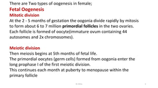
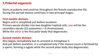
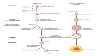
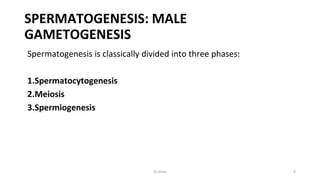
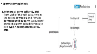
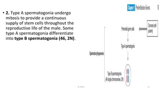
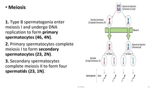
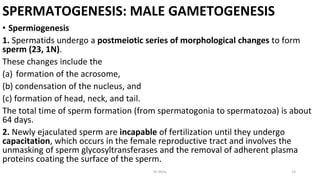
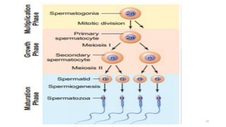
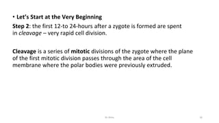
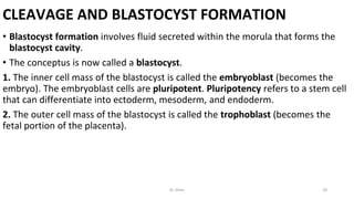
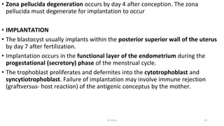
SlideShare a Scribd company logo
OOGENESIS &
SPERMOTOGENESS
EMBROGENESIS
Dr Hauwa Shitu B.
MBchB, Resident (Gen.Surg.)
OUTLINE
Definitions
Oogenesis
Spermatogenesis
Embryogenesis
Dr.Shitu 2
Definitions
• Oogenesis: the process of growth and maturation of the oogonia into ovum.
• Spermatogenesis: is the process by which the male gametes called spermatozoa
(sperms) are formed from the primitive spermatogenic cells (spermatogonia) in
the testis.
• Embryogenesis : the first eight weeks of development after fertilization, is an
incredibly complicated process.
Dr.Shitu 3
Oogenesis
Gametogenesis, in females, oogenesis process of growth and maturation of the
oogonia into ovum.
Begins with the ovarian stem (oogonia) migrating into the ovaries to multiply
during early embryonic development.
Most die prenatally, remaining begin meiosis toward end of gestation called
primary oocytes (still diploid).
Oogenesis is arrested at prophase I of 1st meiotic division during fetal growth
Number of primary oocytes in the ovaries declines from about 2 million in
infant, to approximately 400,000 at puberty, to zero by the end of menopause.
There are Two types of oogenesis in female;
Fetal Oogenesis
Mitotic division
At the 2 - 5 months of gestation the oogonia divide rapidly by mitosis
to form about 6 to 7 million primordial follicles in the two ovaries.
Each follicle is formed of oocyte(immature ovum containing 44
autosomes and 2x chromosomes).
Meiotic division
Then meiosis begins at 5th months of fetal life.
The primordial oocytes (germ cells) formed from oogonia enter the
long prophase I of the first meiotic division.
This continues each month at puberty to menopause within the
primary follicle
Dr.Shitu 5
2.Pubertal oogenesis
Starts at puberty and continue throughout the female reproductive life.
During this period meiosis continues in two principal stages:
First meiotic division:
Begins and is completed just before ovulation
Primary oocyte divides into two daughter haploid cells, one will be the
secondary oocyte (22 autosomes + X chromosome).
While the other is the first polar body that degenerate.
Second meiotic division:
Also begins immediately but its arrested in metaphase II.
And just before ovulation, it is completed only if the mature ovum is fertilized by
a sperm, forming a zygote while the second polar body also degenerates.
Dr.Shitu 6
Dr.Shitu 7
Note:
•In contrast to male who produces spermatogonia and primary spermatocytes
continuously throughout life.
•The female cannot form oogonia after birth, its formed before birth.
•Oogenesis in the female begins during intra-uterine life i.e. before birth (At the
5th month of gestation the ovaries contain about 6 to 7 million oogonia are
formed)
•While in the male spermatogenesis is stopped at the spermatogonia stage and
commences at puberty.
Dr.Shitu 8
SPERMATOGENESIS: MALE
GAMETOGENESIS
Spermatogenesis is classically divided into three phases:
1.Spermatocytogenesis
2.Meiosis
3.Spermiogenesis
Dr.Shitu 9
• Spermatocytogenesis
1.Primordial germ cells (46, 2N)
from wall of the yolk sac arrive in
the testes at week 6 and remain
dormant until puberty. At puberty,
primordial germ cells differentiate
into type A spermatogonia (46,
2N).
Dr.Shitu 10
• 2. Type A spermatogonia undergo
mitosis to provide a continuous
supply of stem cells throughout the
reproductive life of the male. Some
type A spermatogonia differentiate
into type B spermatogonia (46, 2N).
Dr.Shitu 11
• Meiosis
1. Type B spermatogonia enter
meiosis I and undergo DNA
replication to form primary
spermatocytes (46, 4N).
2. Primary spermatocytes complete
meiosis I to form secondary
spermatocytes (23, 2N).
3. Secondary spermatocytes
complete meiosis II to form four
spermatids (23, 1N).
Dr.Shitu 12
SPERMATOGENESIS: MALE GAMETOGENESIS
• Spermiogenesis
1. Spermatids undergo a postmeiotic series of morphological changes to form
sperm (23, 1N).
These changes include the
(a) formation of the acrosome,
(b) condensation of the nucleus, and
(c) formation of head, neck, and tail.
The total time of sperm formation (from spermatogonia to spermatozoa) is about
64 days.
2. Newly ejaculated sperm are incapable of fertilization until they undergo
capacitation, which occurs in the female reproductive tract and involves the
unmasking of sperm glycosyltransferases and the removal of adherent plasma
proteins coating the surface of the sperm.
Dr.Shitu 13
Dr.Shitu 14
EMBRYOGENESIS
• Embryogenesis, the first eight weeks of development after fertilization, is an
incredibly complicated process.
• Step 1: a zygote is the single cell formed when an egg and a sperm cell fuse;
the fusion is known as fertilization
Dr.Shitu 15
• Let’s Start at the Very Beginning
Step 2: the first 12-to 24-hours after a zygote is formed are spent
in cleavage – very rapid cell division.
Cleavage is a series of mitotic divisions of the zygote where the plane
of the first mitotic division passes through the area of the cell
membrane where the polar bodies were previously extruded.
Dr.Shitu 16
CLEAVAGE AND BLASTOCYST FORMATION
2. The process of cleavage eventually forms
a blastula consisting of cells called
blastomeres.
3. A cluster of blastomeres (16–32
blastomeres) forms a morula.
4. Blastomeres are totipotent up to the
eight-cell stage (i.e., each blastomere can
form a complete embryo by itself ).
Totipotency refers to a stem cell that can
differentiate into every cell within the
organism, including extraembryonic tissues.
Dr.Shitu 17
CLEAVAGE AND BLASTOCYST FORMATION
• Blastocyst formation involves fluid secreted within the morula that forms the
blastocyst cavity.
• The conceptus is now called a blastocyst.
1. The inner cell mass of the blastocyst is called the embryoblast (becomes the
embryo). The embryoblast cells are pluripotent. Pluripotency refers to a stem cell
that can differentiate into ectoderm, mesoderm, and endoderm.
2. The outer cell mass of the blastocyst is called the trophoblast (becomes the
fetal portion of the placenta).
Dr.Shitu 18
• Zona pellucida degeneration occurs by day 4 after conception. The zona
pellucida must degenerate for implantation to occur
• IMPLANTATION
• The blastocyst usually implants within the posterior superior wall of the uterus
by day 7 after fertilization.
• Implantation occurs in the functional layer of the endometrium during the
progestational (secretory) phase of the menstrual cycle.
• The trophoblast proliferates and defernites into the cytotrophoblast and
syncytiotrophoblast. Failure of implantation may involve immune rejection
(graftversus- host reaction) of the antigenic conceptus by the mother.
Dr.Shitu 19
Week 2 of Human Development (Days 8–14)
• FURTHER DEVELOPMENT OF THE
EMBRYOBLAST
• The embryoblast FORM epiblast layer
(columnar cells) and the ventral
hypoblast layer (cuboidal cells).
• Together form bilaminar embryonic disk.
• Epiblast will give us amniotic cavity and
Hypoblast will form exocoelomic
membrane,
• Syncytiotrophoblast the outer
multinucleated zone of the trophoblast
where no mitosis occurs (i.e., it arises
from the cytotrophoblast). And continues
its invasion of the endometrium, thereby
eroding endometrial blood vessels and
glands.
Dr.Shitu 20
• Lacunae forms within the
syncytiotrophoblast and become filled
with maternal blood and glandular
secretions.
• Cytotrophoblast. The cytotrophoblast
is mitotically active as new
cytotrophoblastic cells migrate into
the syncytiotrophoblast, thereby
fueling the growth of the
syncytiotrophoblast. In addition,
cytotrophoblastic cells also produce
local mounds called primary chorionic
villi that bulge into the surrounding
syncytiotrophoblast.
Dr.Shitu 21
Embryonic Period (Weeks 3–8)
• GENERAL CONSIDERATIONS
• By the end of the embryonic period, all major organ systems begin
development, although functionality may be minimal.
• During the embryonic period, the uteroplacental circulation cannot satisfy the
increasing nutritional needs of the rapidly developing embryo, so
development of the cardiovascular system is essential.
Dr.Shitu 22
• During the embryonic period, folding of the embryo occurs in two distinct
planes. Craniocaudal folding progresses due to the growth of the central
nervous system (CNS) and the amnion.
• Lateral folding progresses due to the growth of the somites, amnion, and other
components of the lateral body wall.
• Both the craniocaudal folding and lateral folding change the shape of the
embryo from a twodimensional disk to a three-dimensional cylinder.
Dr.Shitu 23
• Both the craniocaudal folding and lateral folding change the shape of the
embryo from a two dimensional disk to a three-dimensional cylinder.
• By the end of week 8, the embryo has a distinct human appearance.
• During the embryonic period, the basic segmentation of the human embryo
in the craniocaudal direction is controlled by the Hox (homeobox) complex
of genes.
Dr.Shitu 24
Gastrulation
• Gastrulation is the process that establishes
the three definitive germ layers of the
embryo (ectoderm, intraembryonic
mesoderm, and endoderm) forming a
trilaminar embryonic disk by day 21 of
development.
• These three germ layers give rise to all the
tissues and organs of the adult.
• Gastrulation is heralded by the formation
of the primitive streak and is caused by a
proliferation of epiblast cells. The primitive
streak consists of the primitive groove,
primitive node, and primitive pit.
Dr.Shitu 25
• The cloacal membrane, located caudal
to the primitive streak. is the future
site of the anus where the epiblast
and hypoblase cells fuse.
The ectoderm, intraembryonic
mesoderm, and endoderm of the
trilaminar embryonic disk are all derived
from the epiblast.
The term intraembryonic mesoderm
describes the germ layer that forms
during week 3 (gastrulation), in contrast
to the extraembryonic mesoderm,
which forms during week 2.
Dr.Shitu 26
• The caudal-most somites eventually
disappear to give a final count of
approximately 35 pairs of somites.
The number of somites is one of the
criteria for determining the age of the
embryo. Somites further differentiate
into the following components:
a. Sclerotome forms the cartilage and
bone components of the vertebral
column.
b. Myotome forms epimeric and
hypomeric muscles.
Dermatome forms dermis and
subcutaneous area of skin
Dr.Shitu 27
Changes involving intraembryonic mesoderm
• 1. Paraxial mesoderm is a thick plate of mesoderm located on each side of the
midline.
• Paraxial mesoderm becomes organized into segments known as somitomeres,
which form in a craniocaudal sequence.
• Somitomeres 1–7 do not form somites but contribute mesoderm to the
pharyngeal arches. The remaining somitomeres further condense in a
craniocaudal sequence to form 42–44 pairs of somites. The first pair of somites
forms on day 20, and new somites appear at a rate of 3 per day.
Dr.Shitu 28
2. Intermediate mesoderm is a longitudinal dorsal ridge of mesoderm located
between the paraxial mesoderm and lateral mesoderm. This ridge develops into
the urogenital ridge, which forms the future kidneys and gonads.
3. Lateral mesoderm is a thin plate of mesoderm located along the lateral sides of
the embryo.
Large spaces develop in the lateral mesoderm and coalesce to form the
intraembryonic coelom.
• The intraembryonic coelom divides the lateral mesoderm into two layers:
a. Intraembryonic somatic mesoderm (also called somatopleure)
b. Intraembryonic visceral mesoderm (also called visceropleure or
splanchnopleure)
Dr.Shitu 29
• Notochord is a solid cylinder of mesoderm extending in the midline of the
trilaminar embryonic disk from the primitive node to the prochordal plate. It has
a number of important functions,
• which include the following:
• a. The notochord induces the overlying ectoderm to differentiate into
neuroectoderm to form
• the neural plate.
• b. The notochord induces the formation of the vertebral body of each of the
vertebrae.
• c. The notochord forms the nucleus pulposus of each intervertebral disk.
Dr.Shitu 30
5. Cardiogenic region is a horseshoe-shaped region of mesoderm located at the
cranial end of the trilaminar embryonic disk rostral to the prochordal plate. This
region forms the future heart.
Dr.Shitu 31
Dr.Shitu 32
Dr.Shitu 33
CLINICAL CONSIDERATIONS
• Ectopic tubal pregnancy (ETP)
1. ETP occurs when the blastocyst implants within the uterine tube due to delayed
transport.
2. The ampulla of the uterine tube is the most common site of an ectopic
pregnancy. The rectouterine pouch (pouch of Douglas) is a common site for an
ectopic abdominal pregnancy.
3. ETP is most commonly seen in women with endometriosis or pelvic
inflammatory disease.
4. ETP leads to uterine tube rupture and hemorrhage if surgical intervention (i.e.,
salpingectomy) is not performed.
Dr.Shitu 34
CLINICAL CONSIDERATIONS
• Caudal dysplasia (sirenomelia) refers to a constellation of syndromes ranging
from minor lesions of lower vertebrae to complete fusion of the lower limbs.
• Caudal dysplasia is caused by abnormal gastrulation, in which the migration of
mesoderm is disturbed.
Dr.Shitu 35
• Human chorionic gonadotropin (hCG) is a glycoprotein produced by the
syncytiotrophoblast, which stimulates the production of progesterone by the
corpus luteum (i.e., maintains corpus luteum function).
• Essential for the maintenance of pregnancy until week 8 prior to placental
production
• hCG can be assayed in maternal blood at day 8 or urine at day 10.
• Low hCG values may predict a spontaneous abortion or indicate an ectopic
pregnancy.
• Elevated hCG values may indicate multiple pregnancy, hydatidiform mole, or
GTD
Dr.Shitu 36
REFERRENCE
• LANGMAN MEDICAL EMBRYOLOGY
• BOARD REVIEW SYSTEMIC EMBRYOLOGY
Dr.Shitu 37

More Related Content

Similar to 1.. DR.Shitu OOGENESISI-SPERM-EMBRY(1).pptx

Physiological changes during pregnancy
Physiological changes during pregnancyPhysiological changes during pregnancy
Physiological changes during pregnancy
Physioaadhar Physiotherapy Services
 
EMBRYOLOGY in Orthodontics and dentofacial orthopaedics
EMBRYOLOGY in Orthodontics and dentofacial orthopaedicsEMBRYOLOGY in Orthodontics and dentofacial orthopaedics
EMBRYOLOGY in Orthodontics and dentofacial orthopaedics
VaishnaviKasarpatil1
 
Basic Embryology(1).pdf
Basic Embryology(1).pdfBasic Embryology(1).pdf
Basic Embryology(1).pdf
YasminZeynu
 
Human embryonic development
Human embryonic developmentHuman embryonic development
Human embryonic developmentt7260678
 
EMBRYOLOGY AND FOETAL DEVELOPMENT-mayu.pdf
EMBRYOLOGY AND FOETAL DEVELOPMENT-mayu.pdfEMBRYOLOGY AND FOETAL DEVELOPMENT-mayu.pdf
EMBRYOLOGY AND FOETAL DEVELOPMENT-mayu.pdf
MayuriGamit2
 
Week 1 3
Week 1 3Week 1 3
Week 1 3kooff
 
02 Fertilization & Implantation.ppt
02 Fertilization & Implantation.ppt02 Fertilization & Implantation.ppt
02 Fertilization & Implantation.ppt
AugustRidlofRiwu
 
Conception and Placental Development
Conception and Placental DevelopmentConception and Placental Development
Conception and Placental Development
NoshirwanGazder
 
fertilization.pptx
fertilization.pptxfertilization.pptx
fertilization.pptx
Sujatha A
 
Embryonic and post-embryonic development in insects :its structure, functions...
Embryonic and post-embryonic development in insects :its structure, functions...Embryonic and post-embryonic development in insects :its structure, functions...
Embryonic and post-embryonic development in insects :its structure, functions...
N.m.c.a
 
Ovulation, fertilization, implantation (1 st week
Ovulation, fertilization, implantation (1 st weekOvulation, fertilization, implantation (1 st week
Ovulation, fertilization, implantation (1 st week
Marami Mustapa
 
4-EMBRYOLOGICAL_DEVELOPMENT_OF_BODY_TISSUES,_ORGANS_AND_SYSTEMS.[1].pptx
4-EMBRYOLOGICAL_DEVELOPMENT_OF_BODY_TISSUES,_ORGANS_AND_SYSTEMS.[1].pptx4-EMBRYOLOGICAL_DEVELOPMENT_OF_BODY_TISSUES,_ORGANS_AND_SYSTEMS.[1].pptx
4-EMBRYOLOGICAL_DEVELOPMENT_OF_BODY_TISSUES,_ORGANS_AND_SYSTEMS.[1].pptx
DunsonNampaso
 
1. Gametogenesis.pptx
1. Gametogenesis.pptx1. Gametogenesis.pptx
1. Gametogenesis.pptx
RabadanHassanMohamed
 
General embryology for PGMW 2014.hgfffypptx
General embryology for PGMW 2014.hgfffypptxGeneral embryology for PGMW 2014.hgfffypptx
General embryology for PGMW 2014.hgfffypptx
MulugetaAbeneh1
 
357 lectures (1-2) a
357 lectures (1-2) a357 lectures (1-2) a
357 lectures (1-2) a
GD_357
 
embryologymo-150405023631-conversion-gate01 (1).pptx
embryologymo-150405023631-conversion-gate01 (1).pptxembryologymo-150405023631-conversion-gate01 (1).pptx
embryologymo-150405023631-conversion-gate01 (1).pptx
Lokesh153390
 
L.12.L13 EMBRYOLOGY 2. FIRST 4 WEEKS.pdf
L.12.L13 EMBRYOLOGY 2. FIRST 4 WEEKS.pdfL.12.L13 EMBRYOLOGY 2. FIRST 4 WEEKS.pdf
L.12.L13 EMBRYOLOGY 2. FIRST 4 WEEKS.pdf
JuliusKauki1
 
EMBRYOLOGY.pptx
EMBRYOLOGY.pptxEMBRYOLOGY.pptx
EMBRYOLOGY.pptx
MaheenBadar3
 
Cleavage, implantation of the embryo and bilaminar
Cleavage, implantation of the embryo and bilaminarCleavage, implantation of the embryo and bilaminar
Cleavage, implantation of the embryo and bilaminar
Robinson Wafula
 
4. Oogenesis.pptxffffffwfvfbhbgg fggbrbg
4. Oogenesis.pptxffffffwfvfbhbgg fggbrbg4. Oogenesis.pptxffffffwfvfbhbgg fggbrbg
4. Oogenesis.pptxffffffwfvfbhbgg fggbrbg
rukkygodsgift
 

Similar to 1.. DR.Shitu OOGENESISI-SPERM-EMBRY(1).pptx (20)

Physiological changes during pregnancy
Physiological changes during pregnancyPhysiological changes during pregnancy
Physiological changes during pregnancy
 
EMBRYOLOGY in Orthodontics and dentofacial orthopaedics
EMBRYOLOGY in Orthodontics and dentofacial orthopaedicsEMBRYOLOGY in Orthodontics and dentofacial orthopaedics
EMBRYOLOGY in Orthodontics and dentofacial orthopaedics
 
Basic Embryology(1).pdf
Basic Embryology(1).pdfBasic Embryology(1).pdf
Basic Embryology(1).pdf
 
Human embryonic development
Human embryonic developmentHuman embryonic development
Human embryonic development
 
EMBRYOLOGY AND FOETAL DEVELOPMENT-mayu.pdf
EMBRYOLOGY AND FOETAL DEVELOPMENT-mayu.pdfEMBRYOLOGY AND FOETAL DEVELOPMENT-mayu.pdf
EMBRYOLOGY AND FOETAL DEVELOPMENT-mayu.pdf
 
Week 1 3
Week 1 3Week 1 3
Week 1 3
 
02 Fertilization & Implantation.ppt
02 Fertilization & Implantation.ppt02 Fertilization & Implantation.ppt
02 Fertilization & Implantation.ppt
 
Conception and Placental Development
Conception and Placental DevelopmentConception and Placental Development
Conception and Placental Development
 
fertilization.pptx
fertilization.pptxfertilization.pptx
fertilization.pptx
 
Embryonic and post-embryonic development in insects :its structure, functions...
Embryonic and post-embryonic development in insects :its structure, functions...Embryonic and post-embryonic development in insects :its structure, functions...
Embryonic and post-embryonic development in insects :its structure, functions...
 
Ovulation, fertilization, implantation (1 st week
Ovulation, fertilization, implantation (1 st weekOvulation, fertilization, implantation (1 st week
Ovulation, fertilization, implantation (1 st week
 
4-EMBRYOLOGICAL_DEVELOPMENT_OF_BODY_TISSUES,_ORGANS_AND_SYSTEMS.[1].pptx
4-EMBRYOLOGICAL_DEVELOPMENT_OF_BODY_TISSUES,_ORGANS_AND_SYSTEMS.[1].pptx4-EMBRYOLOGICAL_DEVELOPMENT_OF_BODY_TISSUES,_ORGANS_AND_SYSTEMS.[1].pptx
4-EMBRYOLOGICAL_DEVELOPMENT_OF_BODY_TISSUES,_ORGANS_AND_SYSTEMS.[1].pptx
 
1. Gametogenesis.pptx
1. Gametogenesis.pptx1. Gametogenesis.pptx
1. Gametogenesis.pptx
 
General embryology for PGMW 2014.hgfffypptx
General embryology for PGMW 2014.hgfffypptxGeneral embryology for PGMW 2014.hgfffypptx
General embryology for PGMW 2014.hgfffypptx
 
357 lectures (1-2) a
357 lectures (1-2) a357 lectures (1-2) a
357 lectures (1-2) a
 
embryologymo-150405023631-conversion-gate01 (1).pptx
embryologymo-150405023631-conversion-gate01 (1).pptxembryologymo-150405023631-conversion-gate01 (1).pptx
embryologymo-150405023631-conversion-gate01 (1).pptx
 
L.12.L13 EMBRYOLOGY 2. FIRST 4 WEEKS.pdf
L.12.L13 EMBRYOLOGY 2. FIRST 4 WEEKS.pdfL.12.L13 EMBRYOLOGY 2. FIRST 4 WEEKS.pdf
L.12.L13 EMBRYOLOGY 2. FIRST 4 WEEKS.pdf
 
EMBRYOLOGY.pptx
EMBRYOLOGY.pptxEMBRYOLOGY.pptx
EMBRYOLOGY.pptx
 
Cleavage, implantation of the embryo and bilaminar
Cleavage, implantation of the embryo and bilaminarCleavage, implantation of the embryo and bilaminar
Cleavage, implantation of the embryo and bilaminar
 
4. Oogenesis.pptxffffffwfvfbhbgg fggbrbg
4. Oogenesis.pptxffffffwfvfbhbgg fggbrbg4. Oogenesis.pptxffffffwfvfbhbgg fggbrbg
4. Oogenesis.pptxffffffwfvfbhbgg fggbrbg
 

Recently uploaded

MANAGEMENT OF ATRIOVENTRICULAR CONDUCTION BLOCK.pdf
MANAGEMENT OF ATRIOVENTRICULAR CONDUCTION BLOCK.pdfMANAGEMENT OF ATRIOVENTRICULAR CONDUCTION BLOCK.pdf
MANAGEMENT OF ATRIOVENTRICULAR CONDUCTION BLOCK.pdf
Jim Jacob Roy
 
ARTIFICIAL INTELLIGENCE IN HEALTHCARE.pdf
ARTIFICIAL INTELLIGENCE IN  HEALTHCARE.pdfARTIFICIAL INTELLIGENCE IN  HEALTHCARE.pdf
ARTIFICIAL INTELLIGENCE IN HEALTHCARE.pdf
Anujkumaranit
 
basicmodesofventilation2022-220313203758.pdf
basicmodesofventilation2022-220313203758.pdfbasicmodesofventilation2022-220313203758.pdf
basicmodesofventilation2022-220313203758.pdf
aljamhori teaching hospital
 
Novas diretrizes da OMS para os cuidados perinatais de mais qualidade
Novas diretrizes da OMS para os cuidados perinatais de mais qualidadeNovas diretrizes da OMS para os cuidados perinatais de mais qualidade
Novas diretrizes da OMS para os cuidados perinatais de mais qualidade
Prof. Marcus Renato de Carvalho
 
Superficial & Deep Fascia of the NECK.pptx
Superficial & Deep Fascia of the NECK.pptxSuperficial & Deep Fascia of the NECK.pptx
Superficial & Deep Fascia of the NECK.pptx
Dr. Rabia Inam Gandapore
 
Are There Any Natural Remedies To Treat Syphilis.pdf
Are There Any Natural Remedies To Treat Syphilis.pdfAre There Any Natural Remedies To Treat Syphilis.pdf
Are There Any Natural Remedies To Treat Syphilis.pdf
Little Cross Family Clinic
 
ANATOMY AND PHYSIOLOGY OF URINARY SYSTEM.pptx
ANATOMY AND PHYSIOLOGY OF URINARY SYSTEM.pptxANATOMY AND PHYSIOLOGY OF URINARY SYSTEM.pptx
ANATOMY AND PHYSIOLOGY OF URINARY SYSTEM.pptx
Swetaba Besh
 
Pharynx and Clinical Correlations BY Dr.Rabia Inam Gandapore.pptx
Pharynx and Clinical Correlations BY Dr.Rabia Inam Gandapore.pptxPharynx and Clinical Correlations BY Dr.Rabia Inam Gandapore.pptx
Pharynx and Clinical Correlations BY Dr.Rabia Inam Gandapore.pptx
Dr. Rabia Inam Gandapore
 
Dehradun #ℂall #gIRLS Oyo Hotel 9719300533 #ℂall #gIRL in Dehradun
Dehradun #ℂall #gIRLS Oyo Hotel 9719300533 #ℂall #gIRL in DehradunDehradun #ℂall #gIRLS Oyo Hotel 9719300533 #ℂall #gIRL in Dehradun
Dehradun #ℂall #gIRLS Oyo Hotel 9719300533 #ℂall #gIRL in Dehradun
chandankumarsmartiso
 
How STIs Influence the Development of Pelvic Inflammatory Disease.pptx
How STIs Influence the Development of Pelvic Inflammatory Disease.pptxHow STIs Influence the Development of Pelvic Inflammatory Disease.pptx
How STIs Influence the Development of Pelvic Inflammatory Disease.pptx
FFragrant
 
Report Back from SGO 2024: What’s the Latest in Cervical Cancer?
Report Back from SGO 2024: What’s the Latest in Cervical Cancer?Report Back from SGO 2024: What’s the Latest in Cervical Cancer?
Report Back from SGO 2024: What’s the Latest in Cervical Cancer?
bkling
 
Light House Retreats: Plant Medicine Retreat Europe
Light House Retreats: Plant Medicine Retreat EuropeLight House Retreats: Plant Medicine Retreat Europe
Light House Retreats: Plant Medicine Retreat Europe
Lighthouse Retreat
 
Phone Us ❤85270-49040❤ #ℂall #gIRLS In Surat By Surat @ℂall @Girls Hotel With...
Phone Us ❤85270-49040❤ #ℂall #gIRLS In Surat By Surat @ℂall @Girls Hotel With...Phone Us ❤85270-49040❤ #ℂall #gIRLS In Surat By Surat @ℂall @Girls Hotel With...
Phone Us ❤85270-49040❤ #ℂall #gIRLS In Surat By Surat @ℂall @Girls Hotel With...
Savita Shen $i11
 
How to Give Better Lectures: Some Tips for Doctors
How to Give Better Lectures: Some Tips for DoctorsHow to Give Better Lectures: Some Tips for Doctors
How to Give Better Lectures: Some Tips for Doctors
LanceCatedral
 
Flu Vaccine Alert in Bangalore Karnataka
Flu Vaccine Alert in Bangalore KarnatakaFlu Vaccine Alert in Bangalore Karnataka
Flu Vaccine Alert in Bangalore Karnataka
addon Scans
 
263778731218 Abortion Clinic /Pills In Harare ,
263778731218 Abortion Clinic /Pills In Harare ,263778731218 Abortion Clinic /Pills In Harare ,
263778731218 Abortion Clinic /Pills In Harare ,
sisternakatoto
 
Tom Selleck Health: A Comprehensive Look at the Iconic Actor’s Wellness Journey
Tom Selleck Health: A Comprehensive Look at the Iconic Actor’s Wellness JourneyTom Selleck Health: A Comprehensive Look at the Iconic Actor’s Wellness Journey
Tom Selleck Health: A Comprehensive Look at the Iconic Actor’s Wellness Journey
greendigital
 
heat stroke and heat exhaustion in children
heat stroke and heat exhaustion in childrenheat stroke and heat exhaustion in children
heat stroke and heat exhaustion in children
SumeraAhmad5
 
Sex determination from mandible pelvis and skull
Sex determination from mandible pelvis and skullSex determination from mandible pelvis and skull
Sex determination from mandible pelvis and skull
ShashankRoodkee
 
Lung Cancer: Artificial Intelligence, Synergetics, Complex System Analysis, S...
Lung Cancer: Artificial Intelligence, Synergetics, Complex System Analysis, S...Lung Cancer: Artificial Intelligence, Synergetics, Complex System Analysis, S...
Lung Cancer: Artificial Intelligence, Synergetics, Complex System Analysis, S...
Oleg Kshivets
 

Recently uploaded (20)

MANAGEMENT OF ATRIOVENTRICULAR CONDUCTION BLOCK.pdf
MANAGEMENT OF ATRIOVENTRICULAR CONDUCTION BLOCK.pdfMANAGEMENT OF ATRIOVENTRICULAR CONDUCTION BLOCK.pdf
MANAGEMENT OF ATRIOVENTRICULAR CONDUCTION BLOCK.pdf
 
ARTIFICIAL INTELLIGENCE IN HEALTHCARE.pdf
ARTIFICIAL INTELLIGENCE IN  HEALTHCARE.pdfARTIFICIAL INTELLIGENCE IN  HEALTHCARE.pdf
ARTIFICIAL INTELLIGENCE IN HEALTHCARE.pdf
 
basicmodesofventilation2022-220313203758.pdf
basicmodesofventilation2022-220313203758.pdfbasicmodesofventilation2022-220313203758.pdf
basicmodesofventilation2022-220313203758.pdf
 
Novas diretrizes da OMS para os cuidados perinatais de mais qualidade
Novas diretrizes da OMS para os cuidados perinatais de mais qualidadeNovas diretrizes da OMS para os cuidados perinatais de mais qualidade
Novas diretrizes da OMS para os cuidados perinatais de mais qualidade
 
Superficial & Deep Fascia of the NECK.pptx
Superficial & Deep Fascia of the NECK.pptxSuperficial & Deep Fascia of the NECK.pptx
Superficial & Deep Fascia of the NECK.pptx
 
Are There Any Natural Remedies To Treat Syphilis.pdf
Are There Any Natural Remedies To Treat Syphilis.pdfAre There Any Natural Remedies To Treat Syphilis.pdf
Are There Any Natural Remedies To Treat Syphilis.pdf
 
ANATOMY AND PHYSIOLOGY OF URINARY SYSTEM.pptx
ANATOMY AND PHYSIOLOGY OF URINARY SYSTEM.pptxANATOMY AND PHYSIOLOGY OF URINARY SYSTEM.pptx
ANATOMY AND PHYSIOLOGY OF URINARY SYSTEM.pptx
 
Pharynx and Clinical Correlations BY Dr.Rabia Inam Gandapore.pptx
Pharynx and Clinical Correlations BY Dr.Rabia Inam Gandapore.pptxPharynx and Clinical Correlations BY Dr.Rabia Inam Gandapore.pptx
Pharynx and Clinical Correlations BY Dr.Rabia Inam Gandapore.pptx
 
Dehradun #ℂall #gIRLS Oyo Hotel 9719300533 #ℂall #gIRL in Dehradun
Dehradun #ℂall #gIRLS Oyo Hotel 9719300533 #ℂall #gIRL in DehradunDehradun #ℂall #gIRLS Oyo Hotel 9719300533 #ℂall #gIRL in Dehradun
Dehradun #ℂall #gIRLS Oyo Hotel 9719300533 #ℂall #gIRL in Dehradun
 
How STIs Influence the Development of Pelvic Inflammatory Disease.pptx
How STIs Influence the Development of Pelvic Inflammatory Disease.pptxHow STIs Influence the Development of Pelvic Inflammatory Disease.pptx
How STIs Influence the Development of Pelvic Inflammatory Disease.pptx
 
Report Back from SGO 2024: What’s the Latest in Cervical Cancer?
Report Back from SGO 2024: What’s the Latest in Cervical Cancer?Report Back from SGO 2024: What’s the Latest in Cervical Cancer?
Report Back from SGO 2024: What’s the Latest in Cervical Cancer?
 
Light House Retreats: Plant Medicine Retreat Europe
Light House Retreats: Plant Medicine Retreat EuropeLight House Retreats: Plant Medicine Retreat Europe
Light House Retreats: Plant Medicine Retreat Europe
 
Phone Us ❤85270-49040❤ #ℂall #gIRLS In Surat By Surat @ℂall @Girls Hotel With...
Phone Us ❤85270-49040❤ #ℂall #gIRLS In Surat By Surat @ℂall @Girls Hotel With...Phone Us ❤85270-49040❤ #ℂall #gIRLS In Surat By Surat @ℂall @Girls Hotel With...
Phone Us ❤85270-49040❤ #ℂall #gIRLS In Surat By Surat @ℂall @Girls Hotel With...
 
How to Give Better Lectures: Some Tips for Doctors
How to Give Better Lectures: Some Tips for DoctorsHow to Give Better Lectures: Some Tips for Doctors
How to Give Better Lectures: Some Tips for Doctors
 
Flu Vaccine Alert in Bangalore Karnataka
Flu Vaccine Alert in Bangalore KarnatakaFlu Vaccine Alert in Bangalore Karnataka
Flu Vaccine Alert in Bangalore Karnataka
 
263778731218 Abortion Clinic /Pills In Harare ,
263778731218 Abortion Clinic /Pills In Harare ,263778731218 Abortion Clinic /Pills In Harare ,
263778731218 Abortion Clinic /Pills In Harare ,
 
Tom Selleck Health: A Comprehensive Look at the Iconic Actor’s Wellness Journey
Tom Selleck Health: A Comprehensive Look at the Iconic Actor’s Wellness JourneyTom Selleck Health: A Comprehensive Look at the Iconic Actor’s Wellness Journey
Tom Selleck Health: A Comprehensive Look at the Iconic Actor’s Wellness Journey
 
heat stroke and heat exhaustion in children
heat stroke and heat exhaustion in childrenheat stroke and heat exhaustion in children
heat stroke and heat exhaustion in children
 
Sex determination from mandible pelvis and skull
Sex determination from mandible pelvis and skullSex determination from mandible pelvis and skull
Sex determination from mandible pelvis and skull
 
Lung Cancer: Artificial Intelligence, Synergetics, Complex System Analysis, S...
Lung Cancer: Artificial Intelligence, Synergetics, Complex System Analysis, S...Lung Cancer: Artificial Intelligence, Synergetics, Complex System Analysis, S...
Lung Cancer: Artificial Intelligence, Synergetics, Complex System Analysis, S...
 

1.. DR.Shitu OOGENESISI-SPERM-EMBRY(1).pptx

  • 1. OOGENESIS & SPERMOTOGENESS EMBROGENESIS Dr Hauwa Shitu B. MBchB, Resident (Gen.Surg.)
  • 3. Definitions • Oogenesis: the process of growth and maturation of the oogonia into ovum. • Spermatogenesis: is the process by which the male gametes called spermatozoa (sperms) are formed from the primitive spermatogenic cells (spermatogonia) in the testis. • Embryogenesis : the first eight weeks of development after fertilization, is an incredibly complicated process. Dr.Shitu 3
  • 4. Oogenesis Gametogenesis, in females, oogenesis process of growth and maturation of the oogonia into ovum. Begins with the ovarian stem (oogonia) migrating into the ovaries to multiply during early embryonic development. Most die prenatally, remaining begin meiosis toward end of gestation called primary oocytes (still diploid). Oogenesis is arrested at prophase I of 1st meiotic division during fetal growth Number of primary oocytes in the ovaries declines from about 2 million in infant, to approximately 400,000 at puberty, to zero by the end of menopause.
  • 5. There are Two types of oogenesis in female; Fetal Oogenesis Mitotic division At the 2 - 5 months of gestation the oogonia divide rapidly by mitosis to form about 6 to 7 million primordial follicles in the two ovaries. Each follicle is formed of oocyte(immature ovum containing 44 autosomes and 2x chromosomes). Meiotic division Then meiosis begins at 5th months of fetal life. The primordial oocytes (germ cells) formed from oogonia enter the long prophase I of the first meiotic division. This continues each month at puberty to menopause within the primary follicle Dr.Shitu 5
  • 6. 2.Pubertal oogenesis Starts at puberty and continue throughout the female reproductive life. During this period meiosis continues in two principal stages: First meiotic division: Begins and is completed just before ovulation Primary oocyte divides into two daughter haploid cells, one will be the secondary oocyte (22 autosomes + X chromosome). While the other is the first polar body that degenerate. Second meiotic division: Also begins immediately but its arrested in metaphase II. And just before ovulation, it is completed only if the mature ovum is fertilized by a sperm, forming a zygote while the second polar body also degenerates. Dr.Shitu 6
  • 8. Note: •In contrast to male who produces spermatogonia and primary spermatocytes continuously throughout life. •The female cannot form oogonia after birth, its formed before birth. •Oogenesis in the female begins during intra-uterine life i.e. before birth (At the 5th month of gestation the ovaries contain about 6 to 7 million oogonia are formed) •While in the male spermatogenesis is stopped at the spermatogonia stage and commences at puberty. Dr.Shitu 8
  • 9. SPERMATOGENESIS: MALE GAMETOGENESIS Spermatogenesis is classically divided into three phases: 1.Spermatocytogenesis 2.Meiosis 3.Spermiogenesis Dr.Shitu 9
  • 10. • Spermatocytogenesis 1.Primordial germ cells (46, 2N) from wall of the yolk sac arrive in the testes at week 6 and remain dormant until puberty. At puberty, primordial germ cells differentiate into type A spermatogonia (46, 2N). Dr.Shitu 10
  • 11. • 2. Type A spermatogonia undergo mitosis to provide a continuous supply of stem cells throughout the reproductive life of the male. Some type A spermatogonia differentiate into type B spermatogonia (46, 2N). Dr.Shitu 11
  • 12. • Meiosis 1. Type B spermatogonia enter meiosis I and undergo DNA replication to form primary spermatocytes (46, 4N). 2. Primary spermatocytes complete meiosis I to form secondary spermatocytes (23, 2N). 3. Secondary spermatocytes complete meiosis II to form four spermatids (23, 1N). Dr.Shitu 12
  • 13. SPERMATOGENESIS: MALE GAMETOGENESIS • Spermiogenesis 1. Spermatids undergo a postmeiotic series of morphological changes to form sperm (23, 1N). These changes include the (a) formation of the acrosome, (b) condensation of the nucleus, and (c) formation of head, neck, and tail. The total time of sperm formation (from spermatogonia to spermatozoa) is about 64 days. 2. Newly ejaculated sperm are incapable of fertilization until they undergo capacitation, which occurs in the female reproductive tract and involves the unmasking of sperm glycosyltransferases and the removal of adherent plasma proteins coating the surface of the sperm. Dr.Shitu 13
  • 15. EMBRYOGENESIS • Embryogenesis, the first eight weeks of development after fertilization, is an incredibly complicated process. • Step 1: a zygote is the single cell formed when an egg and a sperm cell fuse; the fusion is known as fertilization Dr.Shitu 15
  • 16. • Let’s Start at the Very Beginning Step 2: the first 12-to 24-hours after a zygote is formed are spent in cleavage – very rapid cell division. Cleavage is a series of mitotic divisions of the zygote where the plane of the first mitotic division passes through the area of the cell membrane where the polar bodies were previously extruded. Dr.Shitu 16
  • 17. CLEAVAGE AND BLASTOCYST FORMATION 2. The process of cleavage eventually forms a blastula consisting of cells called blastomeres. 3. A cluster of blastomeres (16–32 blastomeres) forms a morula. 4. Blastomeres are totipotent up to the eight-cell stage (i.e., each blastomere can form a complete embryo by itself ). Totipotency refers to a stem cell that can differentiate into every cell within the organism, including extraembryonic tissues. Dr.Shitu 17
  • 18. CLEAVAGE AND BLASTOCYST FORMATION • Blastocyst formation involves fluid secreted within the morula that forms the blastocyst cavity. • The conceptus is now called a blastocyst. 1. The inner cell mass of the blastocyst is called the embryoblast (becomes the embryo). The embryoblast cells are pluripotent. Pluripotency refers to a stem cell that can differentiate into ectoderm, mesoderm, and endoderm. 2. The outer cell mass of the blastocyst is called the trophoblast (becomes the fetal portion of the placenta). Dr.Shitu 18
  • 19. • Zona pellucida degeneration occurs by day 4 after conception. The zona pellucida must degenerate for implantation to occur • IMPLANTATION • The blastocyst usually implants within the posterior superior wall of the uterus by day 7 after fertilization. • Implantation occurs in the functional layer of the endometrium during the progestational (secretory) phase of the menstrual cycle. • The trophoblast proliferates and defernites into the cytotrophoblast and syncytiotrophoblast. Failure of implantation may involve immune rejection (graftversus- host reaction) of the antigenic conceptus by the mother. Dr.Shitu 19
  • 20. Week 2 of Human Development (Days 8–14) • FURTHER DEVELOPMENT OF THE EMBRYOBLAST • The embryoblast FORM epiblast layer (columnar cells) and the ventral hypoblast layer (cuboidal cells). • Together form bilaminar embryonic disk. • Epiblast will give us amniotic cavity and Hypoblast will form exocoelomic membrane, • Syncytiotrophoblast the outer multinucleated zone of the trophoblast where no mitosis occurs (i.e., it arises from the cytotrophoblast). And continues its invasion of the endometrium, thereby eroding endometrial blood vessels and glands. Dr.Shitu 20
  • 21. • Lacunae forms within the syncytiotrophoblast and become filled with maternal blood and glandular secretions. • Cytotrophoblast. The cytotrophoblast is mitotically active as new cytotrophoblastic cells migrate into the syncytiotrophoblast, thereby fueling the growth of the syncytiotrophoblast. In addition, cytotrophoblastic cells also produce local mounds called primary chorionic villi that bulge into the surrounding syncytiotrophoblast. Dr.Shitu 21
  • 22. Embryonic Period (Weeks 3–8) • GENERAL CONSIDERATIONS • By the end of the embryonic period, all major organ systems begin development, although functionality may be minimal. • During the embryonic period, the uteroplacental circulation cannot satisfy the increasing nutritional needs of the rapidly developing embryo, so development of the cardiovascular system is essential. Dr.Shitu 22
  • 23. • During the embryonic period, folding of the embryo occurs in two distinct planes. Craniocaudal folding progresses due to the growth of the central nervous system (CNS) and the amnion. • Lateral folding progresses due to the growth of the somites, amnion, and other components of the lateral body wall. • Both the craniocaudal folding and lateral folding change the shape of the embryo from a twodimensional disk to a three-dimensional cylinder. Dr.Shitu 23
  • 24. • Both the craniocaudal folding and lateral folding change the shape of the embryo from a two dimensional disk to a three-dimensional cylinder. • By the end of week 8, the embryo has a distinct human appearance. • During the embryonic period, the basic segmentation of the human embryo in the craniocaudal direction is controlled by the Hox (homeobox) complex of genes. Dr.Shitu 24
  • 25. Gastrulation • Gastrulation is the process that establishes the three definitive germ layers of the embryo (ectoderm, intraembryonic mesoderm, and endoderm) forming a trilaminar embryonic disk by day 21 of development. • These three germ layers give rise to all the tissues and organs of the adult. • Gastrulation is heralded by the formation of the primitive streak and is caused by a proliferation of epiblast cells. The primitive streak consists of the primitive groove, primitive node, and primitive pit. Dr.Shitu 25
  • 26. • The cloacal membrane, located caudal to the primitive streak. is the future site of the anus where the epiblast and hypoblase cells fuse. The ectoderm, intraembryonic mesoderm, and endoderm of the trilaminar embryonic disk are all derived from the epiblast. The term intraembryonic mesoderm describes the germ layer that forms during week 3 (gastrulation), in contrast to the extraembryonic mesoderm, which forms during week 2. Dr.Shitu 26
  • 27. • The caudal-most somites eventually disappear to give a final count of approximately 35 pairs of somites. The number of somites is one of the criteria for determining the age of the embryo. Somites further differentiate into the following components: a. Sclerotome forms the cartilage and bone components of the vertebral column. b. Myotome forms epimeric and hypomeric muscles. Dermatome forms dermis and subcutaneous area of skin Dr.Shitu 27
  • 28. Changes involving intraembryonic mesoderm • 1. Paraxial mesoderm is a thick plate of mesoderm located on each side of the midline. • Paraxial mesoderm becomes organized into segments known as somitomeres, which form in a craniocaudal sequence. • Somitomeres 1–7 do not form somites but contribute mesoderm to the pharyngeal arches. The remaining somitomeres further condense in a craniocaudal sequence to form 42–44 pairs of somites. The first pair of somites forms on day 20, and new somites appear at a rate of 3 per day. Dr.Shitu 28
  • 29. 2. Intermediate mesoderm is a longitudinal dorsal ridge of mesoderm located between the paraxial mesoderm and lateral mesoderm. This ridge develops into the urogenital ridge, which forms the future kidneys and gonads. 3. Lateral mesoderm is a thin plate of mesoderm located along the lateral sides of the embryo. Large spaces develop in the lateral mesoderm and coalesce to form the intraembryonic coelom. • The intraembryonic coelom divides the lateral mesoderm into two layers: a. Intraembryonic somatic mesoderm (also called somatopleure) b. Intraembryonic visceral mesoderm (also called visceropleure or splanchnopleure) Dr.Shitu 29
  • 30. • Notochord is a solid cylinder of mesoderm extending in the midline of the trilaminar embryonic disk from the primitive node to the prochordal plate. It has a number of important functions, • which include the following: • a. The notochord induces the overlying ectoderm to differentiate into neuroectoderm to form • the neural plate. • b. The notochord induces the formation of the vertebral body of each of the vertebrae. • c. The notochord forms the nucleus pulposus of each intervertebral disk. Dr.Shitu 30
  • 31. 5. Cardiogenic region is a horseshoe-shaped region of mesoderm located at the cranial end of the trilaminar embryonic disk rostral to the prochordal plate. This region forms the future heart. Dr.Shitu 31
  • 34. CLINICAL CONSIDERATIONS • Ectopic tubal pregnancy (ETP) 1. ETP occurs when the blastocyst implants within the uterine tube due to delayed transport. 2. The ampulla of the uterine tube is the most common site of an ectopic pregnancy. The rectouterine pouch (pouch of Douglas) is a common site for an ectopic abdominal pregnancy. 3. ETP is most commonly seen in women with endometriosis or pelvic inflammatory disease. 4. ETP leads to uterine tube rupture and hemorrhage if surgical intervention (i.e., salpingectomy) is not performed. Dr.Shitu 34
  • 35. CLINICAL CONSIDERATIONS • Caudal dysplasia (sirenomelia) refers to a constellation of syndromes ranging from minor lesions of lower vertebrae to complete fusion of the lower limbs. • Caudal dysplasia is caused by abnormal gastrulation, in which the migration of mesoderm is disturbed. Dr.Shitu 35
  • 36. • Human chorionic gonadotropin (hCG) is a glycoprotein produced by the syncytiotrophoblast, which stimulates the production of progesterone by the corpus luteum (i.e., maintains corpus luteum function). • Essential for the maintenance of pregnancy until week 8 prior to placental production • hCG can be assayed in maternal blood at day 8 or urine at day 10. • Low hCG values may predict a spontaneous abortion or indicate an ectopic pregnancy. • Elevated hCG values may indicate multiple pregnancy, hydatidiform mole, or GTD Dr.Shitu 36
  • 37. REFERRENCE • LANGMAN MEDICAL EMBRYOLOGY • BOARD REVIEW SYSTEMIC EMBRYOLOGY Dr.Shitu 37